Keywords
Ganglioneuroblastoma, Embryonal tumor, Juvenile brain tumor
Ganglioneuroblastoma, Embryonal tumor, Juvenile brain tumor
Ganglioneuroblastoma is a tumor of peripheral neuroblastic tissue that arises from the neural ectodermal cells. Ganglioneuroblastoma is a variant of neuroblastic tumor, which encompasses neuroblastoma, ganglioneuroblastoma and ganglioneuroma. The most commonly seen is the neuroblastoma, which contributes 97% of the neuroblastic tumors. Among pediatric cancers, the neuroblastoma is the third most common cancer after brain cancer and leukemias1. Among neuroblastic tumors, neuroblastoma is the least differentiated, ganglioneuroma is the most differentiated and ganglioneuroblastoma is the intermediate between them. Neuroblastoma histology includes primitive neuroblasts, ganglioneuroblastoma includes both primitive neuroblasts and ganglion cells and ganglioneuromas include mature Schwann cells and ganglion cells as the most differentiated variant. Neuroblastic tumors are seen in the adrenal gland, para spinal neural tissue, chest, neck, spine, intra-abdominal areas and the presentation of these tumors is highly dependent on their location and metastasis. Ganglioneuroblastomas are rarely seen in the spinal column2.
The authors present here the first case of primary intraspinal ganglioneuroblastoma of the thoracic and lumbar spine in Pakistan. A histopathological study helped the authors to establish the diagnosis. Surgical excision is the treatment of choice for both cerebral and spinal ganglion cell tumors. Radiotherapy is given for residual disease, recurrence, or progression. We report a 2.5-year-old boy who achieved improvement after surgical excision.
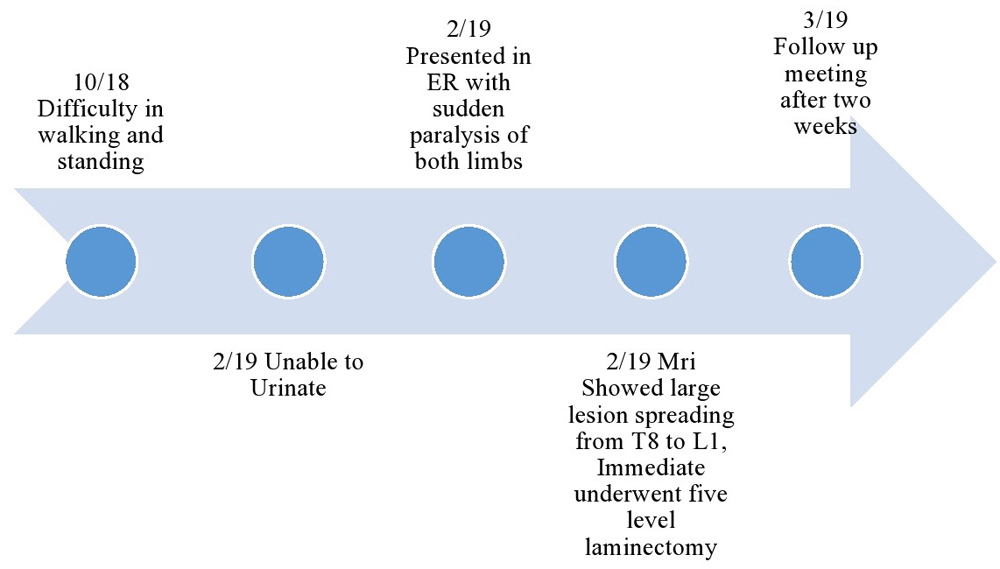
A 2.5-year-old South Asian boy resident of Karachi presented to the emergency room with an inability to urinate for the last seven days. This was followed by sudden paralysis of both lower limbs of two days’ duration., resulting in the patient being completely unable to sit, stand or walk. The patient had a history of difficulty in walking and standing for the prior six months.
A neurological examination revealed sensory loss below the T12 vertebra and a bilateral increase in muscle tone. Using the Medical Research Council scale for muscle power, power was 0/5 on the left side and 1/5 on the right side upon flexion and extension. Babinski reflexes were normal on both sides, indicating an upper motor lesion. The child had no past history of birth asphyxia, hypertension, diabetes mellitus or tuberculosis and their vaccination history was normal, with vaccines administered at routine time points. The child had no past history of trauma or other constitutional symptoms. The child was afebrile. An MRI scan was carried out, which showed a large homogenous enhancing lesion spreading from the T8 to L1 vertebrae. The intra-spinal region was completely adherent to the dural coverings of the dorsal cord and dural sleeves of nerve roots. Moreover, the abdominal region of cord covered the retroperitoneal structures, including the renal fascia, kidneys, bowels and para-spinal area, extending up to the aorta and adjacent vascular structures.
The surgical plan included a five level laminectomy, which was then performed immediately, and the whole segment was reconstructed after complete resection of the spinal lesion. The lesion was badly adherent to the cord roots. Two cord roots were compromised and the cerebrospinal fluid leak was fixed. Operative findings were of a soft elastic, moderately vascularized, exophytic tumor, extending into the extradural region of the spine. The tumor had extradural growth and encircled the tissue around the spinal cord, extending into the retroperitoneal area as well. The biopsy sample was sent for histopathology reporting, which revealed a tumor consisting primarily of nodules and sheets of mature looking ganglion cells with abundant Schwannian stroma with diffusely present round blue cells, hyperchromatic nuclei and scanty cytoplasm. These cells showed increased mitotic activity as well. Hematoxylin and eosin staining showed the tumor to be neurofilament and synaptophysin positive. According to the International Neuroblastoma Staging System, the patient had a stage 4 tumor as metastasis to other organs was found. In conclusion, the histopathology report confirmed the diagnosis of an intermixed ganglioneuroblastoma. and the patient was assigned to intensive modulation radiation therapy with a dose of 18 grays for six months.
At the follow-up appointment two weeks later, a physical examination was carried out and power was found to be improved on the right side, measuring 2/5, and on the left side, measuring 1/5. The patient returned two months later for a follow-up appointment, when a detailed neuro-physical examination was carried out and power was increased, measuring 3/5 on the left side and 2/5 on the right side. A timeline of events from initial symptoms to post-surgery follow up meeting is shown in Figure 1.
ER, emergency room; MRI, magnetic resonance imaging.
Ganglioneuroblastomas are a rare type of neuroblastic tumor that are composed of a varying proportion of undifferentiated neuroblast and mature ganglionic cells3. Ganglioneuroblastoma is a pediatric tumor and is uncommon in the adult population. The histology of ganglioneuroblastomas involves tumor cells derived from the mantle layer of the embryological spinal cord that populate the embryological sympathetic ganglia and adrenal medulla. Ganglioneuroblastoma histology includes undifferentiated small round cells in all stages of neuronal differentiation2,4. These embryological neural crest cells may mature to become Schwann cells or remain undifferentiated, called neuroblasts5,6.
Ganglioneuroblastomas are subdivided into two categories (nodular and intermixed) and both have significance in terms of prognosis, along with age of disease presentation, disease pathogenesis, genetics, nuclear maturation and grading. Ganglioneuroblastomas have both ganglionic and neuroblast cells, which both have the potential to become malignant7. In the nodular type of ganglioneuroblastoma, there is ganglioneuromatous stroma around nodules of neuroblasts and in the intermixed type, there are small numbers of neuroblasts with ganglioneuromatous stroma.
Symptoms of ganglioneuroblastomas are highly dependent on the primary location of the tumor and metastasis. Symptoms include abdominal distension, weight loss, irritability, focal neurological deficits and opsoclonus-myoclonus. The most common presentation of ganglioneuroblastoma is abdominal distension. Survival rates in infancy are almost 91% and decline as the age of presentation increases. Ganglioneuroblastomas show no gender predominance and are seen equally among males and females. Ganglioneuroblastomas predominantly present in the first decade of life7 and are very uncommon among adults, with most affected patients being under five years of age8. Genetics plays an important role in the pathogenesis of neuroblastic tumors, although sporadic cases are also common.
Ganglioneuroblastomas predominantly involve the adrenal gland, although other sites such as retroperitoneal ganglion, posterior mediastinum and pelvis are also involved. Ganglioneuroblastomas are likely to be seen among the sympathetic ganglia and therefore often occur in the paramedian region in the neck, pelvis and head9. Ganglioneuroblastoma arising in the lower thoracic spine is one of the rare presentations of this tumor2,10,11. The least common sites for ganglioneuroblastomas to occur are the thymus, cauda equina, lung, spinal column and kidney. The spinal column is a rare site for ganglioneuroblastomas to occur and it is quite possible that the tumor extended intraspinally from the mediastinum. In the spinal column, ganglioneuroblastomas are seen as extradural lesions with a dumbbell shape.
Ganglioneuroblastomas are associated with various diseases including Von Recklinghausen disease, Hirsch sprung disease, DiGeorge syndrome and Beckwith-Weidman syndrome7,12. The most common site of ganglioneuroblastoma metastasis is the bone, which is called Hutchinson’s disease and presents as limping and unexplained irritability. Other metastatic locations include the liver and skin8. Definitive treatment involves resection of the tumor, and radiotherapy is also included in cases of high-grade tumors.
Ganglioneuroblastoma is a neuroblastic tumor that is mostly seen in the pediatric population. The lower thoracic spine is a rare location for a tumor and it is very important to include ganglioneuroblastoma in the differential diagnosis when investigating lower thoracic spine masses. With proper investigative approaches, ganglioneuroblastoma can be diagnosed early and can be managed accordingly, decreasing morbidity and mortality in patients.
All data underlying the results are available as part of the article and no additional source data are required.
Written informed consent for publication of their clinical details and clinical images was obtained from the patient.
| Views | Downloads | |
|---|---|---|
| F1000Research | - | - |
|
PubMed Central
Data from PMC are received and updated monthly.
|
- | - |
Provide sufficient details of any financial or non-financial competing interests to enable users to assess whether your comments might lead a reasonable person to question your impartiality. Consider the following examples, but note that this is not an exhaustive list:
Sign up for content alerts and receive a weekly or monthly email with all newly published articles
Already registered? Sign in
The email address should be the one you originally registered with F1000.
You registered with F1000 via Google, so we cannot reset your password.
To sign in, please click here.
If you still need help with your Google account password, please click here.
You registered with F1000 via Facebook, so we cannot reset your password.
To sign in, please click here.
If you still need help with your Facebook account password, please click here.
If your email address is registered with us, we will email you instructions to reset your password.
If you think you should have received this email but it has not arrived, please check your spam filters and/or contact for further assistance.
Comments on this article Comments (0)