Keywords
Computed tomography, cardiac imaging, Tetralogy of Fallot, Pulmonary atresia, Blalock Taussig shunt.
Computed tomography, cardiac imaging, Tetralogy of Fallot, Pulmonary atresia, Blalock Taussig shunt.
Tetralogy of Fallot (TOF) is the most frequent form of cyanotic congenital heart disease. Only a few patients come of age without surgical intervention mainly in the extreme form with pulmonary atresia1. Here, we relate the case of a patient who survived until the age of 40 years without surgical curative intervention. The patient had an unusual association of chronic Blalock-Taussig shunt (BTS) thrombosis and huge major aortopulmonary collateral arteries (MAPCAs). This case provides the main role that a cardiac CT scan can lay in understanding the late outcome of these untreated cardiac abnormalities.
A 40-year-old man with “complex” cardiac disease and dyspnea was referred to our department for imaging. Old medical records with conventional angiographic studies inferred the diagnosis of TOF with pulmonary atresia, made at birth, treated by BTS. As the patient was asymptomatic, he had not consulted previously, and had been lost to follow-up. Two years ago, he began to present dyspnea on exertion, and also short cyanosis spells.
Clinical findings on presentation were unspecific except for sinus tachycardia. ECG showed biventricular hypertrophy and incomplete right bundle branch block. Echocardiography confirms the diagnosis of TOF with pulmonary atresia. To reassess cardiac abnormalities, primarily pulmonary trunk morphology and associated intrathoracic malformations, a 128-slice CT scan with ECG synchronization was performed. The scan revealed a large ventricle septal defect with overriding aorta (Figure 1, curved arrow) and right ventricle hypertrophy (Figure 1, asterisks). The pulmonary trunk was atretic, showing characteristic seagull pattern (Figure 2, arrows). The right pulmonary artery had a good diameter, while the left was significantly smaller (Figure 2). Pulmonary blood flow was given by MAPCA connecting blood vessels between the aorta and the pulmonary arteries (Figure 3, arrows). MAPCA was more numerous on the right side supplying the largest pulmonary artery. The BTS was completely thrombosed with a total heterogeneous filling defect and parietal calcifications (Figure 4, arrows); this is compatible with chronic thrombosis.
The mechanism for prolonged survival, despite the old shunt thrombosis, was the expansion of several and huge MAPCA allowing sufficient pulmonary blood flow. Two months after his hospitalization, the patient becom asymptomatic, receiving life-long conservative therapy including anticoagulation with a vitamin K antagonist (acenocoumarol 4 mg once a day) and a low dose of diuretic (Furosemide 40 mg once a day).

Curved arrow shows the ventricular septal defect; arrows show the overriding Ao; and asterisks show the RV hypertrophy. LA, left atrium; RA, right atrium; LV, left ventricle; RV, right ventricle; Ao, aorta.
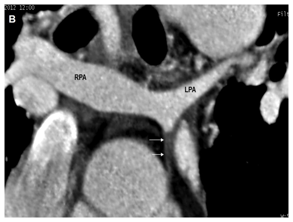
Arrows shows the atretic pulmonary trunk. LPA, left pulmonary artery; RPA, right pulmonary artery.

Arrows show major aortopulmonary collateral arteries.
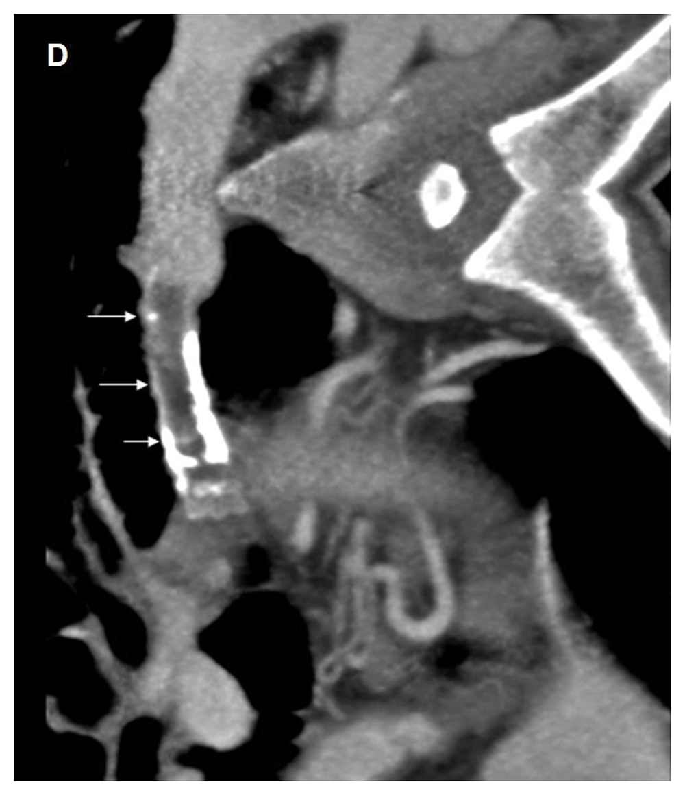
Arrows show a total heterogeneous filling defect of the tube with parietal calcifications.
TOF is the most frequent cyanotic congenital heart disease. It includes four characteristic anatomical findings: sub-arterial ventricular septal defect, aorta straddling of the ventricles, a variable obstruction through the pulmonary outflow tract, and right ventricle hypertrophy2. The spectrum of severity within this lesion depends upon the anatomy of the pulmonary arteries. The different forms of TOF include TOF with a patent foramen oval/atrial septal defect, TOF with absent pulmonary valve, and TOF with pulmonary atresia, which is the extreme form characterized by absence of flow from the right ventricle to the pulmonary arteries1. In the latter event, pulmonary vascularization is derived from patent ductus arteriosus and aortopulmonary collaterals with significant variability from one patient to another2.
MAPCAs are arteries that grow to irrigate lung circulation when native pulmonary arteries are underdeveloped. MAPCAs often arise from the descending aorta but also from the aortic arch and other systemic arteries like subclavian, the carotid, or rarely the coronary arteries. Pulmonary atresia-ventricular septal defects are classified into 3 types3: type A is associated with presence of the native pulmonary arteries with a pulmonary-aortic duct; type B has MAPCAs and native pulmonary arteries, such as in our case; type C has only MAPCAs, which are seen to provide the pulmonary blood without native pulmonary arteries.
Adulthood clinical presentation, as in our case, is extremely rare. Without surgical intervention, most patients die at a young age with a rate of survival of 66% at one year of age, 11% at 20 years, and 3% at 40 years. In contrast, the survival rate after surgical treatment is over 90% at 40 years old2. The surgical option includes palliative forms, such as BTS, which consists in the creation of systemic to pulmonary shunt. Complete repair is the main option and associated with excellent outcome results4. In this case, despite a ductus arteriosus closure and a chronic pediatric shunt thrombosis, the patient survived at the age of 40 due to large and developed MAPCAs. The oldest survivor ever reported in the literature is 59 years old5.
After a cardiac ultrasound, multidetector angiography CT scan is indicated to the assessment of TOF with pulmonary atresia mainly for associated intrathoracic malformations and to establish the precise MAPCA cartography before curative surgery6,7. Reformatted images allow a comprehensive analysis of pulmonary artery anatomy, measurements of the lumen of ascending aorta, analysis of the origins and course of coronary arteries, study location, the size, and flow in prior shunts8. The excellent spatial resolution and fast acquisition of multidetector CT make it a prime imaging tool to demonstrate even distal aortopulmonary collateral anatomy. Radiation should be reduced as low as possible in the pediatric population.
Untreated pulmonary atresia with a ventricular septal defect is uncommon in adults. Most patients die from serious respiratory troubles or congestive cardiac failure very early. An angiography CT scan is the main imaging tool to delineate the pulmonary arterial supply, which is essential for an appropriate surgical approach.
Written informed consent for publication of their clinical details and/or clinical images was obtained from the patient.
All data underlying the results are available as part of the article and no additional source data are required.
| Views | Downloads | |
|---|---|---|
| F1000Research | - | - |
|
PubMed Central
Data from PMC are received and updated monthly.
|
- | - |
Provide sufficient details of any financial or non-financial competing interests to enable users to assess whether your comments might lead a reasonable person to question your impartiality. Consider the following examples, but note that this is not an exhaustive list:
Sign up for content alerts and receive a weekly or monthly email with all newly published articles
Already registered? Sign in
The email address should be the one you originally registered with F1000.
You registered with F1000 via Google, so we cannot reset your password.
To sign in, please click here.
If you still need help with your Google account password, please click here.
You registered with F1000 via Facebook, so we cannot reset your password.
To sign in, please click here.
If you still need help with your Facebook account password, please click here.
If your email address is registered with us, we will email you instructions to reset your password.
If you think you should have received this email but it has not arrived, please check your spam filters and/or contact for further assistance.
Comments on this article Comments (0)