Keywords
pleura, lymphoma, pleural effusion, malignant, neoplasm, remission, case report
pleura, lymphoma, pleural effusion, malignant, neoplasm, remission, case report
Pleural effusions are frequently encountered within the respiratory outpatient setting. There are a wide range of causes and potential interventions to be offered in order to diagnose and treat effusions. Pleural effusions rarely spontaneously resolve, and here we report an interesting case where this rare phenomenon occurred.
A 95-year old Caucasian female was seen in July 2018 in a community respiratory clinic with a 4-month history of increasing breathlessness and a widespread vasculitic-looking rash. She had a background of high blood pressure and stable non-vasculitic chronic kidney disease. She lived alone and was fully independent with all activities of daily living.
The clinical features on examination were in keeping with a left sided pleural effusion.
The patient was followed over the course of 6 months in 2018.
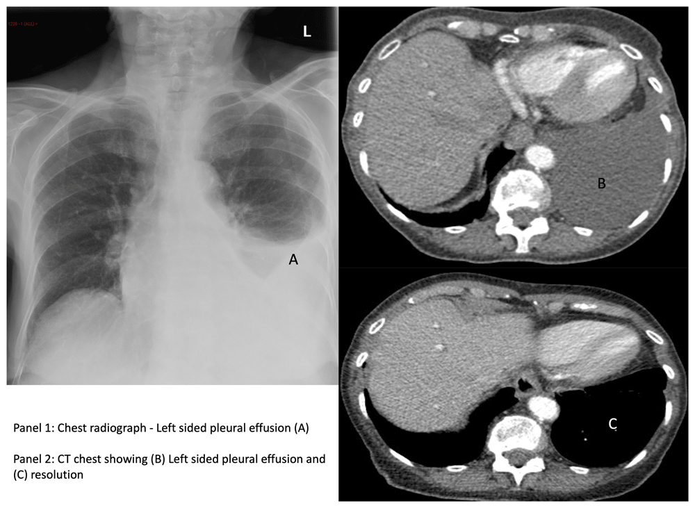
She had a chest X-ray (Figure 1a) which showed a moderate left sided effusion, and subsequent computed tomography (CT) (Figure 1b) confirmed the presence of an effusion, along with mediastinal lymphadenopathy. An echocardiogram showed normal left ventricular function with moderate mitral stenosis and mild mitral regurgitation. A positron emission tomography (PET) CT scan did not suggest any size significant or fluorodeoxyglucose (FDG) avid hilar or mediastinal lymphadenopathy.

Clinical imaging of the patient (A) Chest radiograph showing left sided pleural effusion (B) computed tomography (CT) chest showing left sided pleural effusion (C) CT chest showing resolution of left sided pleural effusion.
Pleural aspiration was performed with 700 millilitres of yellow serous fluid obtained (exudative, protein - 47g/l, lactate dehydrogenase (LDH) - 3072U/l, negative microbiology culture). Other investigations demonstrated a negative vasculitis screen, serum LDH of 392U/l (<250U/l), and a small paraprotein was detected on immunofixation.
Fluid cytology revealed a diffuse population of intermediate to large sized lymphoid cells containing moderate amphophilic cytoplasm and round nucleus with fine chromatin and conspicuous nucleoli, some of them with plasmacytoid appearance. The lesional cells were of lymphoid B-cell lineage as evidenced by strong, diffuse expression of CD45, CD20 and PAX-5. A proportion of these cells co-expressed BCL-6 (90% of cells), BCL-2 (90%) and MUM1 (>80%), but not CD10, CD5, CD56, CD138, TdT, CD30, CD38, Cyclin-D1 or CD23. There was c-myc expression in approximately 80%. The proliferation fraction estimated by Ki-67 marker was high at approximately 80%. TTF-1 and MNF116 were negative. EBER (Epstein Barr virus-encoded small RNA) in situ hybridization for Epstein-Barr virus (EBV) was negative. The appearances were consistent with a high-grade, non-Hodgkin B-cell lymphoma, in keeping with a diffuse large B-cell lymphoma, not otherwise specified (DLBCL NOS). The immunohistochemical appearances were those of a non-germinal centre B-cell-like (non-GCB) subgroup.
While awaiting treatment planning, repeat CT showed improvement in the effusion with resolution 6 months later; without any intervention (Figure 1c). Two years on, the patient remains symptom free, and in apparent remission.
Primary effusion lymphoma (PEL) is a rare subtype of diffuse large B-cell lymphoma (DLBCL) and is universally associated with human herpes virus-8 (HHV-8) that involves body cavities and causes serous effusions without detectable masses or lymphadenopathy1–4. It also occurs in immunocompromised patients infected with human immunodeficiency virus (HIV) and Epstein-Barr virus (EBV)2,4,5. On the other hand, many cases of DLBCL with lymphomatous effusions on serosal surfaces, and no detectable mass lesion like PEL, have been reported. These cases were not regarded as cases of PEL, but proposed to represent a new entity, ‘PEL-like lymphoma (PEL-LL)’6. A single-centre evaluation of 185 consecutive patients with DLBCL presenting with pleural effusions confer an independent predictor of poor survival in Cox regression modelling (hazard ratio 1.9)7.
The frequency of spontaneous regression of cancer has been estimated to be about 1 case per 100,000 patients, and approximately 20 cases are reported each year8. Hypernephroma, melanoma, neuroblastoma, leukaemia, and non-Hodgkin lymphoma are the most commonly reported cancers exhibiting spontaneous regression.
In the case we present, there was spontaneous regression of the effusion, however, the aetiology underlying the development of spontaneous remission of malignancy remains unclear. Proposed mechanisms to explain this phenomenon include immunological, hormonal and genetic factors; concomitant infections; elimination of carcinogens; surgical trauma of the primary tumour and induction of differentiation9,10.
In this case, there were no obvious precipitating factors such as infections. The apparent absence of EBER expression argues against a lymphoma associated with immune senescence. There was no evidence of acquired immunodeficiency, or prior therapeutic interventions. The Ki-67 protein is a cellular marker for proliferation; in this case it was 80%, suggesting the presence of a rapidly proliferative neoplasm11. Taken into consideration previous reports associating primary cavitary DLBCL with a poor prognosis, it is noteworthy that our patient has remained in remission 2 years following her initial diagnosis, without therapeutic intervention. To our knowledge, this is the first case to describe spontaneous remission in a primary cavitary DLBCL complicated by pleural effusion.
In the case of unexplained pleural effusion, suspected to have an underlying neoplastic aetiology, clinicians should consider the possibility of haematological malignancy in the differential diagnoses, and investigate accordingly. They should also be aware that rarely, these have the potential to spontaneously resolve.
Written informed consent for publication of their clinical details and clinical images was obtained from the patient.
| Views | Downloads | |
|---|---|---|
| F1000Research | - | - |
|
PubMed Central
Data from PMC are received and updated monthly.
|
- | - |
Is the background of the case’s history and progression described in sufficient detail?
Partly
Are enough details provided of any physical examination and diagnostic tests, treatment given and outcomes?
Partly
Is sufficient discussion included of the importance of the findings and their relevance to future understanding of disease processes, diagnosis or treatment?
No
Is the case presented with sufficient detail to be useful for other practitioners?
Partly
References
1. Moy MP, Levsky JM, Berko NS, Godelman A, et al.: A new, simple method for estimating pleural effusion size on CT scans.Chest. 2013; 143 (4): 1054-1059 PubMed Abstract | Publisher Full TextCompeting Interests: No competing interests were disclosed.
Reviewer Expertise: Medical Oncology - Thoracic Oncology
Peer review at F1000Research is author-driven. Currently no reviewers are being invited.
Alongside their report, reviewers assign a status to the article:
| Invited Reviewers | |
|---|---|
| 1 | |
|
Version 1 02 Jul 20 |
read |
Provide sufficient details of any financial or non-financial competing interests to enable users to assess whether your comments might lead a reasonable person to question your impartiality. Consider the following examples, but note that this is not an exhaustive list:
Sign up for content alerts and receive a weekly or monthly email with all newly published articles
Already registered? Sign in
The email address should be the one you originally registered with F1000.
You registered with F1000 via Google, so we cannot reset your password.
To sign in, please click here.
If you still need help with your Google account password, please click here.
You registered with F1000 via Facebook, so we cannot reset your password.
To sign in, please click here.
If you still need help with your Facebook account password, please click here.
If your email address is registered with us, we will email you instructions to reset your password.
If you think you should have received this email but it has not arrived, please check your spam filters and/or contact for further assistance.
Comments on this article Comments (0)