Keywords
Hax1, neutrophil, PLB-985, tubulin
This article is included in the Antibody Validations gateway.
Hax1, neutrophil, PLB-985, tubulin
HS1-associated protein X-1 (Hax1) is a 32 kDa protein consisting of 279 amino acids that is ubiquitously expressed1. Hax1 has been demonstrated to be a negative regulator of apoptosis in many immune cell types2–4. Furthermore, Hax1 has been shown to have additional roles in regulating cell motility and adhesion5,6, and is overexpressed in many types of cancer7. Patients with autosomal recessive mutations in the HAX1 gene have a form of severe congenital neutropenia called Kostmann syndrome. Severe congenital neutropenia is characterized by early recurrent bacterial infections and decreased neutrophil counts in the blood stream8.
Because of the recent increase in Hax1 investigations, it is important to identify reliable antibodies directed against Hax1. Using the human neutrophil model cell line PLB-985 cells, which can be terminally differentiated into neutrophil-like cells after treatment with DMSO, we demonstrate the applicability and selectivity of two antibodies against Hax1. A mouse Hax1 monoclonal antibody (BD Biosciences) that is routinely used in publications investigating Hax15,6,9–11 directed against Hax1 amino acids 10-148, and a rabbit polyclonal antibody (Proteintech Group, Inc.) directed against the full length Hax1 protein6.
Details of all reagents used in the Western blotting procedures can be found in Table 1.
Anti-tubulin (beta-) is a mouse monoclonal IgG1 [E7 was deposited to the DSHB by Klymkowsky, Michael (DSHB Hybridoma Product E7)] and was used as a loading control for all Western blots at a dilution of 1:1000 resulting in a final concentration of 45 ng/mL. Rabbit anti-Hax1 (Proteintech Group, Inc, Table 2) is a polyclonal antibody generated to full length Homo sapiens Hax1. The lot number used was 1, and a dilution of 1:1000 was used for all Western blots resulting in a final concentration of rabbit anti-Hax1 of 230 ng/mL. Mouse anti-Hax1 (BD Biosciences) is a mouse monoclonal IgG1 raised against Homo sapiens Hax1 amino acids 10–148. The lot number used was 3266979, and a dilution of 1:1000 was used for all Western blots resulting in a final concentration of 250 ng/mL. Goat anti-rabbit IgG IRDye 680LT and Goat anti-mouse IgG IRDye 800CW (Li-Cor Biosciences, Table 2) were used at a dilution of 1:40,000 (25 ng/mL).
PLB-985 cells were maintained in RPMI 1640 (Mediatech, Inc.) supplemented with 10% fetal bovine serum, 60 μg/mL penicillin, and 100 μg/mL streptomycin (Mediatech, Inc.) at a concentration of 0.1–1 × 10⁶ cells/mL. To differentiate PLB-985 cells into “neutrophil-like” cells 1.25% DMSO (Fisher Scientific) was added to 2 × 10⁵ cells/mL for 6 days. Lentiviral Hax1 shRNA targets were purchased from Open Biosystems. Targets used; Hax1 shRNA (5'-ACAGACACTTCGGGACTCAAT-3') and control shRNA (5'-TGTCTCCGAACGTGTCACGTT-3'). HEK293-FT cells were grown to 70% confluency in a 10cm tissue culture dish for each lentiviral target and transfected using 6μg Hax1, 0.6μg vesicular stomatitis virus (VSV)-G, and 5.4μg cytomegalovirus (CMV) 8.9.1. 72 hour viral supernatant was collected and concentrated using Lenti-X concentrator (Clontech, Inc.) following the manufacturer’s instructions. 1 × 10⁶ PLB-985 cells were infected with viral supernatant for 3 days in the presence of polybrene (4 μg/mL, Santa Cruz Biotechnology). Stable cell lines were generated with puromycin (1 μg/mL, Sigma Aldrich) selection.
Differentiated PLB-985 cells were counted and 0.1 × 10⁶, 0.5 × 10⁶, and 1 × 10⁶ cells were pelleted by centrifugation.
Cells were lysed in Triton X-100 lysis buffer with protease inhibitors (25 mM HEPES, pH 7.5, 150 mM NaCl2, 1% TX-100, 10 mM MgCl2, 1 mM EDTA, 10% glycerol, 1 μg/mL pepstatin A, 2 μg/mL aprotinin, 1 μg/mL leupeptin) on ice for 10 minutes and clarified by centrifugation.
Cellular lysate was then removed and added to Laemmli sample buffer, boiled at 90°C for 5 minutes, and run on 10% SDS-PAGE gels.
Proteins were then transferred to 0.45μm nitrocellulose membranes (Santa Cruz Biotechnology) at 400mA for 1 hour at 4°C.
Following transfer, the membrane was blocked in 5% BSA in 1× T-TS for 1 hour at room temperature with gentle rocking.
Membranes were then probed with mouse anti-tubulin [(beta-) (45 ng/mL)], and either mouse anti-Hax1 (BD Biosciences, 250 ng/mL) or rabbit anti-Hax1 (Proteintech Group, Inc., 230 ng/mL) at room temperature for 1 hour.
After primary antibody incubation the membranes were washed 3 × 5 minutes with 1× Tris-HCL/NaCl saline buffer (1× T-TS), see Table 1.
The membranes were incubated with goat anti-rabbit IgG IRDye 680LT and goat anti-mouse IgG IRDye 800CW (Li-Cor Biosciences, 25 ng/mL).
After secondary antibody incubation the membranes were washed 3 × 5 minutes with 1× T-TS.
Blots were imaged with an infrared imaging system (Odysssey Fc; Li-Cor Biosciences) using a 2-minute exposure time.
To determine the reproducibility and sensitivity of the mouse and rabbit anti-Hax1 antibodies on the PLB-985 cells, we performed Western blot analysis using three separate cell densities, 0.1 × 10⁶, 0.5 × 10⁶, and 1 × 10⁶ cells. In our research using the PLB-985 cell system, we routinely use 1 × 10⁶ – 10 × 106 cells in a Western blot. Using beta-tubulin as a loading control our Western blots illustrate an increasing protein concentration in the three samples as would be expected with increasing cell densities. We found that the mouse anti-Hax1 antibody (BD Biosciences) is visible as low as 0.5 × 10⁶ cells, binding to a protein band at the expected Hax1 size of 32 kDa (Figure 1). In six different experiments (Figure 1 and Figure 4) we found inconsistency in protein detection with the Ms anti-Hax1 antibody. In all blots Hax1 was visible, however with varying degrees of intensity. Conversely, when the rabbit anti-Hax1 antibody (Proteintech Group, Inc.) was used, the antibody gave consistent and robust detection (Figure 2 and Figure 4). In some cases, Hax1 can be detected in as low as 0.1 × 10⁶ cells using the Rb anti-Hax1 antibody (Figure 2C). We do not believe the difference between the two antibodies is due to variations in the cell extract or imaging software because when the same cell extract is immunoblotted on two different blots and scanned simultaneously the difference in sensitivity can be observed (Figure 3A). Using the Odyssey imaging system (Li-Cor Biosciences) to measure the intensity of each band, we calculated the intensity ratio of Hax1 relative to the tubulin loading control (Figure 3B). In both blots the levels of tubulin are similar, however it is evident that the rabbit anti-Hax1 antibody exhibits a stronger signal compared to the mouse monoclonal antibody. However, it should be noted that both antibodies reliably detect Hax1 in differentiated PLB-985 cells.
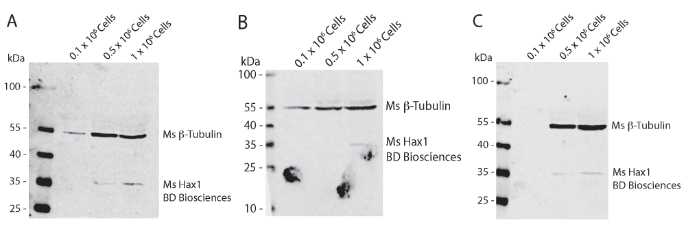
Western blot analysis of differentiated PLB-985 cell lysates from 0.1 × 10⁶, 0.5 × 10⁶, and 1 × 10⁶ cells from three independent replicates (A–C). Mouse anti-tubulin (beta-) is used as a loading control and can be seen present at 55 kDa. Mouse anti-Hax1 detects a band at approximately 32 kDa as predicted. Hax1 can be detected in densities of 0.5 × 10⁶ and 1 × 10⁶ cells.
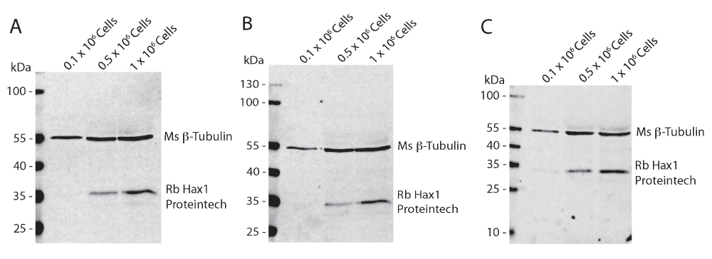
Western blot analysis of differentiated PLB-985 cell lysates from 0.1 × 10⁶, 0.5 × 10⁶, and 1 × 10⁶ cells from three independent replicates (A–C). Mouse anti-tubulin (beta-) is used as a loading control and can be seen present at 55 kDa. Rabbit anti-Hax1 detects a band at approximately 32 kDa as predicted. Hax1 can be detected in densities as low as 0.1 × 10⁶ cells.
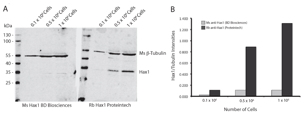
(A) Western blot analysis of differentiated PLB-985 cell lysates from 0.1 × 10⁶, 0.5 × 10⁶, and 1 × 10⁶ cells comparing mouse and rabbit anti-Hax1 antibodies. Lysates from the same cell extractions were run on a single SDS-PAGE gel and blotted onto a single nitrocellulose membrane. After transfer, the membrane was cut and probed with either mouse anti-Hax1 or rabbit anti-Hax1. The membranes were imaged simultaneously. (B) Quantification of the band intensities was measured and the ratios of Hax1 to tubulin were graphed.
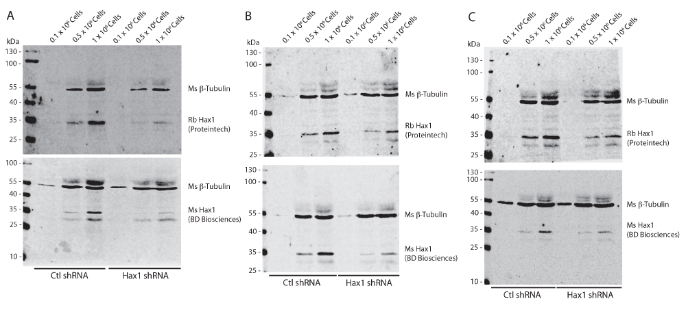
Western blot analysis of differentiated PLB-985 cell lysates from 0.1 × 10⁶, 0.5 × 10⁶, and 1 × 10⁶ cells expressing either control shRNA or Hax1 shRNA from three independent replicates (A–C). Mouse anti-tubulin (beta-) is used as a loading control and can be seen present at 55 kDa. Both mouse and rabbit anti-Hax1 detects a band at approximately 32 kDa as predicted. The specificity of each antibody can be observed by the reduction in intensities in cells expressing Hax1 shRNA.
To demonstrate the specificity of both Hax1 antibodies we generated stably-expressing control shRNA and Hax1 shRNA PLB-985 cells (Figure 4). As described previously using the mouse anti-Hax1 antibody the control shRNA cells show inconsistent staining intensity, however it is clear that in these samples it is more robust than in the wild-type PLB-985 cells. Both the mouse anti-Hax1 and rabbit anti-Hax1 antibodies show reduced detection in the Hax1-deficient PLB-985 cells, which demonstrates that the antibodies are highly specific for Hax1. In many of the experiments we observed additional background bands that could be attributed to the goat anti-rabbit IgG secondary antibody (Figure 5).
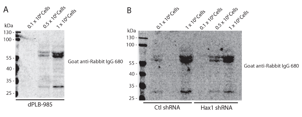
(A) Western blot analysis using goat anti-rabbit IgG 680LT only on cell lysates from 0.1 × 10⁶, 0.5 × 10⁶, and 1 × 10⁶ differentiated PLB-985 cells (dPLB-985), and (B) control shRNA and Hax1 shRNA expressing PLB-985 cells. Two predominant background bands can be observed at approximately 60 and 70 kDa, and one band around 30 kDa. These background bands can also be seen in Figure 1, 2, and Figure 4.
Here we show validation and comparison results of two antibodies generated against HS1-associated protein X-1, an anti-apoptotic protein that has a multi-factorial role in regulating cell proliferation and differentiation, cell motility, and cancer. Homozygous loss-of-function of Hax1 results in severe congenital neutropenia, a life threatening loss of circulating neutrophils in the blood stream. Thus studying the function of Hax1 in primary neutrophils and the neutrophil model cell line PLB-985 will help elucidate the disease pathogenesis of neutropenia syndromes. We demonstrate that mouse anti-Hax1 (BD Biosciences) and rabbit anti-Hax1 (Proteintech Group, Inc.) are both specific for Hax1. Furthermore we show that as little as 0.5 × 10⁶ differentiated PLB-985 cells can be used to reliably detect Hax1 expression with both of the antibodies. We have evidence that the rabbit anti-Hax1 (Proteintech Group Inc.) results in a more robust and consistent detection of Hax1, likely due to the polyclonal nature of the antibody. Finally, lentiviral knockdown of endogenous Hax1 expression results in loss of Hax1 detection by both mouse anti-Hax1 and rabbit anti-Hax1 demonstrating the specificity of each antibody.
In conclusion we recommend the use of either mouse or rabbit anti-Hax1 antibodies shown here for studies using the PLB-985 cells as a neutrophil model cell line. Furthermore, it is our conclusion that a minimum cell density of 0.5 × 10⁶ neutrophils should be used as a starting point for immunoblotting of Hax1, with greater than or equal to 1 × 10⁶ cells being optimal.
F1000Research: Dataset 1. Raw data for Figure 3, 10.5256/f1000research.6516.d4758212
| Views | Downloads | |
|---|---|---|
| F1000Research | - | - |
|
PubMed Central
Data from PMC are received and updated monthly.
|
- | - |
Click here to access the data.
Spreadsheet data files may not format correctly if your computer is using different default delimiters (symbols used to separate values into separate cells) - a spreadsheet created in one region is sometimes misinterpreted by computers in other regions. You can change the regional settings on your computer so that the spreadsheet can be interpreted correctly.
Provide sufficient details of any financial or non-financial competing interests to enable users to assess whether your comments might lead a reasonable person to question your impartiality. Consider the following examples, but note that this is not an exhaustive list:
Sign up for content alerts and receive a weekly or monthly email with all newly published articles
Already registered? Sign in
The email address should be the one you originally registered with F1000.
You registered with F1000 via Google, so we cannot reset your password.
To sign in, please click here.
If you still need help with your Google account password, please click here.
You registered with F1000 via Facebook, so we cannot reset your password.
To sign in, please click here.
If you still need help with your Facebook account password, please click here.
If your email address is registered with us, we will email you instructions to reset your password.
If you think you should have received this email but it has not arrived, please check your spam filters and/or contact for further assistance.
Comments on this article Comments (0)