Introduction
Hydranencephaly is a rare abnormality in which the cerebral hemispheres are almost absent and have been replaced with fluid. It was first described by cruveilher (1892) as “Anencephalie hydrocephalique” or “Hydroanencephalie”1. Crome and Sylvester reviewed the disease and defined it as a congenital condition2. It is a very rare isolated abnormality occurring less than 1 per 10,000 births world wide3. It is said to be present in 0.2% of infant autopsies and approximately 1% of babies are diagnosed clinically as hydrocephalus4. It's important to distinguish early between Hydranencephaly and extreme hydrocephalus, because the latter carries a considerably superior prognosis5. We report a rare case of Hydranencephaly which was diagnosed in late third trimester followed by successful obstetric management.
Case presentation
A 28-year-old woman, gravida 2 para 1 was presented to our tertiary care institution at 38 weeks' gestation because of lower abdominal pain. She and her partner were Sinhalese and unrelated. There was no family history of genetic or congenital anomalies. Her first pregnancy was uncomplicated normal vaginal delivery with normal healthy male child. She had not attended the local antenatal clinic regularly where she was recognized as a noncompliant patient. She had not undergone routine dating scan, anomaly scan or the growth scan of the fetus. She was a nonsmoker and there was no history suggestive of congenital infections or exposure to toxins. The clinical obstetric examination was unremarkable. The cardiotocography of the fetus was normal.
Obstetric ultrasound scan showed fluid filled cranial cavity with absent cerebral cortex, thalami and basal ganglia. The third ventricle dilated and remnants of midbrain structures were seen (Figure 1). The cerebellum and other posterior fossa structures were preserved. The falx cerebri was disrupted (Figure 2). There was no polyhydramnios. Umbilical artery Doppler studies were normal. Sonographicaly it was suggestive of Hydranencephaly. Her blood group was B positive and Infectious disease Antibody test showed negative titers for Rubella, HIV, Hepatitis B and Toxoplasmosis. We planed caesarian section to avoid obstetric complications due to possible cephalopelvic disproportion and minimize the risk for both mother and fetus. Specialized Pediatric team was pre informed. Parents were counseled regarding poor prognosis of the newborn's condition. At 39 week of gestation emergency caesarian section performed due to fetal distress. The newborn was a 2,980g girl with normal physical appearance and normal sized skull (head circumference = 33.5 cm) which was brilliantly transilluminated.
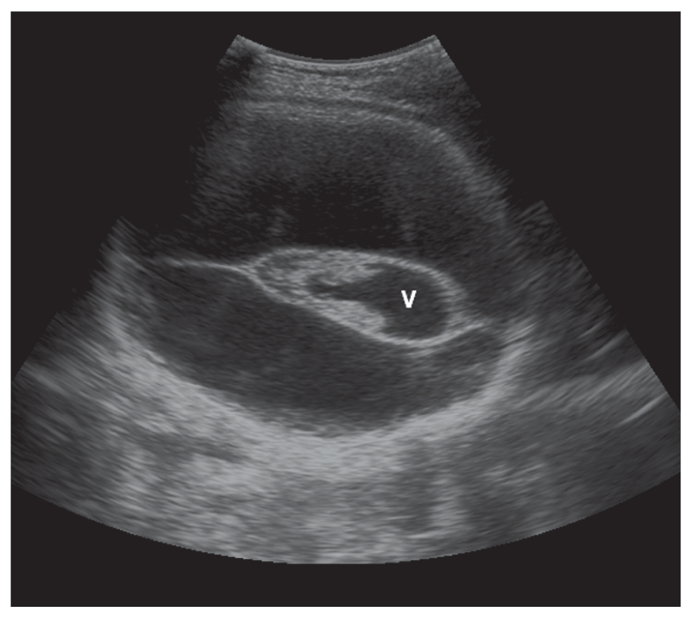
Figure 1. Prenatal ultrasound scans image of the fetus.
Transabdominal transverse section of the fetal head at 38 weeks of gestation shows dilated third ventricle (V) with absent thalami and basal ganglia and cerebral cortex.
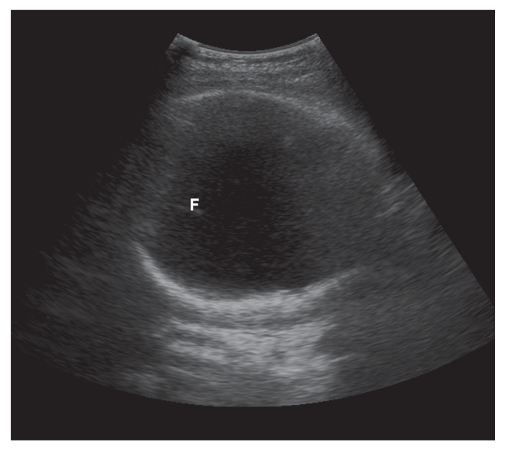
Figure 2. Prenatal ultrasound scans image of the fetus.
Transabdominal transverse section of the fetal head shows disrupted falx (F).
Newborn was transferred to neonatal Intensive care unit because of the respiratory distress. Four days after delivery Computed tomography (CT) of the newborn's head was performed without intravenous contrast. On CT there was no cerebral cortex, thalami or basal ganglia identified. The third ventricle dilated and remnants of midbrain structures were seen (Figure 3). The cerebellum and other posterior fossa structures were preserved with disrupted falx cerebri (Figure 4a, 4b).
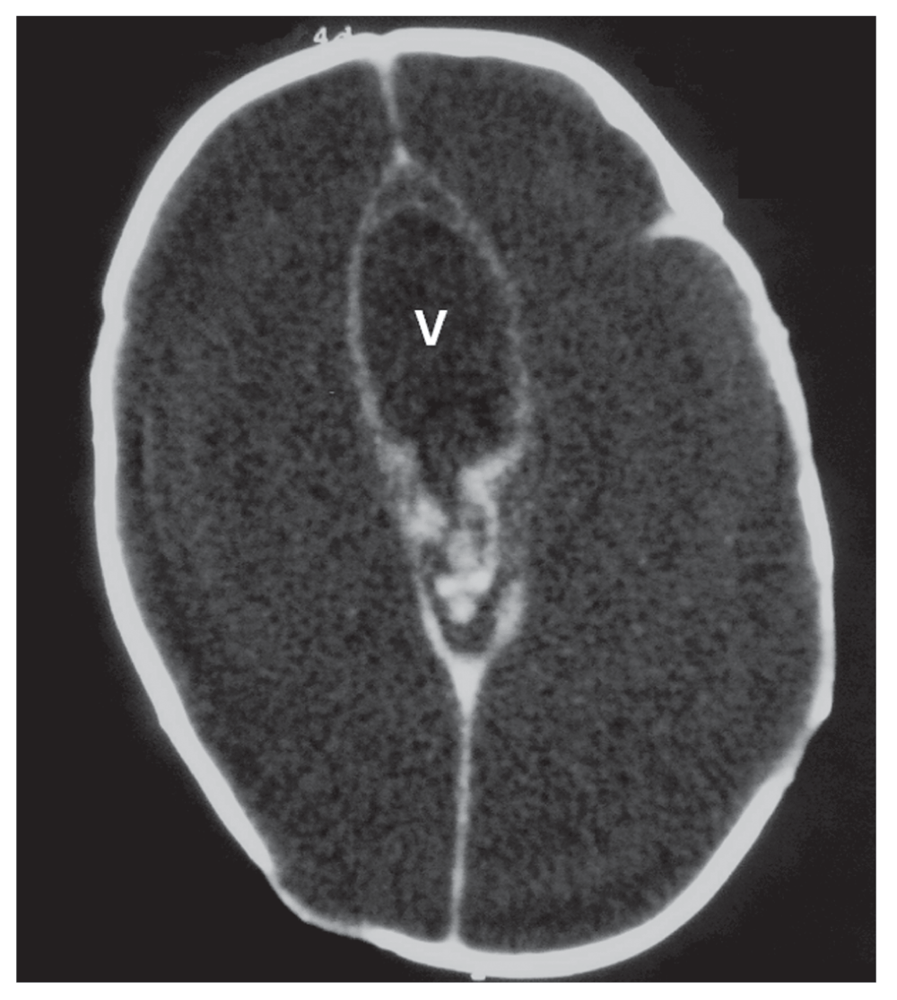
Figure 3. CT image of 2 days old newborns brain.
Dilated third ventricle (V) with absent thalami and basal ganglia and cerebral cortex.
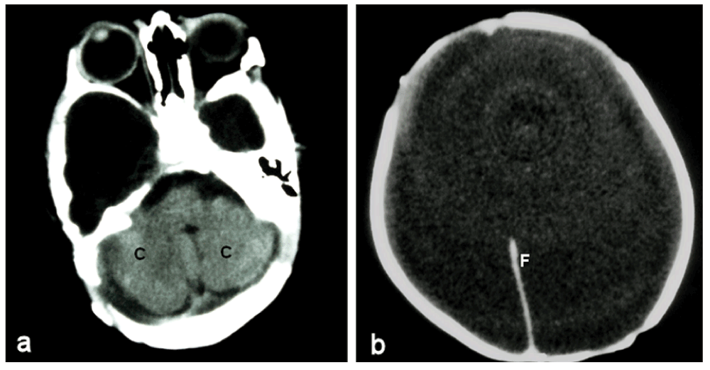
Figure 4. CT images of 2 days old newborns brain.
(a) Intact Cerebellum (C) and other posterior fossa structures. (b) Shows Disrupted Falx cerebri (F).
The CT scan confirmed the diagnosis Hydranencephaly. Baby died after two weeks of birth due to cardiac arrest. The parents declined a postmortem examination.
Discussion
Hydranencephaly is an encephaloclastic abnormality characterized by replacement of cerebral hemisphere with the cerebrospinal fluid and necrotic debris surrounded by leptomeninges such that no cerebral cortex is present but there may be partial preservation of portion of the occipital lobe6. The midbrain, thalamus, basal ganglia, choroid plexuses, cerebellum and brain stem are usually preserved and contained within normal skull. The Falx cerebri is usually present but may be partially or completely absent. The septum pellucidum may be absent.
The aetiopathogenesis of Hydranencephaly is heterogeneous, but several theories have been postulated. It has been suggested that bilateral occlusion of supraclenoid segment of the internal carotid artery or middle cerebral7 arteries before the 24 weeks of gestation cause ischemia, edema, autolysis and disappearance of cerebral hemispheres respectively8. Nevertheless some reports suggest occlusion of arteries due to temporary spasm or compression rather that direct occlusion9. Intrauterine infections also cause necrotizing vasculitis or local destruction of brain. Infections like congenital toxoplasmosis or viral infections (Adenovirus, cytomegalovirus, Enterovirus, Epstein-Barr virus, herpes simplex virus, Parvovirus, and respiratory syncytial viruses) have been implicated in numerous cases3. Fetal hypoxia due to exposure to toxin like Carbon monoxide or butane gas during antenatal period may result diffuse hypoxic ischemic brain necrosis lead to Hydranencephaly10. Thromboplastic material from a deceased co-twin in monochorionic twin pregnancies reported to be implicated11. Hydranencephaly has been described in rare syndromes12.
Ultrasonographic findings include large cystic mass filling entire intracranial cavity with absence of cerebral cortex. The head size may be normal or large13. Appearance of thalami and brainstem protruding in to cystic cavity is characteristic, together with a midline echo from the remnant of falx, the tentorium cerebella and cerebellum14. The third ventricle and choroid plexuses are often visible. Absence of septum pellucidum may give rise to what appears to be a single ventricle in midline15. The major differential diagnosis includes extreme hydrocephaly, alobar holoprosencephaly and porencephaly. In these conditions the above mentioned structures will still be surrounded by rim of cortex. In extreme hydrocephalus, the thin cortical mantle may be hard to identify sonographically and Magnotic resonance imaging (MRI) or rarely intrauterine CT scan might aid diagnosis13. The ultrasound is the best diagnostic tool during the prenatal period and postnatal period it's MRI16–18 or CT. It is important to distinguish Hydranencephaly from other differential diagnosis because they carry better prognosis. The most of the cases detected second half of pregnancy12 and there have been reported cases of Sonographic evaluation of fetal Hydranencephaly in the first trimeste19.
Hydranencephaly has an irretrievably poor prognosis, with merely remaining brainstem functions. Some die at birth, but most infant die within the first year of life20 and if survived they are profoundly retarded. The recurrence risk is negligible14. Because of the poor prognosis, termination of pregnancy is recommended once definitive diagnosis has been established13. If macrocrania is found in late pregnancy cephalocentesis may be indicated (aid delivery)13. Counsel parent regarding poor prognosis and management options are very important.
Ultrasonographic findings, in the case we have described, made a diagnosis of Hydranencephaly particularly likely and this was confirmed by postnatal CT of the fetal head. This is the first published case of Hydranencephaly with absent thalami and basal ganglia along with midbrain. Sonographic evaluation is sufficient for the prenatal diagnosis of Hydranencephaly in most cases, and MRI or intrauterine CT cannot be considered a first-line diagnostic tool.
Conclusions
Early diagnosis is important as an early treatment options avoids obstetrics complications and it may be very useful for giving appropriate advice to parents during the pregnancy and also for preparing the optimal conditions of birth at a unit with available specialized pediatric facilities.
Consent
Written informed consent was obtained from the patient for publication of this case report and any accompanying images. A copy of the written consent is available for review by the Editors.
Author contributions
SKR and BTBW reviewed the manuscript. BTBW and GKR were involved in drafting the manuscript and reviewing the literature. BTBW was a major contributor in revising the manuscript and getting informed consent from patient. GKR, SKR and BTBW were involved in reviewing the literature. All authors were responsible for the diagnosis, treatment and follow-up of the patient. All authors read and approved the final manuscript.
Competing interests
No competing interests were disclosed.
Grant information
The author(s) declared that no grants were involved in supporting this work.
Acknowledgments
We thank Mrs. Janaki Munasinghe for correcting grammar and spellings of the manuscript.
Faculty Opinions recommendedReferences
- 1.
Raybaud C:
Destructive lesion of the brain.
Neuroradiology.
1983; 25(4): 265–291. PubMed Abstract
| Publisher Full Text
- 2.
Crome L, Sylvester PE:
Hydranencephaly (hydrencephaly).
Arch Dis Child.
1958; 33(169): 235–245. PubMed Abstract
| Publisher Full Text
| Free Full Text
- 3.
Kurtz AB, Johnson PT:
Diagnosis please. Case 7: Hydranencephaly.
Radiology.
1999; 210(2): 419–22. PubMed Abstract
- 4.
Halsey JH:
Hydranencephaly. In Handbook of Clinical Neurology. Volume 50: Malformations. Edited by PJ Vinken, GW Bruyn and JL Klawans. Amsterdam: Elsevier 1987; 337–353.
- 5.
Malheiros JA, Trivelato FP, Oliveira MM, et al.:
Endoscopic choroid plexus cauterization versus ventriculoperitoneal shunt for hydranencephaly and near hydranencephaly: a prospective study.
Neurosurgery.
2010; 66(3): 459–64. PubMed Abstract
| Publisher Full Text
- 6.
Jeffrey MC:
Hydranencephaly: Transillumination May Not Illuminate Diagnosis.
Neoreviews.
2012; 13(4): e233–e240. Publisher Full Text
- 7.
Myers RE:
Brain pathology following fetal vascular occlusion: an experimental study.
Invest Ophthalmol.
1969; 8(1): 41–50. PubMed Abstract
- 8.
Govaert P:
Prenatal stroke.
Semin Fetal Neonatal Med.
2009; 14(5): 250–266. PubMed Abstract
| Publisher Full Text
- 9.
Lindenberg R, Swanson PD:
“Infantile Hydranencephaly”--a report of five cases of infarction of both cerebral hemispheres in infancy.
Brain.
1967; 90(4): 839–850. PubMed Abstract
| Publisher Full Text
- 10.
Hoyme HE, Higginbottom MC, Jones KL:
Vascular etiology of disruptive structural defects in monozygotic twins.
Pediatrics.
1981; 67(2): 288–291. PubMed Abstract
- 11.
Larroche JC, Droulle P, Dalezoide AL, et al.:
Brain damage in monozygous twins.
Biol Neonate.
1990; 57(5): 261–78. PubMed Abstract
| Publisher Full Text
- 12.
Alasdair GW Hunter:
Brain. in Human malformations and related anomalies. 2nd Edition. Edited by Roger E Stevenson, Judith G Hall. New York: Oxford University Press, 2006; 639–645. Reference Source
- 13.
Keith CD, Hylton BM, David OC, et al.:
Ultrasound in obstetrics and gynecology. 2nd Edition. Michigan: Churchill Livingstone; 2001; 326–327.
- 14.
David KJ, Philip JS, Carl PW, et al.:
High Risk Pregnancy: Management Options. 3rd Edition. Pennsylvania: Elsevier; 2006; 386–387. Publisher Full Text
- 15.
Gordon IRS, Ross FGM:
Diagnostic radiology in pediatrics. Illustrated edition. Michigan: Butterworths; 1977; 305–306.
- 16.
Barkovich AJ, Kevin RM, Elena G, et al.:
Diagnostic Imaging: Pediatric Neuroradiology. 1st edition: AMIRSYS; 2007; 1-1-152. Publisher Full Text
- 17.
Wagner AL, Rohrer D:
Imaging in hydranencephaly.2008; Accessed September 15, 2012. Reference Source
- 18.
Jones J:
Hydranencephaly. 2010; Accessed September 15, 2012.
- 19.
Lin YS, Chang FM, Liu CH:
Antenatal detection of hydranencephaly at 12 weeks, menstrual age.
J Clin Ultrasound.
1992; 20(1): 62–4. PubMed Abstract
| Publisher Full Text
- 20.
Hadi HA, Mashini IS, Devoe LD, et al.:
Ultrasonographic prenatal diagnosis of hydranencephaly. A case report.
J Reprod Med.
1986; 31(4): 254–6. PubMed Abstract
Comments on this article Comments (0)