Keywords
Capecitabine, radiation recall, ulcer, cancer, ulcerating dermatitis
Capecitabine, radiation recall, ulcer, cancer, ulcerating dermatitis
We have included details of our choice for successive different chemotherapy lines. We have added two figures to illustrate the microscopic appearance of the skin reaction. We have added more details of the clinical evolution of the patient from the diagnosis of recall dermatitis until she passed away more than a year afterwards. We have made slight corrections to the discussion section, adding some details on the response predictive ability of skin toxicity (with the appropriate reference).
See the authors' detailed response to the review by Rita De Sanctis
We report a case of radiation recall phenomenon after the administration of capecitabine, consisting of pain, hyperpigmentation, and ulceration in the field of post-mastectomy irradiation (which the patient received 3 years previously).
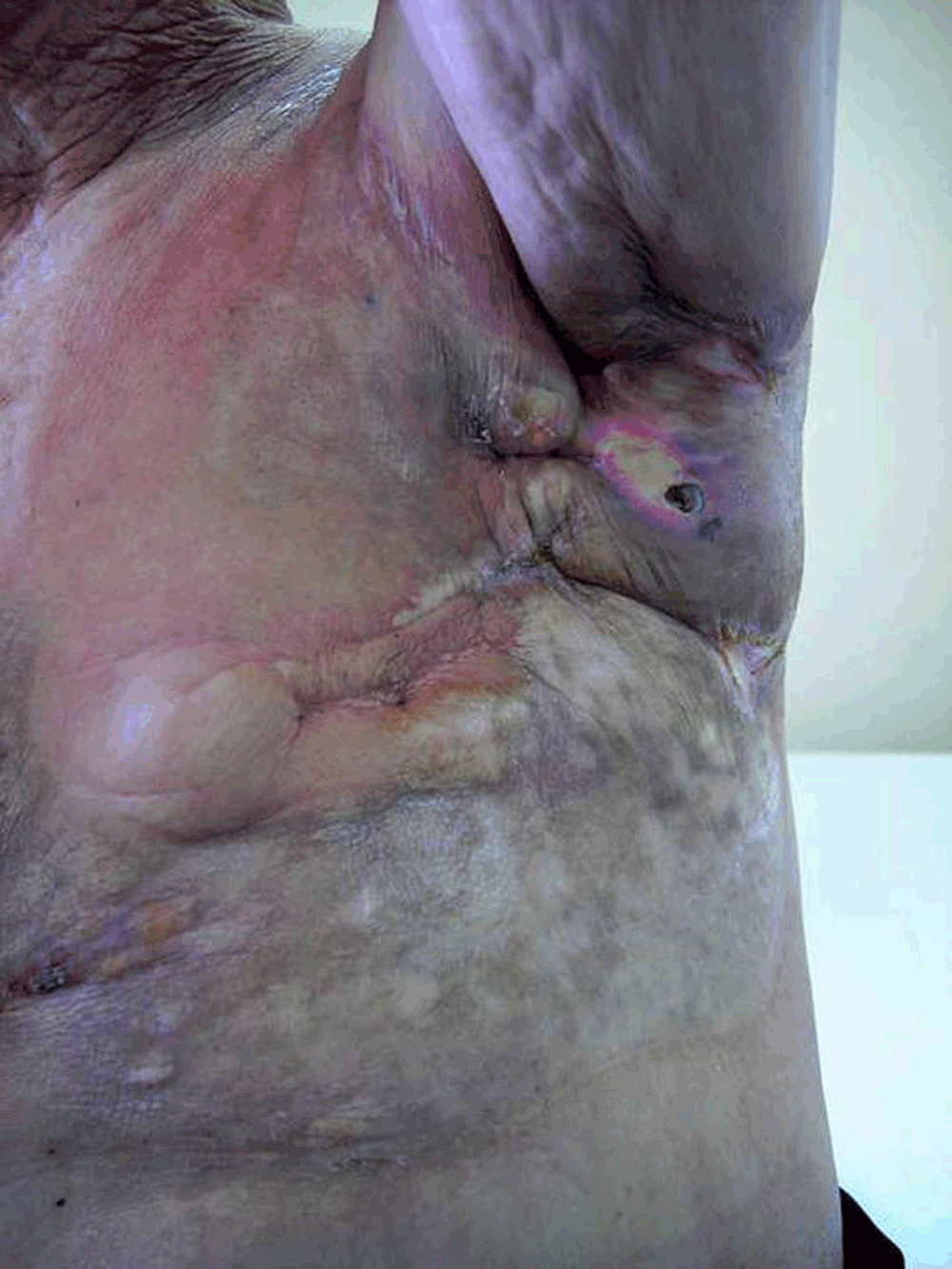
A 78 year old woman allergic to salicylics was diagnosed with a T4dN3M0 (American Joint Committee on Cancer) infiltrating ductal left breast carcinoma (inflammatory breast cancer) in March 2006. Owing to her general condition and advanced local disease, she was initially treated with primary hormonotherapy consisting of letrozole 2.5 mg/d over a period of six months with a good local response as measured by ultrasound scanning. In October 2006, she was operated on and a modified radical mastectomy was performed. Pathology reported a 6 cm in diameter infiltrating ductal carcinoma pT4dN2a positive for both estrogen and progesterone receptors, and Her2-neu negative. After surgery she started on chemotherapy (Taxol 80 mg/m2 on a weekly schedule for four weeks) and adjuvant radiotherapy (50 Gy over left hemithorax and supraclavicular nodes in February 2007). Immediately after initial radiotherapy, in 2007, she developed skin toxicity Radiation Therapy Oncology Group (RTOG) grade 2, which was successfully managed with topical medication (Radiocrem® Rotthafarm SL [tocopheryl acetate, disodium EDTA, silybum marianum, vitis vinifera] three times a day). She started letrozole 2.5 mg/d again in January 2007. In September 2009 she developed neoplasic left pleural involvement and began hormonotherapy with fulvestrant 500 mg/monthly for five months, followed by exemestane 25 mg/d due to clinical and radiological progression. In May 2010, she developed new pleural progression, which was treated with capecitabine at a dose of 1000 mg/m2/12h (three cycles). Three months later in July 2010, she was noted to have rapidly developed (no more than two weeks after the patient felt the first symptoms of skin stiffness and a local burning sensation) a series of ulcers on the previous mastectomy scar, which had changed in colour (hyper and dispigmentation) and elasticity (stiffness and extreme fragility) over the skin of the previously irradiated area in the left hemithorax (Figure 1). A punch-biopsy was performed, pathological changes in the skin consistent with radiodermatitis were observed and carcinoma in the involved skin was ruled out (Figure 2). The diagnosis of a recall radiodermatitis (ulcerating dermatitis, grade 4 RTOG) was thus established in July 2010 and, to mimize the risk of an opportunistic infection, capecitabine was withdrawn and palliative 20 mg tamoxifen started. The skin began to improve after 3–4 weeks following withdrawal of capecitabine and treatment with topical steroids (Menaderm® Menarini; beclomeatsone 0.025 %).

The area with preserved epidermis (arrow 2) shows acanthosis (arrow 3) and parakeratosis with loss of epidermal ridges. Vascular ectasia (arrow 4), hyalinized collagen (arrow 5) and loss of skin adnexa are seen in the dermis. Infiltration by neoplasm was ruled out.
Tamoxifen treatment stabilized the patient’s disease and serological response for four months. Afterwards, pleural progression was diagnosed and vinorelbine started with good response; after eight cycles, the patient suffered a new episode of skin toxicity that was managed with vinorebiline withdrawal and letrozole treatment, which allowed for a nine month stability period. In March 2012, progression was seen (liver metastasis and greater pleural effusion with clinical deterioration) and cyclophosphamide started. All active medication was stopped in May that year and palliative care lasted until the patient passed away a few months afterwards.
Radiation-recall dermatitis is an inflammatory reaction of the skin at the site of previous irradiation. Many chemotherapy drugs have been presumed to cause this phenomenon and a database to collect these rare reaction cases has even been proposed1. It can also affect other anatomical areas such as the digestive system (when abdominal radiotherapy has been used)2. Although a closer time gap is more usual, the time gap between the inflammatory reaction and previous radiation can range from days to several years3.
There are few reported cases of capecitabine-induced radiation recall phenomenon, the first one being authored by Ortmann et al. in 20024. Their hypothesis relied on the pro-drug entity of capecitabine being capable of being activated in previously irradiated tissue.
More recently, Ghosal and Misra have reported a case5 with thoracic hyperpigmentation instead of inflammation being the main clinical finding. Other well known capecitabine side effects are hyperpigmentation associated with palmar-plantar erythrodisesthesia6 or even Stevens-Johnson syndrome7. Some authors have suggested that skin toxicity might be a predictor of response though it has been better related to anti-epidermal growth factor receptor monoclonal antibodies8.
To our knowledge, this is the first reported case of capecitabine-induced RTOG grade 4 (ulcerating dermatitis) recall skin toxicity of previously irradiated skin. We suggest it is relevant for differential diagnosis with other entities such as local cancer relapse or even surgical site infection. In case of any doubt, such as in our case, punch biopsy can help. Clinicians should be aware of this phenomenon, even if a long period of time has elapsed since the previous radiation therapy.
Written informed consent for publication of the clinical details and clinical images was obtained from the patient.
| Views | Downloads | |
|---|---|---|
| F1000Research | - | - |
|
PubMed Central
Data from PMC are received and updated monthly.
|
- | - |
Provide sufficient details of any financial or non-financial competing interests to enable users to assess whether your comments might lead a reasonable person to question your impartiality. Consider the following examples, but note that this is not an exhaustive list:
Sign up for content alerts and receive a weekly or monthly email with all newly published articles
Already registered? Sign in
The email address should be the one you originally registered with F1000.
You registered with F1000 via Google, so we cannot reset your password.
To sign in, please click here.
If you still need help with your Google account password, please click here.
You registered with F1000 via Facebook, so we cannot reset your password.
To sign in, please click here.
If you still need help with your Facebook account password, please click here.
If your email address is registered with us, we will email you instructions to reset your password.
If you think you should have received this email but it has not arrived, please check your spam filters and/or contact for further assistance.
Comments on this article Comments (0)