Keywords
Pediatrics, Child, Teenager, Refraction, School vision
Pediatrics, Child, Teenager, Refraction, School vision
The new version includes minor changes:
- Correction of a spelling error and removal of a sentence in the methods section.
- Removal of a statistical data in the abstract and in the results section
See the authors' detailed response to the review by Carla Costa Lança
See the authors' detailed response to the review by Pedro M. Serra
Anisometropia is an ocular disorder characterized by an interocular difference (IOD) in refractive error. It represents a specific refractive condition where the two eyes of an individual, can have asymmetric eye growth.1 This condition can occur in situations of myopic, hyperopic or astigmatic asymmetry and is strongly associated with the development of other eye changes such as aniseikonia, amblyopia, diplopia and strabismus.2,3
Although there is no uniformly defined dioptric value for its clinical classification, an IOD in the spherical equivalent (SE) of 1 diopter or more is accepted as the threshold, for most authors.1,4–8 However, even using this limit, the scientific literature presents a significant variation in the prevalence values of anisometropia in terms of age, gender and ethnicity.4,5,8,9 Factors associated with lifestyle and educational level have also been referred to as risk factors for anisometropia.7,8,10
Early detection and early treatment is crucial to prevent permanent visual loss. Although it is not clear what is the ideal age to perform the correction, in order to guarantee an ideal visual development and maturation, the early correction of anisometropia is important.11 This prevents the development of other changes such as aniseikonia, amblyopia and strabismus,3,12,13 and even in small degrees (<1D) facilitates emmetropization.11 It also improves quality of life, reducing or eliminating symptoms of visual discomfort. In this way, visual screening at a young age is useful in identifying who is most likely to benefit from early optical correction or preventive treatment.11,14,15
The clinical methods used to characterize anisometropia are refractive techniques, with autorefraction, using Plusoptix, one of the most recommended techniques for screening activities.16,17 This instrument allows quantifying the refractive error in which it is possible to obtain a very similar value to that obtained by cycloplegic refraction17–19 and with excellent precision in anisometropia signalling.19
Studies on the prevalence of anisometropia focus on children or adults, with less research being found in adolescence.
The aim is to estimate the prevalence of anisometropia (spherical and astigmatic) and to analyse its pattern of variation in a sample of children and adolescents, from preschool education (from three to six years old) to the various cycles of basic education (from the 1st to the 9th school year in Portugal, from six to fifteen years old).
This is an observational cross sectional study to evaluate the prevalence of anisometropia in children and adolescents. Data from the Portuguese Census 2011, showed a population of 319284 from 0 to 14 years old in the central region.20 For a confidence level of 95% and a 5% margin of error, taking into account that the prevalence of the studied condition is unknown, it was fixed at 50% to obtain a large enough sample size. The result was a minimum of 384 subjects.
The data collection took place in five schools of the central region of Portugal, including all students that were authorized to participate by their legal guardians, between October 2018 and February 2019. Participants were 749 children and adolescents aged between three and sixteen years. Students data for which it was not possible to obtain refraction were excluded, due to technical issues associated with the performance of the instrument (presence of strabismus, opacities, retinal anomalies or when the refractive error exceeded the instrument's measurement limit - spherical measurement range or cylindrical from −7.00 to +5.00D) or due to lack of cooperation from the participant. Only two participants were excluded.
The refractive error was measured with the paediatric auto refractometer model A09 by PlusOptix, Nuremberg, Germany, by the average of three consecutive measurements, binocular and without the use of a cycloplegic. The PlusOptix allows measuring the refractive error in open field simultaneously in both eyes and under the same conditions, in a fast, easy, safe, non-invasive way, at a distance of one meter from the subject's eyes.
In Portugal the compulsory education includes basic education, which is divided into 3 cycles, followed by the secondary education. The 1st cycle of basic education includes the 1st to 4th school year (ages 6 to 10), the 2nd cycle includes the 5th and 6th year (ages 10 to 12) and the 3rd cycle includes the 7th to the 9th year (ages 12 to 15). The sample under study had students from preschool to the 3rd cycle of basic education and was characterized according to gender, area of residence and cycle of studies. Rural and urban areas classification was based on the information provided by the municipalities, considering the residence area of each participant. Regarding the use of glasses, 177 participants wore glasses or contact lenses and 572 did not use any type of optical correction. Table 1 summarizes the characteristics of the sample.
In order to calculate the average of the three refractive measurements, the power was converted from its spherical-cylindrical form to its power vector representation, described by Thibos,21 using the following expressions:
Where SE represents the spherical equivalent; J0 represents Jackson's crossed cylinders on the 90° or 180° axis, which stands for the amount of direct or indirect astigmatism; J45 represents Jackson's crossed cylinders on the 45° or 135° axis, which stands for the amount of oblique astigmatism; S, C and α represent the spherical, cylindrical (in negative form) and cylinder axis component, respectively, of the auto refractometer measurement.
The participants were classified in emmetropes, myopes, hyperopes, astigmats or anisometropes, according to the average value of the auto refractometer. In order to carry out this classification, the criteria recommended for the auto refractometer used were applied (Table 2).
| Refractive state | < 6 years old | ≥6 years old |
|---|---|---|
| Emmetrope | −1.00D < SE < +1.25D | −1.00D < SE < +1.00D |
| Myope | SE ≤ −1.00D | |
| Hyperope | SE ≥ +1.25D | SE ≥ +1.00D |
| Astigmat | |C|≥1.00D | |C|≥1.25D |
| Anisometrope | |IOD|≥1.25 (SE or C) | |
The absolute value of IOD of the refractive error in terms of SE was designated as spherical anisometropia (SA), the absolute IOD in astigmatism was designated as meridional anisometropia (MA),
When there was no IOD in the spherical equivalent and the absolute IOD was only in the astigmatic component, it was designated by simple meridional anisometropia (sMA). The anisometropia classification was done according to the cutoff points referred to in Table 2. The presence of at least one of the previous conditions was designated as total anisometropia (TA). Low anisometropia was considered for IOD values below 2.00D, high anisometropia for values between 2.00D and 6.00D and very high anisometropia for IOD values above 6.00D.
According to the type of refractive error, anisometropia was classified as myopic, when both eyes were myopic or when one eye was myopic and the other was emmetropic; hyperopic, when both eyes were hyperopic or when one eye was hyperopic and the other emmetropic; antimetropic, when one eye was myopic and the other hyperopic; simple meridional anisometropia when there was no SA, but there was MA.
A descriptive statistical analysis was carried out, using SPSS version 26 package, characterizing the sample in the variables of interest, sociodemographic and refractive, presenting means and standard deviations, frequencies and percentages both in the whole of the sample and also according to several stratifications to which it was subjected.
In all the sociodemographic factors in which the sample was categorized, groups with a large size (n > 30) were obtained. The proportion of subjects with anisometropia was analysed according to gender, area of residence and school cycle, and through the Chi-square test, it was evaluated whether these variables were associated with the occurrence of anisometropia in the studied population.
All the results of the statistical inference tests were interpreted to a 95% confidence level, that is, the significance level of 0.05 was used.
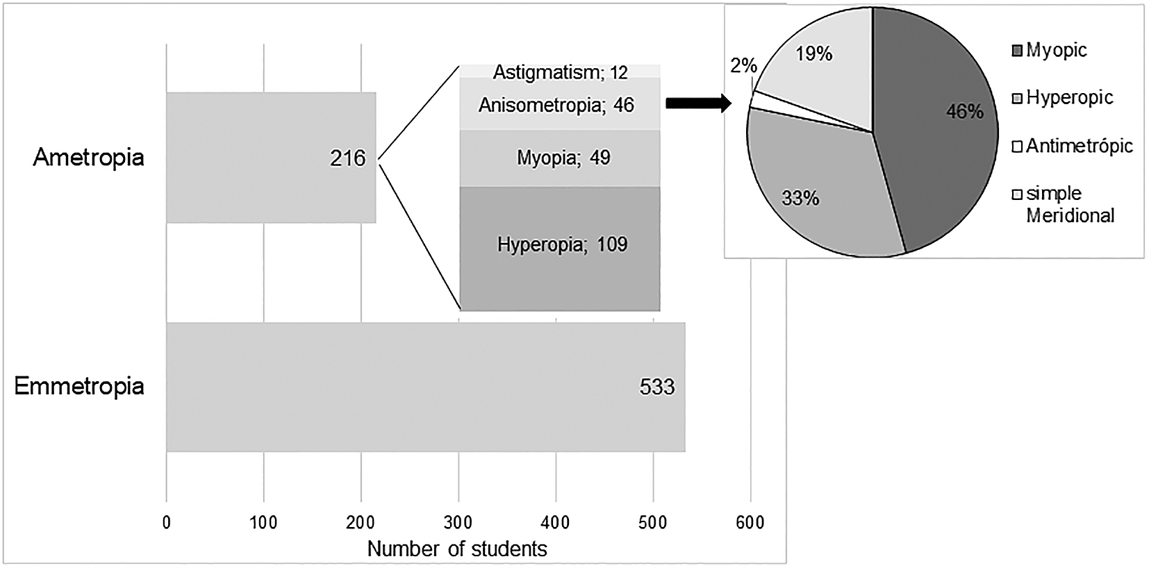
According to the classification criteria previously defined for the classification of refractive state, it was concluded that in the study sample 71.16% (n = 533) were emmetropic. Among subjects with significant refractive error (n = 216), it was found that hyperopia was the most prevalent refractive error (n = 109, corresponding to 14.6% in the studied population) followed by myopia (n = 49, corresponding to 6.5% in the population), anisometropia (n = 46, corresponding to 6.1% in the population) and astigmatism (n = 25 where 12 were cases of simple astigmatism and 13 compound astigmatism). Figure 1 shows graphically the distribution of the different refractive states in the study sample. The representation of astigmatism, refers only to the occurrence of simple astigmatism, without a significant SE. The mean values of refractive errors, according to the previous classification, are shown in Table 3.
| Refractive group | Wearing glasses (n) | Spherical equivalent (D) | Cylindrical component (D) | |||
|---|---|---|---|---|---|---|
| Yes | No | Mean ± SD | Range [min; max] | Mean ± SD | Range [min; max] | |
| Myopia | 44 | 5 | −2.67 ± 1.32 | −5.79; −0.92 | −0.39 ± 0.33 | −1.29; 0.00 |
| Hyperopia§ | 22 | 85 | +1.46 ± 0.59 | +1.00; +5.25 | −0.41 ± 0.47 | −2.50; 0.00 |
| Simple astigmatism | 6 | 6 | +0.29 ± 0.55 | −0.83; +1.21 | −1.50 ± 0.33 | −2.07; −1.14 |
| Anisometropiaϯ | 37 | 8 | Interocular difference | |||
| 1.53 ± 0.82 | 0.00; 3.79 | 0.78 ± 0.85 | 0.00; 3.9 | |||
Anisometropia was identified in 46 participants (6.1% of the studied population). Regarding the use of glasses, it was observed that eight cases did not use any type of optical correction.
According to the magnitude of the refractive error, no child was found with very high anisometropia, 15 were registered with high anisometropia and 31 with low anisometropia, that is, of the anisometrope subjects, most (67.4%) had low anisometropia. The dispersion of interocular diference in SA and MA is shown in Table 3.
In the classification of anisometropia according to the type of refractive error, 37 subjects (corresponding to 4.9% in the studied population) were found with SA, integrating 21 with myopia, 15 with hyperopia and 1 with antimetropia; 15 subjects (corresponding to 2% in the population) with MA, and only 9 of these did not have SA, and were classified as simple meridional anisometropia (sMA). This distribution is represented graphically in Figure 1. It is possible to observe that myopic anisometropia was the most prevalent (46%), followed by hyperopic anisometropia (33%) and sMA (19%). Antimetropic anisometropia was the least prevalent (2%).
The study of the influence of sociodemographic variables in the occurrence of anisometropia is presented in Table 4. The Chi-square test indicates that there was no association between MA and any of the factors under analysis.
| Gender | Chi-square test | ||||
|---|---|---|---|---|---|
| Female (350) | Male (399) | ||||
| N | % | N | % | P | |
| TA | 25 | 7.1 | 21 | 5.3 | 0.29 |
| SA | 21 | 6 | 16 | 4 | 0.21 |
| MA | 8 | 2.3 | 7 | 1.8 | 0.60 |
| Area of residence | Chi-square test | ||||
|---|---|---|---|---|---|
| Rural (320) | Urban (423) | ||||
| N | % | N | % | p | |
| TA | 22 | 6.8 | 23 | 5.4 | 0.42 |
| MA | 7 | 2.2 | 8 | 1.9 | 0.78 |
| SA | 18 | 5.6 | 18 | 4.3 | 0.39 |
| School stage | Chi-square test | ||||||||
|---|---|---|---|---|---|---|---|---|---|
| Preschool (103) | 1st cycle (231) | 2nd cycle (181) | 3rd cycle (234) | ||||||
| N | % | N | % | N | % | N | % | p | |
| TA | 3 | 2.9 | 10 | 4.3 | 11 | 6.1 | 22 | 9.4 | 0.06 |
| MA | 2 | 1.9 | 3 | 1.3 | 6 | 3.3 | 4 | 1.7 | 0.52 |
| SA | 1 | 1.0 | 9 | 3.9 | 7 | 3.9 | 20 | 8.6 | 0.012* |
| p(adjust) | 0.36 | <0.001* | <0.001* | 0.015* | |||||
No significantly different occurrence of anisometropia was found according to gender or area of residence (p > 0.05). In relation to the school, there was a pattern of variation that increased with the cycle of studies, ranging from 2.9% in preschool education to 9.4% in the 3rd cycle of studies, however this association was only statistically significant for SA (p = 0.012), where there was a rate of 1% in preschool education and 8.6% in the 3rd cycle. It should be noted that the study cycle is dependent on age.
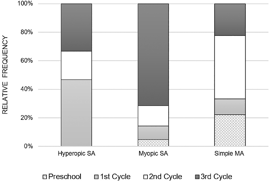
Figure 2 illustrates the distribution of anisometropia according to the type of refractive error, for each school cycle. It is observed that myopic anisometropia was present in all study cycles, registering a considerable increase from the beginning of school, that is, from the 1st cycle to the 3rd cycle. On the other hand, hyperopic anisometropia was manifested on a larger scale in children of the 1st cycle, with a lower occurrence in the following cycles. As for the simple MA, it was present in all study cycles and there was no specific pattern in its variation with the study cycle progress.
An anisometropia rate of 6.1% was found, considering the spherical and meridional anisometropia. It was found that this rate varies with the cycle of studies, showing an increase as the level of education advances, ranging from 2.9% in preschool education to 9.4% in the 3rd cycle of basic education. No statistically significant differences were found in the distribution of anisometropia either between genders or between areas of residence. It was also found that myopic anisometropia was the most prevalent (46%), with a considerable increase in the 3rd cycle of studies.
Table 5 summarises worldwide epidemiological data regarding studies in children and adolescents, which used the same criterion for anisometropia definition (IOD ≥ 1D).
TA - Total Anisometropia; SA - Spherical Equivalent Anisometropia; MA - Meridional Anisometropia.
| Author/year | Location | Age (years) | Sample | Refractive test | Definition | Prevalence (%) | |
|---|---|---|---|---|---|---|---|
| Ohlsson/200322 | America | Monterrey, Mexico | 12-13 | 1035 | Retinoscopy | AT ≥ 1.00 | 15 |
| Dobson/200813 | Toronto, Canada | 4-13 | 1041 | Cycloplegic Autorefraction | SA ≥ 1.00 MA ≥ 1.00 TA ≥ 1.00 | 6.7 15 18.1 | |
| Deng/20125 | Boston, USA | 5 12-15 | 395 312 | Retinoscopy | AS ≥ 1.00 | 1.3 5.8 | |
| Quek/200423 | Asia | Singapore | 15-19 | 946 | N/Cycloplegic Autorefraction | SA ≥ 1.00 | 11.2 |
| Yekta/201024 | Shiraz, Iran | 7-15 | 1872 | N/Cycloplegic Autorefraction | SA ≥ 1.00 | 2.6 | |
| Hu/20167 | Shandong, China | 4-18 | 6025 | Cycloplegic Autorefraction | SA ≥ 1.00 MA ≥ 1.00 | 7 | |
| Lee/20178 | Taiwan, China | 8 | 23114 | Cycloplegic Autorefraction | SA ≥ 1.00 | 3.7 | |
| Alrahili/201725 | Medina, Saudi Arabia | 3-10 | 1893 | N/Cycloplegic Autorefraction | TA ≥ 1.00 | 7.4 | |
| Hendriks/20094 | Europe | Maastricht, Netherlands | 11-13 | 520 | N/Cycloplegic Autorefraction | SA ≥ 1.00 | 4.6 |
| Flitcroft/202011 | Ireland | 6-7 | 362 | Cycloplegic Autorefraction | SA ≥ 1.00 | 6.9 | |
| Huynh/200626 | Oceania | Sydney, Australia | 6 | 1765 | Cycloplegic Autorefraction | AS ≥ 1.00 AM ≥ 1.00 | 1.6 1.0 |
Comparing with others studies, some authors point to a frequency similar to the one found on this research,4,7,8,11,25 others point to a lower frequency24 and others still refer to a higher frequency.22,23 Studies that included only children aged five and six years report rates of 1.3% and 1.6% (SA).5,26 The frequency of SA, found in the present study, in preschool education (children from three to six years old) was 1%, this value being closer to those studies. Other studies included participants from 15 to 19 years old and report rates of 11,2% for SA.23 For the 3rd cycle of studies (average between 12 and 15 years of age), the present study indicated a frequency of 8.6% (SA) and according to the observed variation pattern, at a more advanced age and school stage, a higher rate is expected.
Geographical and methodological issues make it difficult to compare prevalence studies. There is a pattern of greater variability in studies on the Asian continent, where for an identical age group, there are records ranging from 2.5%24 to more than 10%;7 however this is the continent where more studies on the subject are found. In Europe, more similar results are found, 4,6% and 6,9%.4,11 In Portugal, a study in children from 6 to 11 years old found a 0.7% rate (5 in 672) of uncorrected anisometropia,27 similar to the 1.1% (8 in 749) found in the present study.
In the literature, the pattern of variation of anisometropia as a function of age and during the school period is not clear, presenting discordant results. Although many studies focus on children, it is common to have relatively wide age ranges. Most authors conclude that anisometropia varies with age,4,5,7,28 although there are also studies where this relationship has not been found.10,13 Studies on the subject, at school age, which showed an increasing prevalence with age were carried out in populations with a high frequency of myopia, a condition whose prevalence increases in adolescence.28 On the other hand, longitudinal studies show that the prevalence of anisometropia increases after children start attending school.5,7,25 Given these two lines that justify the variation of anisometropia during school age, it appears that they are related to each other, since the progress in the school path is accompanied by increasing age. In the present study, the anisometropia prevalence was also found to increase with advancement in the level of education. Consequently the age factor is also contributing to this situation, with myopic anisometropia being the one that most contributes to this variation pattern.
Regarding the influence of gender on anisometropia, contradictory data are found in the scientific literature. While in one study it is reported that the prevalence found was higher in males28 in others it is reported that the prevalence rates are higher in females.10,23 The results of the present study revealed a higher frequency in females (7.1%) than in males (5.3%), however these differences were not statistically significant. This finding is in line with the results of other authors.9,13,25,26
The influence of living in rural or urban areas has also been the object of study by several researchers, considering the development of myopia and, consequently, myopic anisometropia, which is more pronounced in urban areas.7,8,10 The present study did not prove whether the area of residence and the frequency of anisometropia were related. This parameter is highly dependent on the way in which each author classifies the area of residence, as rural or urban, and the limits of these regions are sometimes difficult to define. Also, living in a rural area and working in an urban area means that the daily experience of some populations turns out to be more urban, in both environments. Some studies show that lifestyle parameters, such as reading and writing habits and time spent indoors, contribute to the prevalence of anisometropia variation.7,8,10,11
This study has several strengths. The study was carried out in children and adolescents in a school environment. This allows minimizing the potential bias that occurs in the sampling process and avoids overestimation of the problem, when the investigation is carried out in a clinical environment. For this study, spherical anisometropia and astigmatic anisometropia were considered. The latter being disregarded in several studies and there is a record that this anisometropia is the one that most varies in terms of ethnicity.9
Certain limitations can be pointed out in this study. Firstly, the use of autorefraction without cycloplegia is highlighted, which is not the gold standard method of refraction. This fact limits the analysis of the distribution of the different types of refractive errors found in the population studied. Despite the use of an open field auto refractometer, recognized as an instrument with good agreement with cycloplegic retinoscopy and which eliminates the need for cycloplegia in children17,18 tends to underestimate hyperopia.17,19 However, for the present study, this situation does not weaken the conclusions, as the refraction was evaluated in an open field and binocularly, both eyes are exposed to the same conditions. In addition to that studies by other authors with the same methodology, point out a sensitivity of 100% for the diagnosis of anisometropia.19 Secondly, the choice of the cut-off point for the classification of anisometropia is pointed out. The literature recommends considering an IOD of at least 1.00D, however the sensitivity studies of the auto refractometer used, recommend considering 1.25D.16 Thirdly, the signalling of the area of residence of the participants as rural or urban is reported, since the limits of these regions were at times difficult to define. Noting also that being a study carried out in an area of the inner country, whose territorial classification is of low density, it is expectable that habits and behaviours are more uniform between rural and urban areas. This wouldn’t be the case if the same study had been carried out in an area of greater population density.
For future studies, data regarding reading habits and time spent outdoors will be collected, since these can be risk factors for the development of refractive errors.
The present study estimated the prevalence of anisometropia in Portuguese children from preschool to the 3rd cycle of basic education finding an occurrence rate of 6.1%. The results of this work also showed that the level of the study cycle and the spherical anisometropia are related, verifying that it is low in preschool education and higher in the 3rd cycle of basic education. It was also found that, hyperopic anisometropia was more prevalent in younger children and that with the progress of the school path, myopic anisometropia predominated. Regarding anisometropia, 17% of the subjects were uncorrected, which can be associated to an absence of visual related symptoms due to a possible amblyopia. Taking into account the cases of uncorrected anisometropia, in our opinion the implementation of visual screening programs is essential for the timely detection and correction of possible eye problems. This course of action will lead to better development, learning and school outcomes.
Dryad: Portuguese Children Refractive data - VER+ Project, https://doi.org/10.5061/dryad.h44j0zpm5.29
This project contains the following underlying data:
Data are available under the terms of the Creative Commons Zero “No rights reserved” data waiver (CC0 1.0 Public domain dedication).
| Views | Downloads | |
|---|---|---|
| F1000Research | - | - |
|
PubMed Central
Data from PMC are received and updated monthly.
|
- | - |
Provide sufficient details of any financial or non-financial competing interests to enable users to assess whether your comments might lead a reasonable person to question your impartiality. Consider the following examples, but note that this is not an exhaustive list:
Sign up for content alerts and receive a weekly or monthly email with all newly published articles
Already registered? Sign in
The email address should be the one you originally registered with F1000.
You registered with F1000 via Google, so we cannot reset your password.
To sign in, please click here.
If you still need help with your Google account password, please click here.
You registered with F1000 via Facebook, so we cannot reset your password.
To sign in, please click here.
If you still need help with your Facebook account password, please click here.
If your email address is registered with us, we will email you instructions to reset your password.
If you think you should have received this email but it has not arrived, please check your spam filters and/or contact for further assistance.
Comments on this article Comments (0)