Keywords
Pancreatic adenocarcinoma, AOSPD, acute suppurative pancreatic ductitis, malignancy, pancreas, chronic pancreatitis, obstruction, sphincterotomy
This article is included in the Oncology gateway.
Pancreatic adenocarcinoma, AOSPD, acute suppurative pancreatic ductitis, malignancy, pancreas, chronic pancreatitis, obstruction, sphincterotomy
AOSPD, first described in 1995, is a rare consequence of chronic pancreatitis characterized by an acute infection of the pancreatic ducts while sparing pancreatic parenchyma. The distinguishing feature is a lack of pseudocyst, abscess, or necrosis formation. Literature remains sparse in the domain of AOSPD, with a recent literature search only mentioning this entity in over 29 cases to date1–12. Pancreatic malignancy is also known to promote AOSPD by obstructing pancreaticobiliary secretion outflow ensuing in subsequent infection2,9. We describe a case of AOSPD precipitated by underlying pancreatic adenocarcinoma.
A 71-year-old female with a past medical history of chronic alcohol use, chronic pancreatitis and advanced dementia, presented to our tertiary care setup after a fall with confusion, a day after being discharged from an outpatient facility where she had undergone a colonoscopy. Vitals were remarkable for tachycardia (105 beats per minute), blood pressure 90/70 mmHg, temperature of 100.4F and respiratory rate of 18 per minute. On admission, her blood work-up was significant for an elevated white blood count of 12.4k/µL with urinalysis, positive for nitrites, elevated white blood cells, and bacteria. Blood and urine cultures were sent as part of workup for suspected sepsis, and the patient was started on ceftriaxone (2 grams) to cover for suspected sepsis secondary to urinary tract infection, which was considered as a cause of fall and altered mental status at presentation. Ceftriaxone was continued as urine cultures returned positive with the growth of Klebsiella pneumoniae and Escherichia coli with improvement in mentation 24 hours after admission.
However, two days later, the patient developed new onset of progressively worsening, dull epigastric pain with no alleviating or aggravating factors. The pain did not correlate with breathing, position, or food intake. The patient reported that she had for had previous similar abdominal pain episodes six months prior. Magnetic resonance cholangiopancreatography (MRCP) was performed, which showed chronic pancreatitis and a 1 cm calculus in the main pancreatic duct; however, we pursued no intervention. She again had an episode two months prior with elevated white cell count, which responded to a short antibiotic course, but no infection source was found. The patient had no fever or hemodynamic instability. Her labs were significant for AST 156, ALT 182, ALP 949, GGT 2859, Bilirubin 1.1, lipase 68. It was noted that AST, ALT, and ALP were elevated at the previous encounter where she had the first episode of abdominal pain, six months before current admission. Blood cultures at the time of presentation had been negative to date.
Due to epigastric pain, an abdominal ultrasound performed showed a 14mm stone in the pancreatic head with a dilated pancreatic, common hepatic and common bile duct (CBD), measuring 11mm, 9mm, and 11mm respectively (Figure 1 and Figure 2). On day 5, the patient’s hemodynamic parameters worsened with a surge in leukocytosis, and thus, transferal to intensive care with a change in antibiotic regimen to vancomycin and piperacillin-tazobactam was made. Abdominal computed tomography (CT) performed to look for a possible abscess revealed a 3cm obstructive mass in the pancreatic head, suspicious for malignancy with dilation of CBD and pancreatic duct (PD) measuring 16mm and 11mm respectively. CT also demonstrated extensive pancreatic desiccation consistent with the diagnosis of chronic calcific pancreatitis.
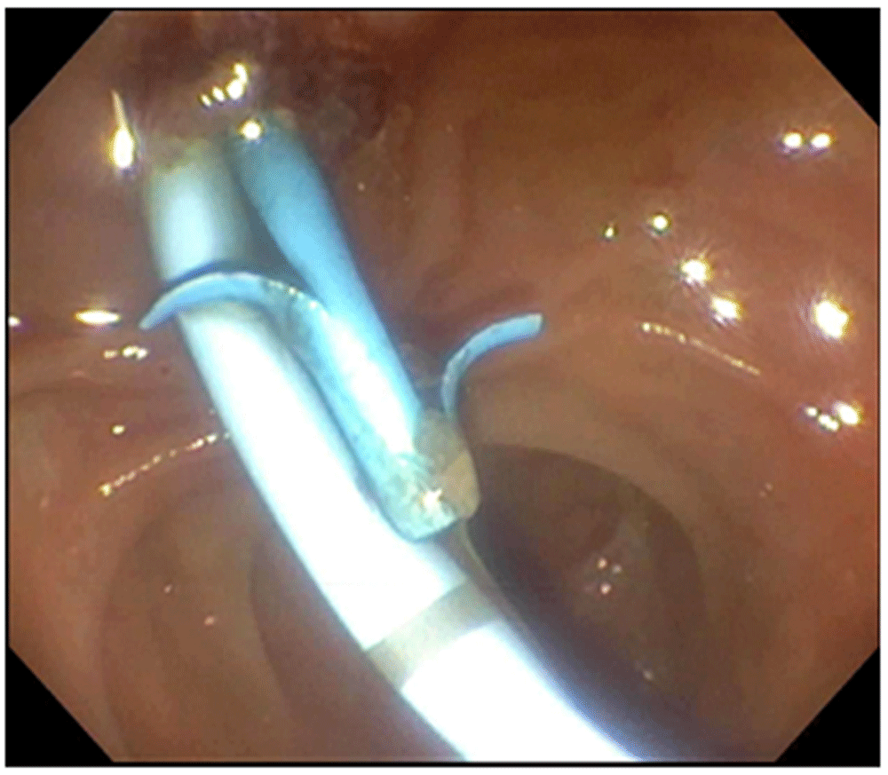
Endoscopic retrograde cholangiopancreatography (ERCP) was performed for suspected acute cholangitis secondary to obstruction from pancreatic mass, showing high-grade distal CBD and PD stricture. Sphincterotomy was performed first for the pancreatic duct, then the distal CBD and the stricture in CBD and PD, dilated by using a 4 mm × 4 cm dilating balloon catheter. Upon 4 mm dilation of the PD, a significant amount of pus with debris and stone fragments were extruded. The dilating balloon catheter was removed and a 9–12 mm injecting below retrieval balloon catheter was used to sweep the PD, yielding more pus, debris, and stone fragments. Brushings were obtained from the PD stricture followed by pediatric forceps biopsy of the CBD stricture to rule out malignancy. Subsequently, a 10 Fr × 9 cm biliary stent was placed into the CBD and a 7 Fr × 7 cm stent in the PD with prompt drainage from both stents consistent with the diagnosis of AOSPD (Figure 3 and Figure 4).

The patient’s sepsis resolved after the pancreatobiliary drainage with prompt resolution of leukocytosis, transaminitis, and total bilirubin levels. Piperacillin-tazobactam was continued for ten days. Biopsy and brushings confirmed hepatobiliary primary invasive adenocarcinoma. Given the grave prognosis, palliative care was proposed. The patient opted for home hospice care.
AOSPD is a very rare entity and its mention in literature is sparse. AOSPD presentation overlaps with cholangitis and this mimicry makes misdiagnosis or non-recognition as a separate clinical entity difficult. AOSPD presentation varies from acute to chronic, the most common symptom being abdominal pain1–5,8–12. Less commonly, it may also present as sepsis or septic shock1–4. However, the presence of infection does not always correlate with clinical manifestation. This was evident from cases of asymptomatic presentation, where lab work uncovered the underlying infection incidentally6,7. Pancreatic imaging plays a pivotal role. The main pancreatic duct diameter was significantly higher in AOSPD with Chronic Pancreatitis (CP) than Acute-on-Chronic Pancreatitis patients10. AOSPD as the initial presentation of pancreatic malignancy is reported only once before, besides our case10.
The presence of pancreatic ductal stones precipitated due to pancreatic secretion stasis also serves as a nidus of infection generation leading to septicemia1–5. Besides enteric pathogens, respiratory pathogens have been increasingly reported in association with AOSPD, especially Klebsiella3,5. In our case, the pancreatic fluid and urinary cultures grew Klebsiella of the same species; however, no bacteremia was present.
The mainstay of AOSPD diagnosis is through ERCP detection of purulent discharge from the main pancreatic duct, seen in the absence of associated lesions like an abscess or pancreatic pseudocyst. MRCP presumptively diagnosed one case by demonstrating pancreatic duct dilation. However, the diagnosis was still confirmed after ERCP lead to symptom resolution2.
Treatment is through source control via ERCP drainage and appropriate antibiotic administration. Notably, endoscopic naso-pancreatic drainage (ENPD) was additionally performed following ERCP in one reported case11. At this time, there are no established guidelines for the duration of therapy. Broad-spectrum antibiotic with enteric anaerobic coverage (vancomycin with piperacillin-tazobactam, ceftazidime with gentamicin) given for 7–14 days, is the mainstay of therapy1–6.
AOSPD is a rare and potentially fatal diagnosis that requires expeditious recognition and treatment with referral to a facility with ERCP capabilities and expert infectious disease management. Future reviews and studies should work toward universal guidance in management.
Written informed consent for publication of clinical details and clinical images was obtained from the patient.
All data underlying the results are available as part of the article.
| Views | Downloads | |
|---|---|---|
| F1000Research | - | - |
|
PubMed Central
Data from PMC are received and updated monthly.
|
- | - |
Is the background of the case’s history and progression described in sufficient detail?
Partly
Are enough details provided of any physical examination and diagnostic tests, treatment given and outcomes?
Yes
Is sufficient discussion included of the importance of the findings and their relevance to future understanding of disease processes, diagnosis or treatment?
Yes
Is the case presented with sufficient detail to be useful for other practitioners?
Yes
Competing Interests: No competing interests were disclosed.
Reviewer Expertise: Pancreatic surgery
Is the background of the case’s history and progression described in sufficient detail?
Yes
Are enough details provided of any physical examination and diagnostic tests, treatment given and outcomes?
Yes
Is sufficient discussion included of the importance of the findings and their relevance to future understanding of disease processes, diagnosis or treatment?
Yes
Is the case presented with sufficient detail to be useful for other practitioners?
Yes
Competing Interests: No competing interests were disclosed.
Reviewer Expertise: Internal Medicine and Neurology
Alongside their report, reviewers assign a status to the article:
| Invited Reviewers | ||
|---|---|---|
| 1 | 2 | |
|
Version 1 10 Mar 21 |
read | read |
Provide sufficient details of any financial or non-financial competing interests to enable users to assess whether your comments might lead a reasonable person to question your impartiality. Consider the following examples, but note that this is not an exhaustive list:
Sign up for content alerts and receive a weekly or monthly email with all newly published articles
Already registered? Sign in
The email address should be the one you originally registered with F1000.
You registered with F1000 via Google, so we cannot reset your password.
To sign in, please click here.
If you still need help with your Google account password, please click here.
You registered with F1000 via Facebook, so we cannot reset your password.
To sign in, please click here.
If you still need help with your Facebook account password, please click here.
If your email address is registered with us, we will email you instructions to reset your password.
If you think you should have received this email but it has not arrived, please check your spam filters and/or contact for further assistance.
Comments on this article Comments (0)