Keywords
Elbow, Arthritis, Tuberculosis, Delayed treatment
Elbow, Arthritis, Tuberculosis, Delayed treatment
AFB: acid fast-bacteria
AP: anteroposterior
CKD: chronic kidney disease
CT: computed tomography
ESR: erythrocyte sedimentation rate
HIV: human immunodeficiency virus
PA: posteroanterior
TB: tuberculosis
WHO: World Health Organization
Extrapulmonary tuberculosis (TB) is known to occur in joints with a percentage of approximately 1-3% of all TB cases, 2-5% of which are rare cases that occur in the elbow joints.1 TB is an endemic disease with the total number of cases approximating 845,000 in Indonesia.2 Elbow dysfunction is the result of progressive erosion and destruction of bone and joint, therefore early diagnosis and treatment are needed to prevent this outcome. Diagnosis is quite challenging and often late due to non-specific symptoms3 Joint TB is rarely detected because joint pain is not commonly considered to be a symptom of joint TB, especially if there are no respiratory complaints. Thus, diagnosis and treatment are often delayed. Here, we report a rare case of a patient with TB of the elbow joint, who received delayed treatment because he chose to undergo traditional treatment with massage therapy.
A 24-year-old Indonesian male who worked in an internet rental shop came to the orthopedic department of Bhayangkara Brimob hospital (Depok, Indonesia) with left arm pain and left elbow joint swelling. Physical examination revealed skin perforation with yellowish discharge on the left elbow. The patient experienced fever on the first few days as the left elbow became swollen, weight loss, and a decreased appetite, but no respiratory complaints.
Chronologically, one year prior to coming to the hospital, the patient noticed another pain in his left arm both in the upper and lower arm. He then chose to undergo regular traditional massage therapy every week for almost one year instead of seeking for medical treatments. At the first hospital visit, the elbow pain had gotten more severe and it became swollen. Within a month, discharge emerged from a small skin perforation located on the inner side of the left elbow. The patient finally went to the orthopedic department and underwent surgery. The patient had a history of undergoing reflexology massages between the fingers of his left hand to alleviate his toothache.
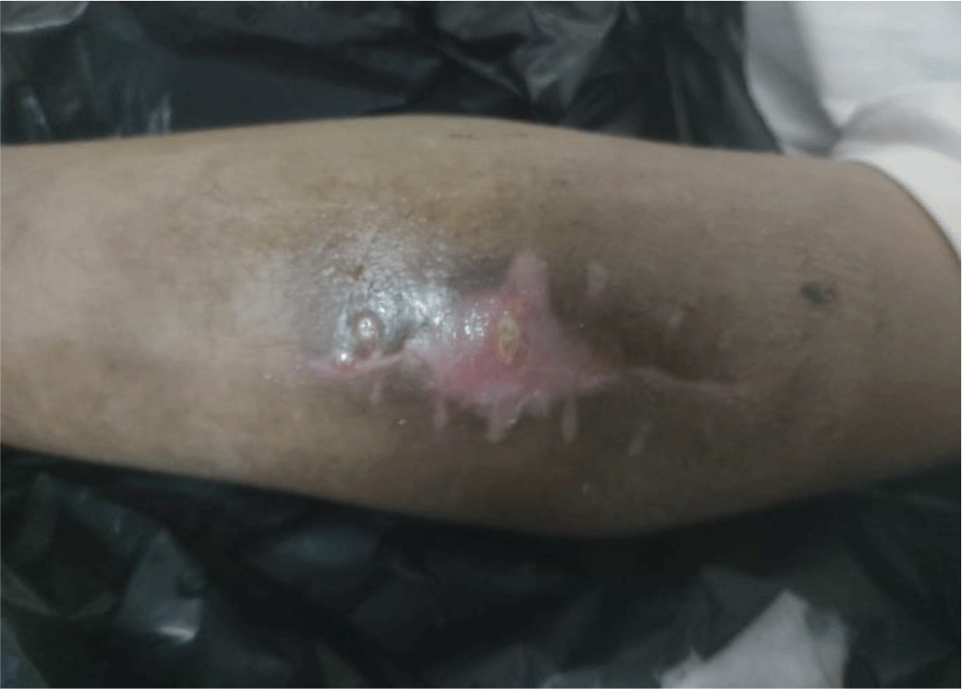
Upon physical examination, the left elbow joint appeared swollen and discharge was exuding from the perforated skin, as depicted in Figure 1. The patient could not lift his left arm because it was painful. Flexion and extension were also difficult due to the severity of the pain. The patient’s social environment has a culture of seeking help from local traditional massage therapists who are known to be uncertified to treat various health problems, and instead of recovering, the patient showed symptoms of worsening.
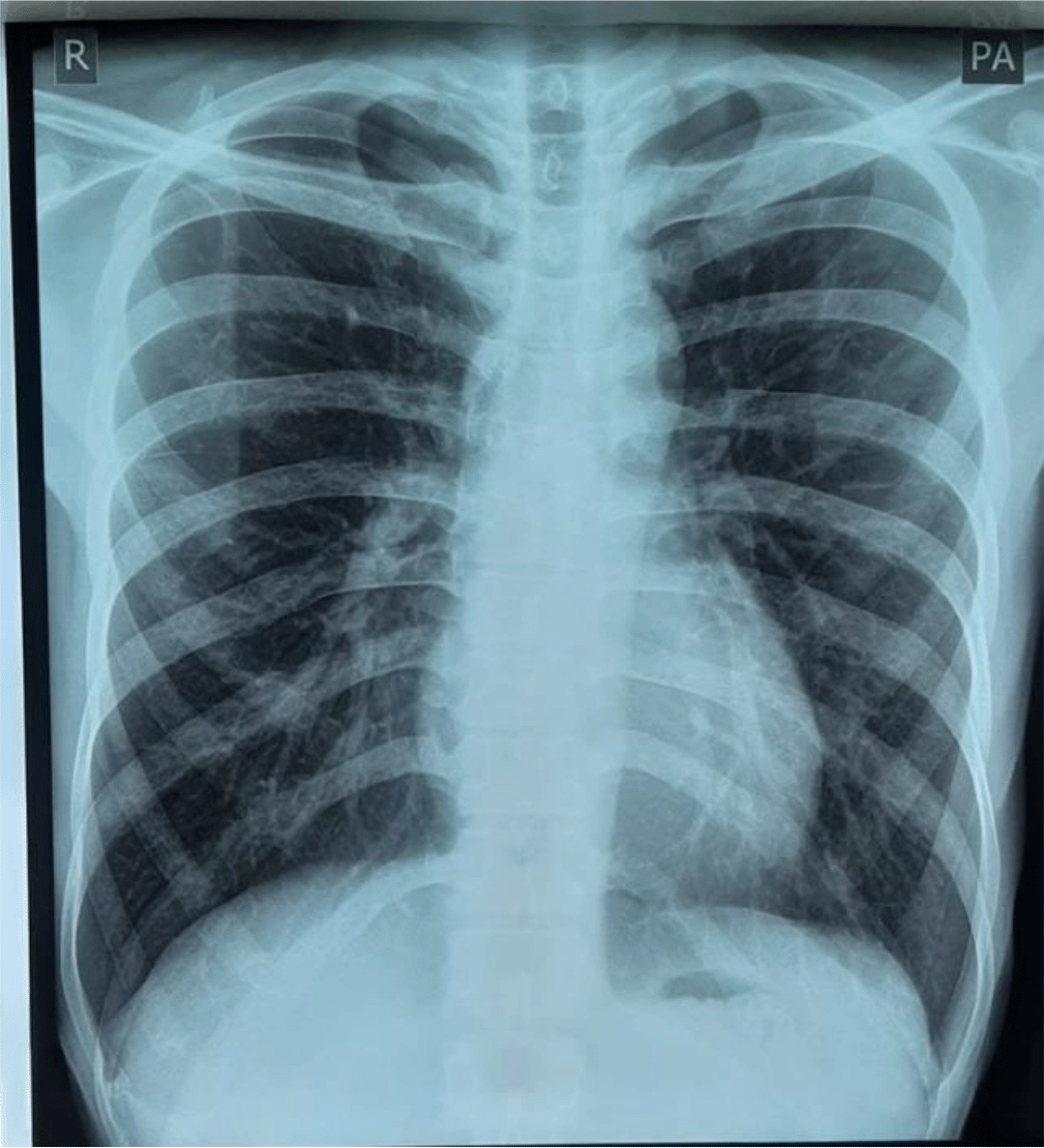
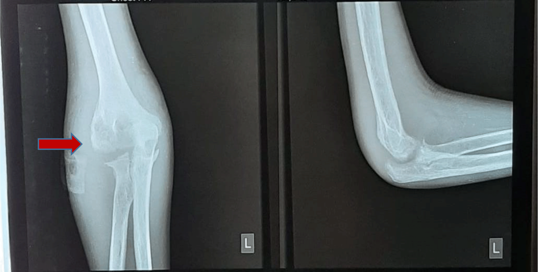
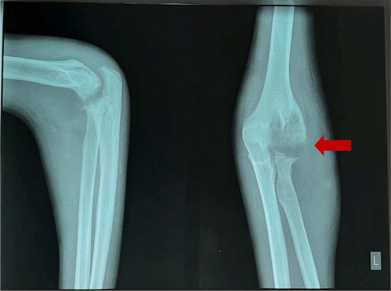
Laboratory examination revealed a leukocyte count of 15,000 (normal range: 5000-10,000 cells/μl), erythrocyte sedimentation rate (ESR) of 40 mm/hour (normal range: 0-10 mm/hour), eosinophils 9% (normal range: 1-3%), and monocytes 10% (normal range: 2-6%). Radiological examination by posteroanterior (PA) chest X-ray did not show any abnormality (Figure 2), anteroposterior (AP) and lateral projection of the left elbow joint radiographs showed erosion of the distal cortex of the humerus and radial bone, destruction of the distal cortex, and swelling of the soft tissue of the left elbow area (Figure 3).
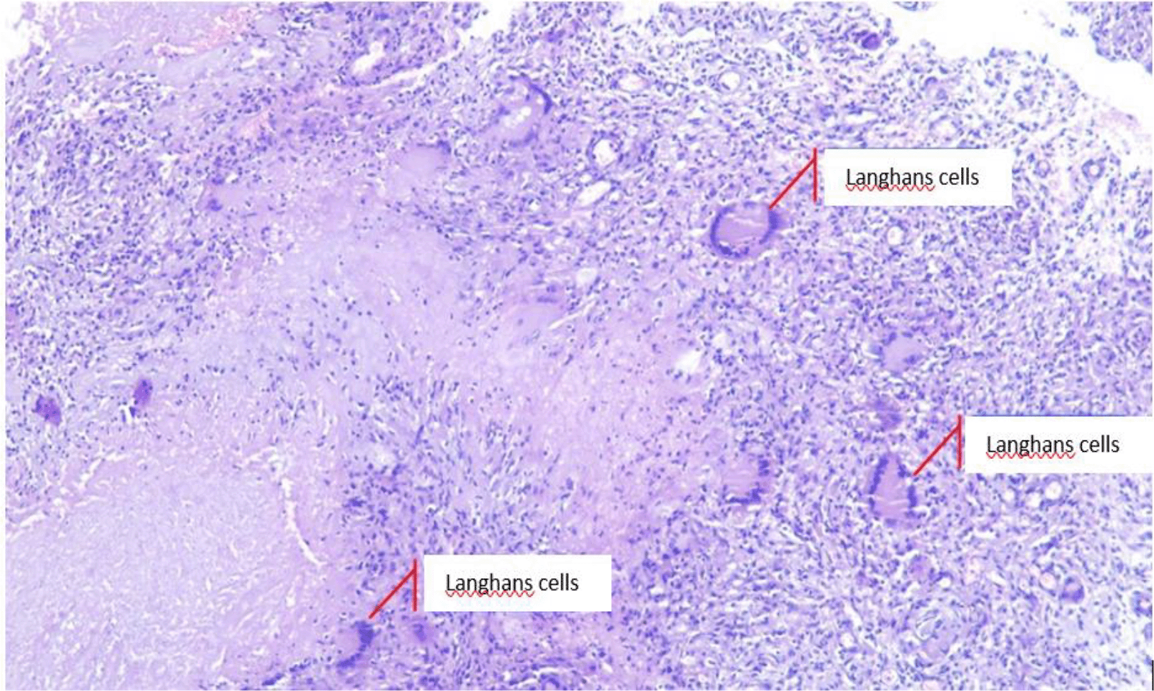
The patient was subsequently diagnosed with TB of the elbow joint. He then underwent left elbow arthrotomy and synovial fluid aspiration. The surgery was performed with the patient supine under general anesthesia. Incisions were made layer by layer on the posterior region of cubiti sinistra. White granulation tissue and thick yellow intra- and extra-articular pus were evacuated. Histopathological examination was also performed. The wound was irrigated with 2 L of 0.9% NaCl and hecting was performed layer by layer subsequently. Specimens were collected and sent for microbiological and pathological analyses. Acid-fast bacteria (AFB) smear and culture showed Mycobacterium tuberculosis. Histopathological examination showed granulomatous inflammation, swollen connective tissues containing epithelioid tubercle nests with necrotization, and datia Langhans cells (Figure 4). The results were consistent with TB. Furthermore, the anti-human immunodeficiency virus (HIV) test was negative.
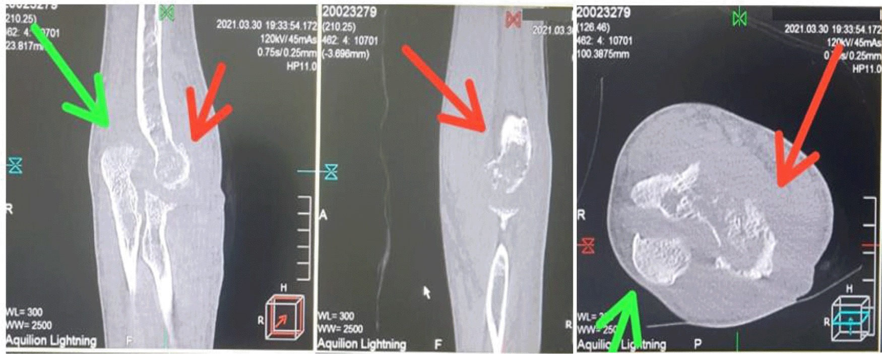
The patient was given a standard first-line oral regimen of extrapulmonary TB treatment; an intensive phase for two months with rifampicin 450 mg once daily, isoniazid 300 mg once daily, pyrazinamide 1000 mg once daily, and ethambutol 1000 mg once daily (2HRZE) and seven months of a continuation phase with rifampicin 450 mg once daily and isoniazid 300 mg once daily (7HR). The patient has been undergoing continuation phase of the treatment and his condition has been showing improvements, including decreased pain, increased appetite, and weight gain. However, flexion and extension are restricted. The patient reported clinical improvement and discharge was decreased. Left elbow joint radiographs showed minimal improvement (Figure 5). Computed tomography (CT) scan results showed destruction of the lateral epicondylus of the humeral bone and the processus olecranon of ulna bones, after two months of the treatment (Figure 6).
Picture was edited with photoshop CS4 version 11.0 to remove specific details of dates of patient care and patient’s identity.
Musculoskeletal TB occurs in about 10% of all cases of extrapulmonary TB, which commonly affects weight-bearing joints such as the spine (51%), pelvis (12%), hip,femur (10%), knee and tibia (10%). Reported cases of non-weight-bearing joints such as elbow joint TB are still relatively rare, and the diagnosis often neglected.1 Diagnosis of musculoskeletal TB requires the clinician’s ability to pay attention to joint swelling and chronic pain, as well as their effects on joint function4
Usually, respiratory and systemic symptoms are absent or only briefly present. In this report, only a history of fever was identified. Radiological examination of the lungs showed no abnormality. The complaints for joint TB are often non-specific, hence a late diagnosis1
The findings in this report are consistent with several previous studies. A study by Yazici et al. (2016) reported a TB of the elbow joint case in which there were no signs and symptoms of respiration. The results of chest radiographs were still within normal limits. The diagnosis was confirmed by AFB and histopathology examinations.5 Another study by Guan & Zeng (2021) reported osteoarticular TB with a picture of swelling and pain that was previously diagnosed as osteoarthritis. Although these cases are rare, they are difficult to diagnose and can cause pain and impaired function.6
Radiographic changes of the joints may suggest multiple osteolytic lesions and there may be erosions of the joints and swelling of the soft tissues.7 Unfortunately, this patient did not undergo a magnetic resonance imaging examination due to the limited available facilities. Definite diagnosis required synovial fluid aspiration. Microscopic examination and culture of fluid aspiration were very helpful, followed by histopathological results showing the caseous granuloma.8 These non-specific signs and symptoms often delay the diagnosis as skeletal TB, as reported in one study that showed the time lag from the onset of complaints until the diagnosis was confirmed as approximately 4-11 months. Additionally, some cases of skeletal TB occasionally were not detected by AFB and culture.9,10
Clinicians should not neglect to explore the history of exposure and factors that increase the risks of TB infection, such as close contact with confirmed TB patients, immunocompromised patients (e.g. HIV infection), diabetes mellitus, and having comorbid diseases such as chronic kidney disease. Therefore, it is necessary to screen the patient for the co-infectious diseases listed above. Other risk factors are old age, poor nutrition, and receiving immunosuppressive treatments6 Regarding this case, the risk factors are not clear.
In summary, the significance of this case is the recognition of risk factors for skeletal TB and chronic symptoms, so that they can be treated properly. Early diagnosis and treatment can be achieved through careful anamnesis that does not ignore the history of close contact with confirmed cases of TB patients, risk factors for TB infection, physical/clinical, radiological, and laboratory examinations. It is important for clinicians, especially those who work in an area endemic to TB, to suspect chronic joint pain whose clinical symptoms do not improve with conventional treatment as skeletal TB in the differential diagnosis. The specific AFB smear and culture tests are still important, although they can occasionally show false negative results. Extrapulmonary TB can be deceptive because it does not always cause typical symptoms and pulmonary involvement. Prompt diagnosis and treatment are essential to prevent joint damage and impaired function.
Left-arm and elbow pain, swelling, and immobility made me suffer. I knew that I had to go to the hospital for further treatment. However, I was afraid of surgery and at the suggestion of my family, I underwent traditional medicine with massage therapy for almost 1 year. I didn't expect that my illness would get worse and I had to have surgery immediately and take long-term medication. Now I feel better, my arm pain and swelling of my left elbow have decreased, even though I haven't been able to move my arm to its full potential.
| Views | Downloads | |
|---|---|---|
| F1000Research | - | - |
|
PubMed Central
Data from PMC are received and updated monthly.
|
- | - |
Provide sufficient details of any financial or non-financial competing interests to enable users to assess whether your comments might lead a reasonable person to question your impartiality. Consider the following examples, but note that this is not an exhaustive list:
Sign up for content alerts and receive a weekly or monthly email with all newly published articles
Already registered? Sign in
The email address should be the one you originally registered with F1000.
You registered with F1000 via Google, so we cannot reset your password.
To sign in, please click here.
If you still need help with your Google account password, please click here.
You registered with F1000 via Facebook, so we cannot reset your password.
To sign in, please click here.
If you still need help with your Facebook account password, please click here.
If your email address is registered with us, we will email you instructions to reset your password.
If you think you should have received this email but it has not arrived, please check your spam filters and/or contact for further assistance.
Comments on this article Comments (0)