Keywords
Phacoemulsification, Corneal astigmatism, Clear corneal incision, Opposite clear corneal incision, Vector analysis.
This article is included in the Eye Health gateway.
Phacoemulsification, Corneal astigmatism, Clear corneal incision, Opposite clear corneal incision, Vector analysis.
Currently cataract surgery is considered to be a refractive surgery, with the main therapeutic goal of achieving emmetropia; as a result, correction of corneal astigmatism becomes essential for such an operation.1,2 The prevalence of corneal astigmatism of more than 1 diopter is as high as 45% of those who undergo cataract surgery.3
It is possible to reduce pre-existing corneal astigmatism by creating a clear corneal incision at the steep meridian of the cornea; however, astigmatism correction is limited to a maximum of 1 diopter (D) when corrected by a small incision. A possible limitation is the difficulty in performing the operation which is caused by the location of the steep meridian, such as the difficulty of creating superonasal or inferonasal incisions at the left eye. This approach is usually sufficient for correcting astigmatism less than 1 D in most eyes.1,4 An opposite side clear corneal incision (OCCI) could enhance the flattening effect on the cornea.5
The current work aims to evaluate the efficacy and safety of on-axis corneal incision with or without OCCIs to correct preoperative corneal astigmatism during uncomplicated phacoemulsification surgeries. The current work is based on the fact that the cornea is not a perfect sphere and many patients present with corneal astigmatism in addition to cataracts. In one procedure, phacoemulsification, cataracts and corneal astigmatism can be corrected based on the principle of releasing incision.
Written informed consent was obtained from each participant for participation and publication of clinical information. The study was approved by the institutional regulation board committee of Faculty of Medicine – Baghdad University (Ref#0122-2019). The study followed the Declaration of Helsinki 2008 for research on human subjects.
This trial was registered with ClinicalTrials.gov (NCT04418986) on 5th June 2020. The trial was registered retrospectively, since mandatory clinical trial registration was only recently introduced to research policies in Iraq so after the study, we registered the trial to ensure transparency of the study.
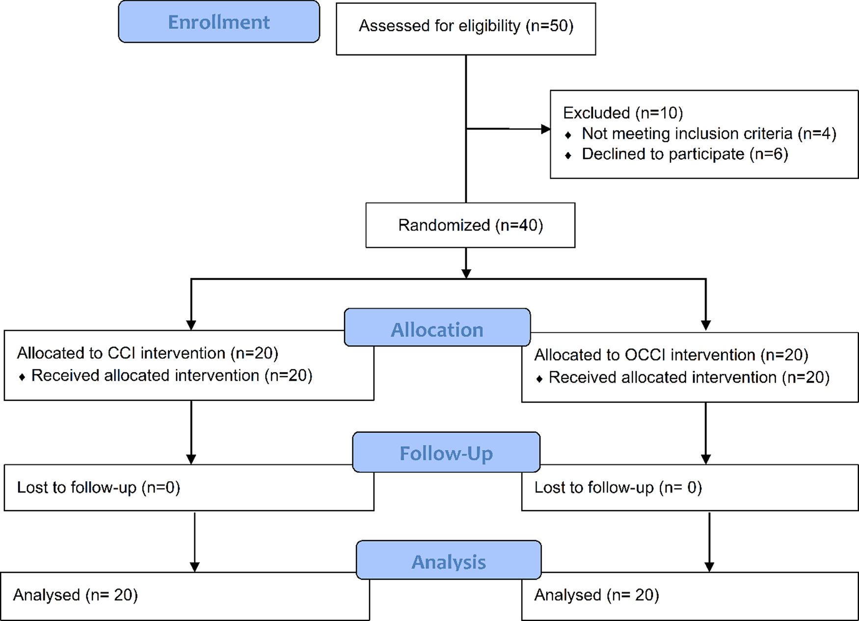
This study was a randomized, parallel two-arm interventional study. The study included 40 eyes with visually significant cataract and preoperative corneal regular astigmatism between 0.75 and 2 D undergoing phacoemulsification surgery.
Patients were divided into two groups in a 1:1 ratio: 20 patients as controls underwent phacoemulsification with on-axis incision (CCI group), and 20 patients underwent phacoemulsification with OCCIs (OCCI group).
The study was carried out in the ophthalmology clinic in Ghazi al-Hariri Surgical Specialties Hospital. The recruitment period was three months, from July 2019 until September 2019. All procedures took place in in the ophthalmology clinic in Ghazi al-Hariri Surgical Specialties Hospital, and all the procedures were carried out by NKM.
The participants were approached by the investigators during their visit to the ophthalmology clinic of Ghazi al-Hariri Surgical Specialties Hospital. The primary investigator, Dr. Najah K. Mohammad, examined each participant at admission to the center and conducted an initial interview to check the adherence of the patients to the inclusion and exclusion criteria, specifically age, previous ocular surgeries, and systemic diseases, and if the cataract intervention met their requirements. After obtaining their informed consent and confirming adherence of the participants to the inclusion and exclusion criteria they entered the study. Since this was a pilot study, the sample size was not determined by formal power analysis.
Computer-based randomization was used in which the patients were randomly divided into two groups. After the initial interview, the patients were numbered consequently, then randomized into two groups using the online software Research Randomizer.
Negative history of ocular surgery, clear cornea, and central corneal thickness (CCT) less than 640.
Patients with lenticular astigmatism, corneal opacities or pathology like Fuch’s endothelial dystrophy, previous ocular surgeries like glaucoma surgery or PKP or pterygium excision, posterior segment diseases and pathology, and complicated phacoemulsification were excluded from this study.
All the patients had their complete history taken and underwent ophthalmic examination that included: manifest refraction; uncorrected distance visual acuity (UCDVA); best corrected distance visual acuity (BCDVA); intraocular pressure (IOP) measurement by non-contact air puff tonometry; detailed examination of the anterior segment using slit lamp biomicroscopy; posterior segment detailed examination using indirect ophthalmoscopy; corneal topography, mesopic pupil size and corneal pachymetry were done using Sirius Topographer (CSO, Italy); and ocular biometry to measure axial length, anterior chamber depth, and horizontal white to white (WTW) distance were done using partial coherence interferometry (PCI), the ZEISS IOLMaster® 500 (Carl Zeiss Meditec AG, Germany).
CCI, clear corneal incision; OCCI, opposite clear corneal incision.
The procedure is similar to our previous study by Eliwa et al.,6 in which the steep meridian of the corneal topography was marked using a Whitehouse Gravity Axis Marker. This was done before local anesthesia was administered. Peribulbar anesthesia (lidocaine 2% [5 ml], bupivacaine 0.5% [5ml], and hyaluronidase 1,500 IU/mL [2 ml], all given via the peribulbar route) associated with mild sedation with midazolam was used in all cases.
Coaxial small incision cataract surgery was performed for all cases of both groups using a 2.8 mm keratome placed at steep meridian and 1-mm paracentesis was made 90 degrees apart with a 20-gauge microvitrectomy blade (Alcon Laboratories, Inc., Fort Worth, TX). Surgery was performed with a 30-degree, 0.9-caliper phacoemulsification tip (microtip) with the divide-and-conquer technique using the INFINITI vision system (ALCON laboratories INC).
The AcrySof SN60WF (Alcon Laboratories, Inc.) intraocular lens (IOL) was loaded in cartridge C and then inserted using a Royale injector (ASISCO LLC, Westmont, IL). The tip of the cartridge was introduced into the external part of the incision, after which the IOL was injected into the capsular bag.
In the OCCI group, a single penetrating incision was created with a 2.8 mm keratome in clear cornea, 1.5 mm anterior to limbal blood vessels, centered over the steep meridian and opposite the phacoemulsification incision.
Postoperative topical therapy included a combination of topical antibiotics (moxifloxacin drops 2% [1 – 2 drops every six hours for a two-week duration]), and steroid eye drops (prednisolone acetate 1% [1 – 2 drops every six hours for a two week duration, than tapered to twice daily for two weeks] and eye lubricants (Systane Ultra drops, 1 – 2 drops every six hours for four weeks).
Primary outcomes
Change in surgically induced astigmatism (SIA) one month after the phacoemulsification. Corneal astigmatism was examined by corneal topography, preoperatively and four weeks after the surgery, by the same technician, and changes in astigmatism were assessed by Alpins vector analysis comparing the SIA pre- and postoperatively.
Secondary outcomes
To investigate the differences between CCI and OCCI regarding the changes in corneal astigmatism assessed by corneal topography and SIA.
The Anderson-Darling test was used to assess the adherence of variables to normality, the paired t-test (or Wilcoxon median rank test if data did not follow normal distribution) was used to analyze the difference between preoperative and postoperative outcomes and the independent t-test (or Mann–Whitney U test if data did not follow normal distribution) was used to analyze the difference between two independent variables. The p-value was considered to be significant if <0.05 and all analysis was carried out using SPSS version 20.0.1 (IBM Corp, Armonk, NY).
The study included 40 eyes (Figure 1). The mean age of participants was 57.9 ± 13.25, and 52.5% were males (21/40).16 These cases were divided into two groups: CCI and OCCI groups. There was no statistically significant difference between the two groups regarding patient demographic data, as illustrated in Table 1.
Mean corneal power significantly reduced after one month postoperatively in the OCCI group, while the CCI group showed a slight reduction in mean corneal power postoperatively. The UCVA significantly improved in both groups after the procedure, from 0.114 ± 0.048 and 0.146 ± 0.102 preoperatively in CCI and OCCI groups, respectively (approximately 6/60, 1 LogMAR), to 0.35 (approximately 6/18, 0.48 LogMAR) in the CCI group and 0.45 (approximately 6/12, 0.3 LogMAR) in the OCCI group postoperatively. Mean BCVA significantly increased from 0.283 ± 0.097 (approximately 6/24, 0.60 LogMAR) preoperatively to 0.438 ± 0.146 (approximately 6/12, 0.3 LogMAR) postoperatively in the CCI group, and the OCCI group showed a statistically significant increase in BCVA from 0.375 ± 0.150 (approximately 6/18, 0.48 LogMAR) preoperatively to 0.564 ± 0.125 (approximately 6/9, 0.18 LogMAR) postoperatively, as illustrated in Table 2.
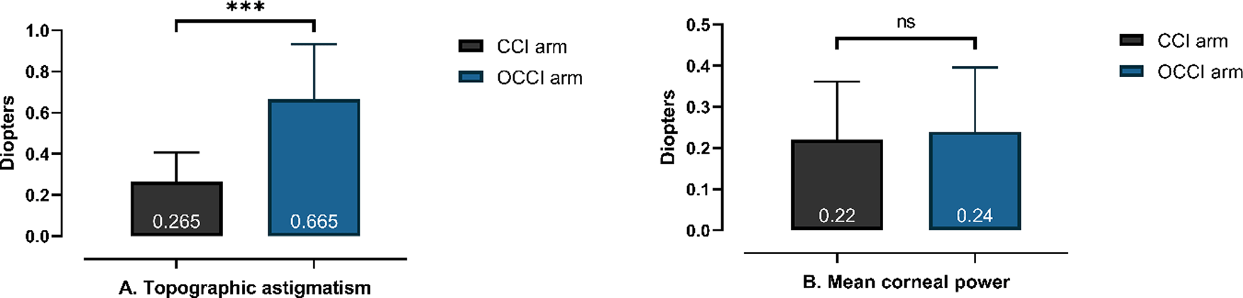
The mean change in topographic astigmatism was statistically significantly higher in the OCCI group (0.665 ± 0.268 D) than the CCI group (0.265 ± 0.142 D) (p-value < 0.001; Figure 2A), while there was no statistically significant difference between the two groups regarding the change in mean corneal power (see Figure 2B).
CCI, clear corneal incision; OCCI, opposite clear corneal incision.
Mean magnitude of target induced astigmatism vector (TIA) of both groups was 1.37 D, which was similar to preoperative topographic astigmatism. Most of the parameters (surgical induced astigmatism, magnitude of error, and correction index) were significantly higher in OCCI group compared to the CCI group (p-value < 0.01), as illustrated in Table 3.
Cataract surgery is considered to be a refractive procedure aiming for postoperative emmetropia, which required correction of spherical and astigmatic errors.2 Corneal astigmatism of more than 1 D has been reported in up to 45% of cataract surgery candidates,3 while 11.6% of eyes undergoing cataract surgery have more than 2 D of corneal astigmatism.7
Since the astigmatic effect of CCIs on the steep meridian is well documented, it is reasoned that adding an CCI opposite to the first incision (OCCI) would enhance the flattening effect, providing a good method to manage the preexisting corneal astigmatism during cataract surgery. OCCIs are simple to perform, do not require additional skills or instruments, and since the additional incision is self-sealing, particularly when it is not used to introduce instruments, the added risk to the surgery is negligible compared to its benefit of astigmatism reduction.5,8
The topographic corneal astigmatism in the CCI group decreased by 0.265 ± 0.142 diopters, while OCCI reduced the topographic corneal astigmatism by 0.665 ± 0.268 diopters, which was in agreement with Bhalla’s study, which reported a reduction of 0.28 and 0.7 D in topographic astigmatism in CCI and OCCI groups, respectively,9 but these results were different to other studies.
In 2000, Lever and Dahn performed cataract surgery on 33 eyes of 26 patients with an OCCI ranging from 2.8 to 3.5 mm in length and they reported a mean corneal astigmatism correction of 2.06 D with this technique.5 Qammar and Mullaney in 2005 reported a mean astigmatic correction of 1.23 ± 0.49 D.10 In 2006, Khokhar and colleagues11 reported that a 3.2 mm CCI reduced topographic astigmatism by 0.59 ± 0.43 D at three months follow-up, which is more than our CCI group, which showed a 0.265 ± 0.142 D reduction in topographic astigmatism. The OCCI group of Khokhar’s study showed a reduction in preoperative corneal astigmatism of 1.60 ± 0.45 D versus 0.665 ± 0.268 D in our study.
On the other hand, our results were higher than Tadros’ study, which reported a 0.5 ± 0.73 D reduction in preoperative corneal astigmatism.12 They were also higher than Bazzazi’s study, which reported the mean topographic astigmatism change to be 0.003 D and 0.036 D in superior and temporal CCI groups, respectively, which was less than our CCI group (0.265 D), while in the OCCI groups topographic astigmatism was reduced by 0.505 D and 0.557 D in superior and temporal groups, respectively, versus 0.665 D in our study.13 In addition, Nemeth’s study found a 0.13 D reduction in topographic astigmatism in their CCI group, which was less than our CCI group (0.265 D), while Nemeth’s OCCI group showed a reduction in topographic astigmatism of 0.2 D versus 0.665 D in our study.14
Using vector analysis,15 we also evaluated the predictability of the astigmatic correction. The topographic data were analyzed. The mean magnitude of TIA was 1.370 ± 0.384 D in the CCI group and 1.370 ± 0.505 D in the OCCI group that was similar to preoperative topographic astigmatism. The mean magnitude of SIA was 0.403 ± 0.183 D in our CCI group, which was less than Khokhar’s study (0.85 ± 0.75 D)11 and Nemeth’s study (0.61 ± 0.43 D)14 but more than Bazzazi’s study (0.036 ± 0.51 D)13 and Bhalla’s study (0.33 ± 0.22 D).9 Regarding the OCCI group of our study, the mean magnitude of SIA was 0.849 ± 0.519 D, which was less than that of Lever and Dahn’s study (2.25 D),5 Tedros’ study (1.57 D),12 Qammar and Mullaney’s study (2.10 D),10 Nemeth’s study (0.99 D)14 and Khokhar’s study (1.66 D).11 On contrary, the mean SIA of our OCCI was higher than that of Bazzazi’s study (0.505 D), and Bhalla’s study (0.76 D).9
All the above differences in the resulted mean change in astigmatism and SIA could be the result of using 2.8 mm incisions in our study compared to 3.2 mm incisions used in most of the studies that corrected a higher astigmatism than we did, or may be due to different sample sizes, postoperative topographic astigmatism (1.37 D in our study versus 3.6 D in other studies), and follow up period (one month versus 2-3 months in others).
Both incisional methods are useful methods for correction of preoperative corneal astigmatism but OCCIs correct a higher amount of astigmatism than the on-axis CCIs. In addition, using a 3.2 mm incision in both ways may correct a higher amount of astigmatism than using a 2.8 mm incision.
Zenodo: Corneal Astigmatism - excel. https://doi.org/10.5281/zenodo.4907691.16
This project contains the study data in XLSX format.
Zenodo: Incisional Correction Of Corneal Astigmatism During Phacoemulsification-proposal. https://doi.org/10.5281/zenodo.4907717.17
This project contains a copy of the study protocol in DOCX format.
Zenodo: CONSORT checklist for “Incisional correction of corneal astigmatism during phacoemulsification – a randomized trial”. http://doi.org/10.5281/zenodo.5028589.18
Data are available under the terms of the Creative Commons Attribution 4.0 International license (CC-BY 4.0).
| Views | Downloads | |
|---|---|---|
| F1000Research | - | - |
|
PubMed Central
Data from PMC are received and updated monthly.
|
- | - |
Provide sufficient details of any financial or non-financial competing interests to enable users to assess whether your comments might lead a reasonable person to question your impartiality. Consider the following examples, but note that this is not an exhaustive list:
Sign up for content alerts and receive a weekly or monthly email with all newly published articles
Already registered? Sign in
The email address should be the one you originally registered with F1000.
You registered with F1000 via Google, so we cannot reset your password.
To sign in, please click here.
If you still need help with your Google account password, please click here.
You registered with F1000 via Facebook, so we cannot reset your password.
To sign in, please click here.
If you still need help with your Facebook account password, please click here.
If your email address is registered with us, we will email you instructions to reset your password.
If you think you should have received this email but it has not arrived, please check your spam filters and/or contact for further assistance.
Comments on this article Comments (0)