Keywords
ApoB100/ApoAI ratio, ApoB100, ApoAI, Hepatic lipase, Glucose
ApoB100/ApoAI ratio, ApoB100, ApoAI, Hepatic lipase, Glucose
Atherosclerotic coronary artery occlusion is the most frequent cause of coronary heart disease (CHD); numerous epidemiologic studies and randomized clinical trials have established that lipoprotein metabolism is a major contributor to CHD.1 Conventionally, it was thought that increases in plasma low-density lipoprotein cholesterol (LDL-C) and decreases in high-density lipoprotein cholesterol (HDL-C) were the major factors causing CHD.2 However, numerous studies have suggested that another factor in CHD risk might be the apolipoprotein B100/apolipoprotein AI (ApoB100/ApoAI) ratio.3–7 ApoB100 is a large surface protein present on low-density lipoprotein (LDL) and serves as a ligand for the LDL receptor, which facilitates its clearance from the plasma. Apolipoprotein AI (ApoAI) is the major protein constituent on high-density lipoprotein (HDL) and plays a central role in reverse cholesterol transport by stabilising the HDL particle, interacting with the ATP-binding cassette transporter I, activating lecithin cholesterol acyl transferase and acting as a ligand for the hepatic scavenger receptor.2
Hepatic lipase (HL) is synthesized and secreted by the liver, and is found extracellularly in the liver and in steroidogenic organs, primarily bound to proteoglycans. As a member of the lipase gene family, HL is a key enzyme catalyzing the hydrolysis of triglycerides (TG) and phospholipids (PLs), and it is involved in the remodeling of remnant LDL and HDL.8 It is also notable that HL facilitates the uptake of chylomicron remnant-like particles, not only as a lipolytic enzyme but also as a ligand, by bridging the lipoproteins to specific receptors or heparan sulphates on the hepatocytes.9
However, it remains unclear whether HL is pro or anti-atherogenic in lipoprotein metabolism. Prior evidence indicates that it functions as a double-edged sword. On one hand, HL accelerates atherosclerosis by hydrolyzing large LDL particles to dense LDL lipoproteins. On the other hand, HL delays atherosclerosis by hydrolyzing HDL2 to smaller HDL3.10 In order to understand the mechanism of how HL modulates atherosclerosis at the molecular level, an in vitro cell assay model was built in the current study. HL activity and extracellular expression of ApoB100 and ApoAI were measured in cultured HepG2 cells. This research may provide the targets of many novel therapeutics and is an area with great potential for the prevention and treatment of CHD.
It has been shown that a high-carbohydrate (high-CHO) diet can reduce the risk of CHD. Further analysis found that this high-CHO diet interacted with different HL promoter polymorphisms to affect the ApoB100/ApoAI ratio instead of the HDL-C related ratios.6,7,11 In the current study, various amounts of glucose were used to stimulate the secretion of HL, ApoB100 and ApoAI in cultured HepG2 cells. It was hypothesized that the differential expression of HL could modulate ApoB100/ApoAI ratio in vitro.
HepG2 cells (American Type Culture Collection, ATCC, Rockville, MD, USA) were routinely cultured in DMEM + 10% FBS (contains 4.5 g/L glucose, normal glucose group) at 37°C in a 5% CO2 incubator up to 70% confluency. For ApoB100 and ApoAI expression assays, cell culture media that contained different concentrations of glucose (1 and 10g/L) were then added to the cultured cells in order to make the low glucose group and high glucose group besides the original normal glucose group. For the HL activity assay, 25U/ml heparin was added to the above low, normal and high glucose cell culture media used for ApoB100 and ApoAI expression assays to make the low, medium and high glucose groups for HL activity assay. All the cell groups then continued to be cultured for eight hours before the assays.
The culture media of HepG2 cells were collected. ApoB100 and ApoAI expression were measured with commercial sandwich enzyme-linked immunosorbent assay (ELISA) kits (cat. no. H0124 and H0123) from ShangHai MEIXUAN Biological Science and Technology Ltd (Shanghai, China). The microplate provided in this kit had been pre-coated with a monoclonal antibody specific for ApoB100 or ApoAI. Standards or samples were then pipetted into the microplate wells, and ApoB100 or ApoAI present in the samples or standards binds to antibodies adsorbed to the microplate wells. To quantitatively determine the amount of ApoB100 or ApoAI present in the samples, the horseradish peroxidase (HRP)-conjugated polyclonal antibody specific for ApoB100 or ApoAI was added to each well. The microplate was incubated for one hour, and then the wells were thoroughly washed to remove any unbound components. The substrate solutions A and B were respectively added to each well. After the enzyme (HRP) and substrate reacting over a short period, this reaction was stopped by addition of a sulphuric acid solution and the color change is measured at a wavelength of 450 nm. The proportion to amount of ApoB100 or ApoAI bound in the initial step develops in the color change. Color intensities were measured using a MK3 microplate reader (Thermo Fisher, MA, USA).
HL activities were determined using the Lipoprotein Lipase (LPL)/Hepatic Lipase (HL) Detection Kit (cat. no. A067) from Nanjing Jiancheng Bioengineering Institute (Nanjing, China). LPL and HL can catalyze the hydrolysis of triglyceride and the product of the reaction is glycerol and free fatty acid (FFA). The amount of free fatty acid can be determined in the presence of reagents from the kit and thus the activity of LPL and HL can be calculated. As a kind of glycoprotein, the activity of HL need not to be activated by apolipoprotein C-II or other ions and the activity will not be inhibited by protamine or high concentration of salt. According to this, the LPL activity and HL activity can be measured separately. The culture media of HepG2 cells were collected. Reagents I, III and VI from the kit were added, blend the mixture thoroughly for 2-5 minutes. The blending is not completed until the bottom of the tube appears to be cream color and the mixture will not separate immediately after stopping the blending. Centrifuge at 3,500 rpm for 10 minutes after the completion of blending. The result mixture should be layered clearly. Otherwise, the mixture should be re-blended and centrifuged again. Remove the supernatant and the solidified layer in middle with a glass pipette. Transfer the solution at the bottom layer with a glass pipette. The glass pipette should be washed 2-3 times with absolute ethanol followed by 2-3 times cleanings by reagent VI. Warm at 37°C for 1-2 minutes if turbidity is observed. Extract 1 ml solution obtained from step 5 and add chromogenic agent. The cuvettes should be washed by absolute ethanol followed by reagent VI. All methods are carried out with glass equipment. No plastic equipment is allowed. Color intensities were measured using a MK3 microplate reader (Thermo Fisher, MA, USA) at 550 nm with 0.5 cm path length. One enzyme activity unit is defined as 1μmol fatty acid generated with 1ml reaction volume within an hour.
Experimental results were expressed as the mean ± standard deviation (SD). Statistical analyses were performed using one-way ANOVA to determine significant differences among groups. Pearson’s correlation coefficient was used to determine the correlation between the variables of ApoAI and HL, ApoB100 and HL, and ApoB100/ApoAI ratio and HL. IBM SPSS Statistics version 22 was used for data analysis.
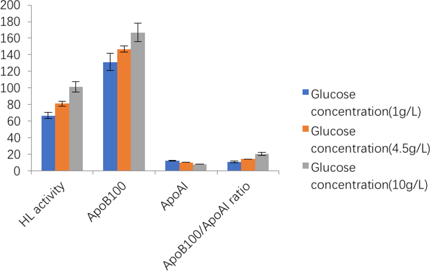
To explore the impacts of glucose on ApoB100 and ApoAI secretion over different concentrations, HepG2 cells were exposed to low (1 g/L), medium (4.5 g/L) and high (10 g/L) concentrations of glucose. As shown in ELISA revealed that with the glucose concentration increase, ApoB100 concentrations were significantly increased (1 g/L < 4.5 g/L < 10 g/L), while ApoAI concentrations were significantly decreased (1 g/L > 4.5 g/L > 10 g/L). ApoB100/ApoAI ratios were significantly increased (1 g/L < 4.5 g/L < 10 g/L). ANOVA showed that the differences are significant (p < 0.001).21
As shown in Table 1 and Figure 1, significantly increased HL activities were observed with the glucose concentration increase (1 g/L < 4.5 g/L < 10 g/L). ANOVA showed that the differences are significant (p < 0.001).
Hepatic lipase activity, ApoB100, ApoAI and ApoB100/ApoAI ratio in HepG2 cell cultured in low, normal and high glucose concentration.
| Low glucose concentration (1 g/L) | Normal glucose concentration (4.5 g/L) | High glucose concentration (10 g/L) | F | P | |
|---|---|---|---|---|---|
| HL activity (U/mg) | 66.71 ± 3.83 | 81.09 ± 2.76** | 101.44 ± 6.28**## | 88.863 | <0.001 |
| ApoB100 (g/ml) | 131.31 ± 10.39 | 146.77 ± 3.82** | 167.02 ± 10.84**## | 24.052 | <0.001 |
| ApoAI (μg/ml) | 12.06 ± 0.47 | 10.31 ± 0.19** | 8.18 ± 0.31**## | 190.710 | <0.001 |
| ApoB100/ApoAI (×106) | 10.92 ± 1.15 | 14.24 ± 0.23** | 20.49 ± 1.95**## | 82.211 | <0.001 |
Hepatic lipase activity, ApoB100, ApoAI and ApoB100/ApoAI ratio in HepG2 cell cultured in low, normal and high glucose concentration.
Pearson’s correlation coefficient results showed positive and significant correlations between ApoB100 and HL activity (r = 0.782) (p < 0.001), and positive and significant correlations between ApoB100/ApoAI and HL activity (r = 0.890) (p < 0.001). Pearson’s correlation coefficient results also showed a negative and significant correlation between ApoAI and HL activity (r = −0.956) (p < 0.001) (Table 2).
While studying the glucose-induced HL, ApoB100 and ApoAI expression in cultured HepG2 cell in vitro, in the culture media that contained a higher concentration of glucose, HL activities, ApoB100 concentrations and ApoB100/ApoAI ratios were found to be significantly increased, and ApoAI concentrations were significantly decreased. In the culture media that contained a lower concentration of glucose, however, HL activities, ApoB100 concentrations and ApoB100/ApoAI ratios were found to be significantly decreased, and ApoAI concentrations were significantly increased. This result is consistent with the author’s previous finding in an in vivo human cohort study that HL activity affects the ApoB100/ApoAI ratios instead of the HDL-C related ratios.6,7,11
However, a high-CHO diet was found to have generally favorable effects on the ApoB100/ApoAI ratio, reducing CHD risk, in a previous human cohort study in vivo,6 while the higher concentration of glucose induced an undesirable ApoB100/ApoAI ratio change, increasing CHD risk, in the present in vitro study. We can reach a reasonable explanation to this seemingly contradictory data if we look at what is happening inside the body after the meal; the rich viscous fiber contained in the high-CHO diet we administered in our cohort study has been shown to significantly reduce postprandial glucose excursions,12–14 which means the cells inside the body were actually exposed to a lower level of glucose after the high-CHO diet. Thus, the results we obtained from the lower concentration of glucose in the HepG2 cell culture experiment in vitro should correspond to the results we obtained from the high-CHO diet experimental group in vivo.
This study supports our hypothesis that HL regulates lipoprotein metabolism by modulating ApoB100/ApoAI ratio. HL is considered to be a key kinase in lipoprotein metabolism, which regulates multiple cell functions, but its functions in cell signal transduction pathways are still not fully understood. This study aimed to elucidate the effect of HL on ApoB100/ApoAI ratio. HL may act as a double edge sword in the regulation of both ApoB100 and ApoAI functions in response to metabolic change such as glucose fluctuation, and the ultimate effects of HL can be measured by ApoB100/ApoAI ratio. It has already been shown in our current research that ApoB100/ApoAI ratio has positive and significant correlations with HL activity. Further studies are needed to determine the details of the series of events that take place in a cell.
Lipoproteins are spherical particles that carry lipids in the body. These particles contain both lipids and proteins. LDL and HDL are the two main types of lipoproteins. LDL delivers fat molecules to cells. A high LDL level means too much LDL cholesterol in the blood. This extra LDL, along with other substances, forms plaque which can build up in the arteries to form atherosclerosis. Plaque can be built up in the arteries, causing heart disease. HDL-C carries cholesterol from other parts of the body back to the liver. The liver then processes the cholesterol for excretion to reduce the risk of heart disease. It is well known that both LDL and HDL consist of heterogeneous particles of different size and density.15 ApoB100 is the primary protein in LDL and ApoAI is the primary protein in HDL. Normally, it is proteins instead of lipids which perform essential functions within organisms, including catalyzing metabolic reactions, providing structure to cells and organisms, and transporting molecules from one location to another. It is likely HL regulates lipoprotein metabolism by directly modulating their protein components like ApoB100 and ApoAI rather than their lipid components like triglycerides, phospholipids, and cholesterol molecules. Although previous in vivo studies have already indicated this,6,7,11 further in vitro studies are needed to prove it on a molecular level. The current study is the first to explore this issue in an in vitro cell culture system, and the result positively supports this hypothesis.
For many years, the ApoB100/ApoAI ratio rather than ApoB100 or ApoAI alone has been extensively reported as a risk factor of CHD.16–18 Atherosclerosis, the process involved in LDL oxidization within the walls of arteries, might be due to the more powerful function of ApoB100 to deliver lipids to the cells than ApoAI to transport lipids out of the cells and take them back to the liver. The dynamic equilibrium of these two proteins, which can be described in their ratios, is essential to our health.
This finding helps to explain why the existing clinical treatments based on the regulation of lipoproteins themselves cannot achieve the desired results in many clinical cases. For example, although statins have documented efficacy in reducing clinical events and angiographic disease progression in patients with coronary atherosclerosis, the results of subsequent large prospective clinical trials using different types of statins clearly demonstrate that statins do not have a short-to-medium term effect on prevention of restenosis after successful conventional percutaneous transluminal coronary angioplasty.19 Current available data also indicate that increased HDL-C levels do not always correlate with enhanced HDL functions because HDL is highly heterogeneous and there are different HDL subpopulations.20
In conclusion, in vitro HepG2 cell culture revealed correlations between ApoB100, ApoAI, ApoB100/ApoAI ratios and HL activity. Higher amounts of glucose can induce significantly increased HL activity, ApoB100 levels and ApoB100/ApoAI ratio and significantly decreased ApoAI levels, while lower amounts of glucose can induce significantly decreased HL activity, ApoB100 levels, ApoB100/ApoAI ratio and significantly increased ApoAI levels. The result suggests a new and central cell signal transduction pathway in lipoprotein metabolism and might provide molecular targets for clinical diagnoses and treatments of CHD.
Harvard Dataverse: Replication Data for: Glucose induced hepatic lipase expression and ApoB100/ApoAI ratio changes in cultured HepG2 cells in vitro, https://doi.org/10.7910/DVN/CUBA3F.21
This project contains the following underlying data:
Data are available under the terms of the Creative Commons Zero “No rights reserved” data waiver (CC0 1.0 Public domain dedication).
| Views | Downloads | |
|---|---|---|
| F1000Research | - | - |
|
PubMed Central
Data from PMC are received and updated monthly.
|
- | - |
Provide sufficient details of any financial or non-financial competing interests to enable users to assess whether your comments might lead a reasonable person to question your impartiality. Consider the following examples, but note that this is not an exhaustive list:
Sign up for content alerts and receive a weekly or monthly email with all newly published articles
Already registered? Sign in
The email address should be the one you originally registered with F1000.
You registered with F1000 via Google, so we cannot reset your password.
To sign in, please click here.
If you still need help with your Google account password, please click here.
You registered with F1000 via Facebook, so we cannot reset your password.
To sign in, please click here.
If you still need help with your Facebook account password, please click here.
If your email address is registered with us, we will email you instructions to reset your password.
If you think you should have received this email but it has not arrived, please check your spam filters and/or contact for further assistance.
Comments on this article Comments (0)