Keywords
Osteosarcoma, intercalary reconstruction, vascularized fibula, immunohistochemistry, vascular analysis, Endomucin
We describe the case report of an Osteosarcoma patient, with a Li-Fraumeni Syndrome, presenting with a pathological femoral fracture. The patient was treated with a multidisciplinary approach associating neoadjuvant and adjuvant chemotherapy with excisional surgery. The femoral reconstruction consisted of a ``Capasquelet'' reconstruction combining an induced membrane and a vascularized fibula allograft allowing a good functional result with an early weight-bearing. We managed to complete our histological analysis in this patient, in order to evaluate the tumor vascularization. Indeed, using the syngeneic osteosarcoma MOS-J mouse model, we highlighted previously that CD31+/\ensuremath{\alpha}-SMA+ vessels may be indicators of vasculature normalization and therefore may be used as specific markers of a good therapeutic response. Thus, we search for its interest in this specific case as preliminary work. The aim was to assess the feasibility and technical validity of the vascularization analysis of a human osteosarcoma tumor specimen. Therefore, we propose an immunohistochemistry methodology with multiplexed immunofluorescence to assess the vascularization as a promising marker in human osteosarcoma tissue.
Osteosarcoma, intercalary reconstruction, vascularized fibula, immunohistochemistry, vascular analysis, Endomucin
According to the first reviewer comment we have checked on TCGA expression of EMCN and aSMA in osteosarcoma.
We have modified our discussion
See the authors' detailed response to the review by Mo-Fan Huang
Osteosarcoma care remains a challenge in various ways, such as determining chemotherapy response on biopsy, but as well with challenging reconstruction, especially in intercalary bone tumor resections. We propose in this case report to put highlight on these two points, describing an original technique of massive bone loss reconstruction, and focusing on innovative immunohistochemestry multiplexing techniques in order to better assess vascularization of osteosarcoma as a possible prognosis factor.
Massive diaphyseal bone defects are a reconstruction challenge, and several surgical methods have been described1–3 (vascularized fibula, autograft, bone transport, massive endoprosthesis, etc.). There is no gold-standard, and indications are a matter of debate, with variable results. We here described an innovative technique for managing a critical femoral bone defect of 24 cm using the Capasquelet procedure.4 This original two-stage technique combine the allograft technique with a centromedullary inlaid vascularized fibula (i.e., “Capanna technique”)5 and the induced membrane technique (i.e., “Masquelet technique”).6 The combination of these, called the “Capasquelet” technique get several advantages: the biological receptacle with the induced membrane prevents graft resorption and has also an osteoinductive role7; it also decreases infection risks, and avoids a long one-stage surgery. This case report describes this hybrid surgical technique on a massive femur defect. We assessed the bone healing, the delay to complete weight-bearing, and a functional score analysis in this patient.
On the other hand, predicting the chemotherapy response of osteosarcoma patients, with treatment such as high-dose methotrexate or doxorubicin cocktail, remains a challenge.8–10 Moreover, the prognostic value of induced necrosis analysis is questionable, and is only available after neo-adjuvant treatment is done on resection piece. New markers of prognostic response are urgently needed, focusing on tumor microenvironment.11 As we showed in our preclinical mouse model, using the syngeneic osteosarcoma MOS-J mouse model, vascular analysis of tumor depicted with immunohistochemestry fluorescent multiplexing might play a role as being a potential prognosis factor of tumor response.10,12 Our results may have suggested that the presence of CD31+/α-SMA+ vessels could be considered to be indicators of mature and functional vasculature able to deliver drugs adequately, and may be used as specific markers of a good therapeutic response. Nonetheless, no human tissue analysis using this method has been made until now. And before proposing a quantitative analysis, we propose in this case report to describe the feasibility of this method and a preliminary observation of the vascular heterogeneity analysis with Endomucin+, CD31+, and α-SMA+ immunohistochemistry.
The 28-year-old patient presented to the ER department with a femur fracture occurring while playing football. The lytic aspect of the fracture was considered suspect, a CT-scan, an MRI and an open surgical biopsy were performed confirming the pathological fracture (Figure 1). Osteosarcoma primitive tumor was suspected in this case on biopsy samples, and as its sister already had been treated for a proximal tibia Osteosarcoma, a Li-Fraumeni Syndrom was diagnosed due to its family anteriority13 (Table 1).
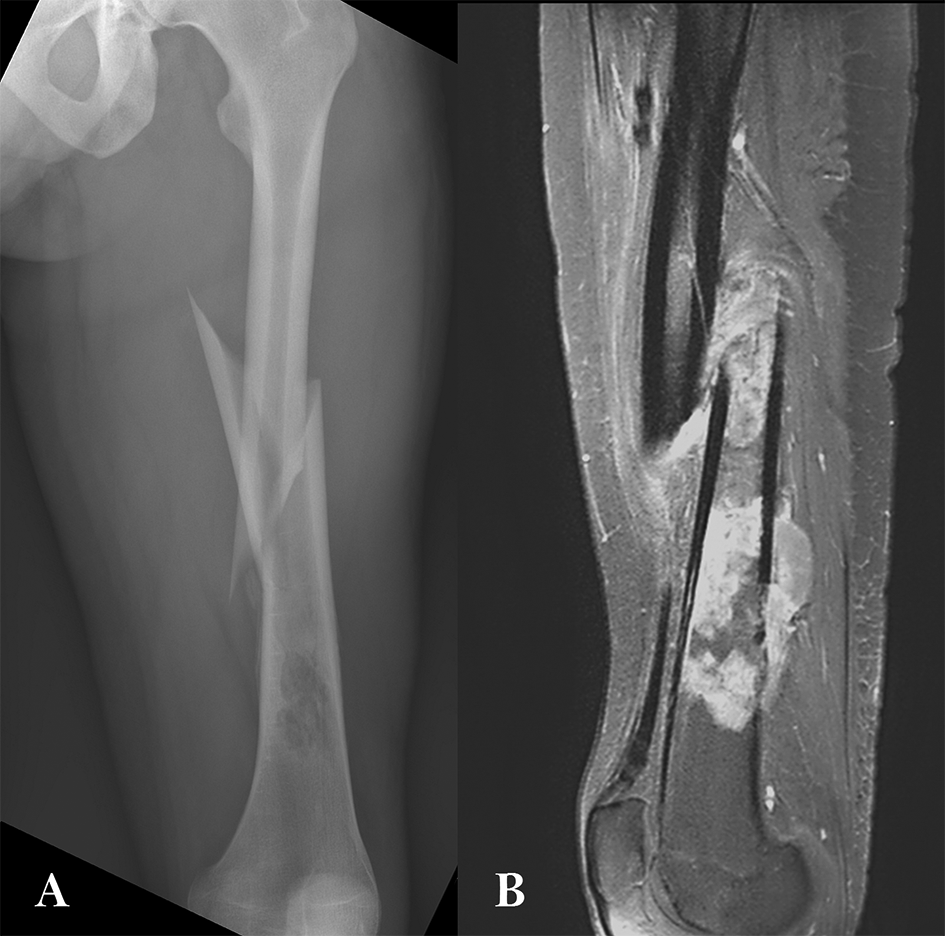
A: AP X-Ray view with osteolytic lesions; B: MRI T2sagittal view showing tumor invasion.
| Epidemiology data | |||||
|---|---|---|---|---|---|
| Gender | Age (years) | BMI (kg/m2) | Etiology | Fracture stabilization | Neo-adjuvant treatment |
| Male | 28 | 22.3 | Osteosarcoma (Li-Fraumeni Syndrom) | Long leg cast | API-AI protocol* |
First stage
The patient underwent initial wide margin (R0) oncologic resection, with a 20 cm diaphysis resection, at this time the femoral fracture has healed during neo-adjuvant CT (Table 2). The femur was prepared with a clean cut transversally. A cement spacer using high-viscosity Heraeus Palacos® R + G cement (Hanau, Germany) was modelled; it fills the bone loss as fully as possible. Due to pathological fracture and limb length discrepancy, the spacer was made 2 cm longer than the resection (22 cm). We also preferred to oversize it somewhat in width as the soft tissue coverage was not an issue, as it increases reconstruction space and facilitate induced membrane closure for the second stage. The spacer was covering the bone–host interfaces as recommended in the Masquelet technique.6 It was enhanced on a locked intramedullary nailing, positioned back and forth (Figure 2). Patient was considered good responder in pathology analysis with 1.5% residual viable cells (Table 3).
| Surgical data | |||||
|---|---|---|---|---|---|
| Spacer stabilization | Total operative time (T1; T2) (minutes) | Fibula graft length (mm) | Graft stabilization | Delay T1 and T2 (weeks) | Allograft length (mm) |
| IMN | 774 (295; 479) | 280 | Single plate | 22 | 220 |
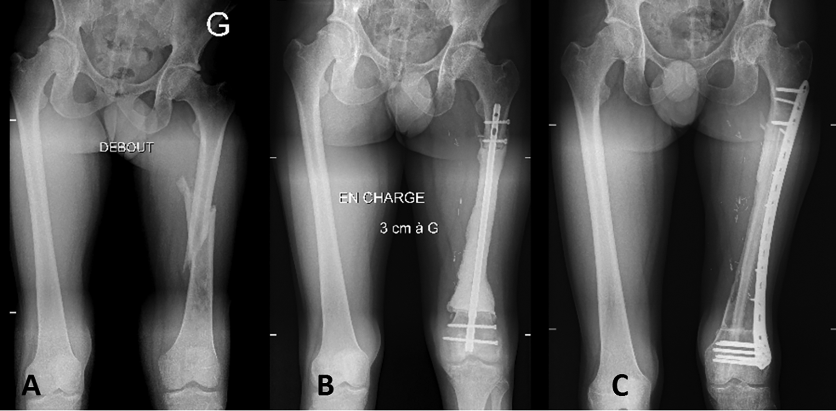
Interstage planification
The femoral allograft dimension was determined to be as close as possible of the patient femur; the diaphyseal shaft width must allow the fibula to slide into it. We used a cryopreserved allograft, in compliance with the criteria of the French Agency of Biomedicine, from the Nantes Multi-Tissue Bank. The delay between the two stages was of 22 weeks due to adjuvant chemotherapy.
Second stage
The length aimed (24 cm) and the rotational axes were determined before the spacer was removed. The vascularized fibula graft required microsurgical technique for harvesting and vascular anastomoses. The fibula graft was 28 cm in our case, and it was four centimeters longer than the planned bone allograft length. Graft harvesting was done from the contralateral limb by a second surgical team with microsurgical expertise in order to decrease the surgery duration. The harvesting was performed under a tourniquet, ensuring at least 8 cm14 of the distal fibula was spare in order to limit malleolar instability risk. A lateral surgical approach was used to raise the osseous graft. The bone was vascularized by an artery from the peroneal artery that entered the middle-third of the fibula. The section must include this area. After identification of the artery and its origin from the tibio-fibular trunk, the vessel was dissected.15
The allograft was warmed for one hour in a hot physiological serum bath and then cut with an oscillating saw with transverse cut similar to the bone defect aimed. One specific team was devoted to set the hybrid graft: the allograft was filled with the fibula after a progressive reaming, care was taken to not hurt the vessels of the fibula, a bone window was performed on the allograft for allowing its mobilization and sliding in the allograft. 2 cm of fibula were planned for overlapping the bone interfaces. Then, the hybrid graft was stabilized and compressed with a 4.5mm locking compression plate, taking care to orientate the fibula vessels towards anastomosis area. Lastly, the microsurgical anastomosis was performed at the terminal branches of the profunda femoris artery (Figure 2).
Postoperative management
Early mobilization and progressive weight-bearing were allowed postoperatively. Strictly limited weight-bearing was prescribed for the first six weeks, with progressive full weight-bearing depending on the patient’s pain and condition (Table 3). Full weight bearing was obtained at twelve weeks, with a functional quality of life of 75% on the EQ 5D score. Adjuvant chemotherapy was then performed with two doxorubicin-ifosfamide and two cisplatin-ifosfamide courses.
Long term follow up
At last follow-up (38 months following tumor resection), no local complication nor local recurrence occurred, with full weight bearing on his lower limb, allowing walking without cane. Nonetheless, despite beeing a good responder with R0 resection, the patient relapsed with pulmonary metastasis at 13 months follow up, needing surgical excision. Early pulmonary recurrence make us introduce a complementary targeted therapy with cabozantinib and chemotherapy ifosfamide and VP16.
On bone biopsy we first suspected a bone undifferentiated pleomorphic sarcoma (UPS), due to the low level of osteoïd matrix on the sample; the diagnosis of Li-Fraumeni Syndrom was also made due to oncological family anteriorities. Nonetheless, an osteosarcoma chemotherapy protocol was performed with an API-AI protocol. On bone tumor resection piece, osteosarcoma pathology diagnosis was made due to visualization of many osteoïd matrix on various places among the tumor. The unusual pejorative oncological evolution, despite an initial good pathology response and good margins was unusual in this case, thus, we performed a complementary histologic analysis.
Our aim was to assess the vascularization of patient osteosarcoma on biopsy prior to any chemotherapy treatment, and also to assess technical feasibility of our previous methodology (described on preclinical mouse model) on multiplexing analysis.12 Indeed, on a preclinical osteosarcoma MOS-J syngenic mouse model we identified two different kinds of vessels on confocal multiplexing analysis: CD31+/α-SMA+ elements and CD31+/Endomucin+elements. The CD31+/α-SMA+ elements exhibited a structured and straight architecture, with a sizeable diameter, and they resembled mature vessels. On the other hand, the CD31+/Endomucin+ elements were much more sinusoidal, with a more serpiginous and unorganized structure. They also had a smaller diameter and they look like immature sinusoidal vessels, which might be linked to tumor neo-angiogenesis (Figure 3).
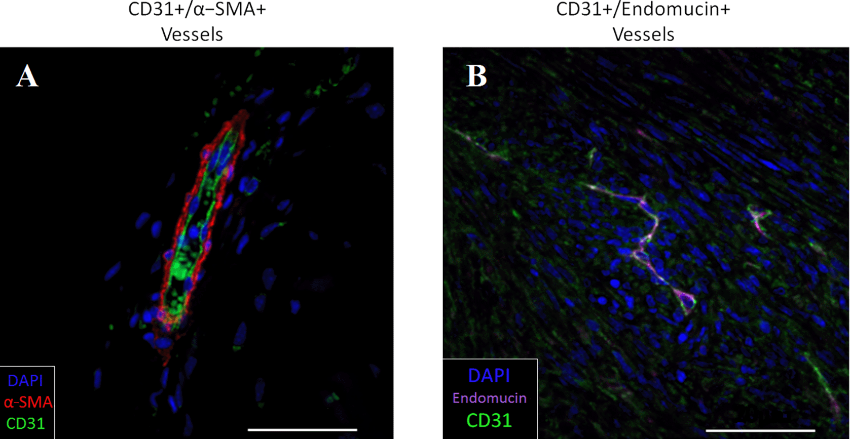
A: CD31+/α-SMA+ elements. B: CD31+/Endomucin+ elements. The images are Z-stack projections at 63× magnification, the scale bars =20 μm. DAPI: nuclear staining (from Crenn et al.12).
Immunohistochemestry tissue processing
The patient biopsy was collected surgically under complete anesthesia through a direct open approach, as soon as the pathological fracture was suspected. Samples were immediately fixed in 4% buffered formalin for 24 hours. The sample was then decalcified in 4.13% EDTA, 0.2% paraformaldehyde, in 1× pH 7,4 PBS buffer for 10 to 15 days at 50°C in the KOS microwave tissue processor (Milestone, Michigan, USA). It was finaly dehydrated through graded ethanol baths, cleared in 2-butanol and embedded in paraffin. Serial 5-μm-thick sections were cut from each sample and stained for H&E and Masson trichrome. Other sections were immunostained for α-SMA, Endomucin, and CD31 markers.
Three-color immunofluorescence histochemistry
Sections were deparaffinized, subjected to 20 h antigen retrieval in Tris-EDTA buffer (1 mM Tris, 0.5 mM EDTA, pH 9.0) at 60°C, and blocked with 2% normal donkey serum and 1% BSA in 1× Tris-buffered saline with 0.05% Tween 20 pH 7.4 to reduce unspecific binding. Sections were first immunostained with rabbit to CD31 (1:40, Abcam, ab28364) antibody for an hour at room temperature. After three washes in 1×TBS Tween 0.05% pH 7.4, sections were then incubated for an hour with biotinylated donkey anti-rabbit (1:200; Jackson ImmunoResearch 711-065-152), followed with Alexa Fluor 488-conjugated streptavidin (1:200; Jackson ImmunoResearch 016-540-084) another hour. To avoid non specific binding of both second primary rabbit antibody and secondary donkey anti-rabbit antibody, sections were then blocked with 5% normal rabbit serum in 1× TBS Tween 0.05% pH 7.4 for 30 min at room temperature followed by a blocking step with donkey anti-rabbit Fab fragments (1:10; Jackson ImmunoResearch 711-007-003) in 1× TBS Tween 0.05% pH 7.4 overnight at 4°C.
The next day, after three washes in 1× TBS Tween 0.05% pH 7.4, sections were then incubated with rabbit to Endomucin (1:100; Thermo Fisher PA5-21395), and mouse to α-SMA (1:1000; R&D Biotechne, MAB1420) antibodies for an hour at room temperature. After three washes in 1× TBS Tween 0.05% pH 7.4, the following secondary antibodies were applied for another hour: Alexa Fluor 594-conjugated donkey anti-rabbit (1:200; Jackson ImmunoResearch 711-585-152), and Alexa Fluor 647-conjugated donkey anti-mouse (1:200; Jackson ImmunoResearch 715-605-150). Finally, nuclei were counterstained with DAPI (1:1000; ThermoFisher Scientific, D1306) and coverslipped with Prolong Gold Antifade reagent (ThermoFisher Scientific, P36930).
Acquisition and histomorphometry
Confocal images were obtained by using a Nikon AI N-SIM confocal microscope and subsequently treated by smoothing and noise reduction of each channels, following by a Z-projection treatment on ImageJ software.16
All histomorphometric analysis were conducted on scans of the immunostained sections. Acquisitions of sections 4-7 were made with a NanoZoomer Slide Scanner (Hamamatsu, Japan), then pictures were saved using the NDP viewer software (Hamamatsu, version 2.2.6) at ×5 and export at ×10 using tiff format and analysed with ImageJ software. Each of the chromogenics immunostained sections were individually analyzed on 3 specific region of interest (ROI): one was defined by a representative 0.5-mm thick ROI which contained the tumors tissue along the bone; the other was a full representative picture of the core of the tumor; the last one was defined by a representative 0.5-mm thick ROI, that was contained the tumoral growth front. All analysis was carried out on each of the four levels of all samples. Briefly, pictures were subjected to color deconvolution using [H DAB] vector, thresholded, and unspecific noise was reduced. The results were shown as the mean value for each compartment of the number of positive elements per square millimeter.
For the triple-immunostaining sections (section 3), analysis was achieved using two levels that was considered, according results of ones that had been obtained on the chromogenics immunostained sections, as enough to give consistent values. Quantification of CD31+/α-SMA+ (mature vessels), and CD31+/Endomucin+ (sinusoid vessels) were realized using the virtual microscope (NDP viewer) at a selected magnification of ×20 on a representative field of 0.366 mm2. Double-positive vessels were counted in the same manner as up described in the different compartment by selecting the two fluorochromes channels corresponding of the two markers of interest. Results were shown as the mean value of each level of each sample of double positive elements per square millimeter.
HE and Masson trichrome staining in the patient biopsy lesion
Immunohistochemistry analysis was performed on the biopsy lesion with Masson’s trichrome staining (Figure 4), and showed a highly cellular malignant tumoral tissue, with a lack of osteoid matrix. We also observed some vessel-like structures.
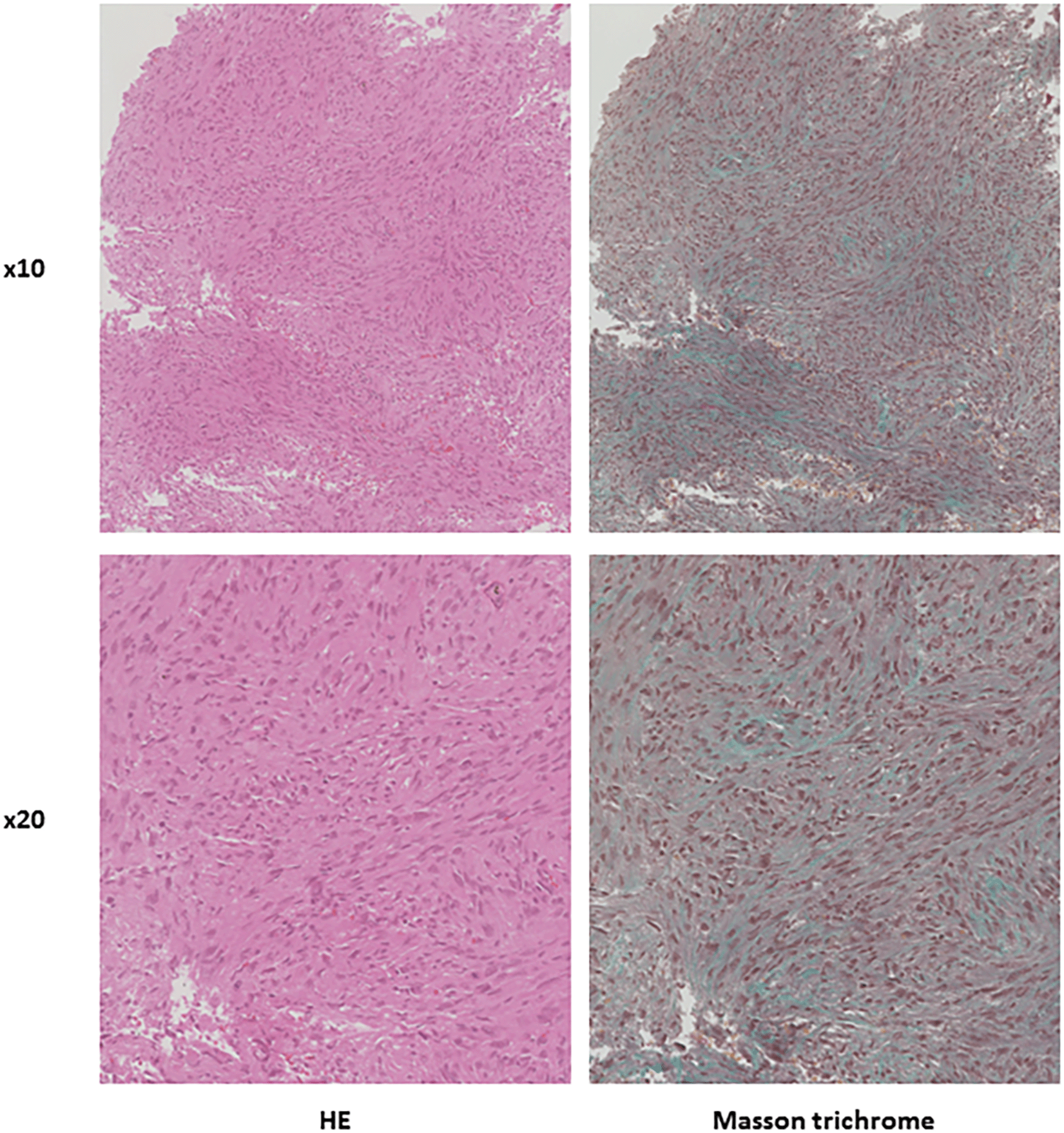
Confocal analysis of CD31+, α-SMA+ and Endomucin+ and costaining in the patient biopsy lesion
Immunohistochemistry analysis on the biopsy lesion (Figure 5) showed CD31+ elements and α-SMA+ elements which looked like vascular mature structures. On the other hand, tumor cells seem to express endomucin with a diffuse staining along a rare expression of endothelial endomucin.
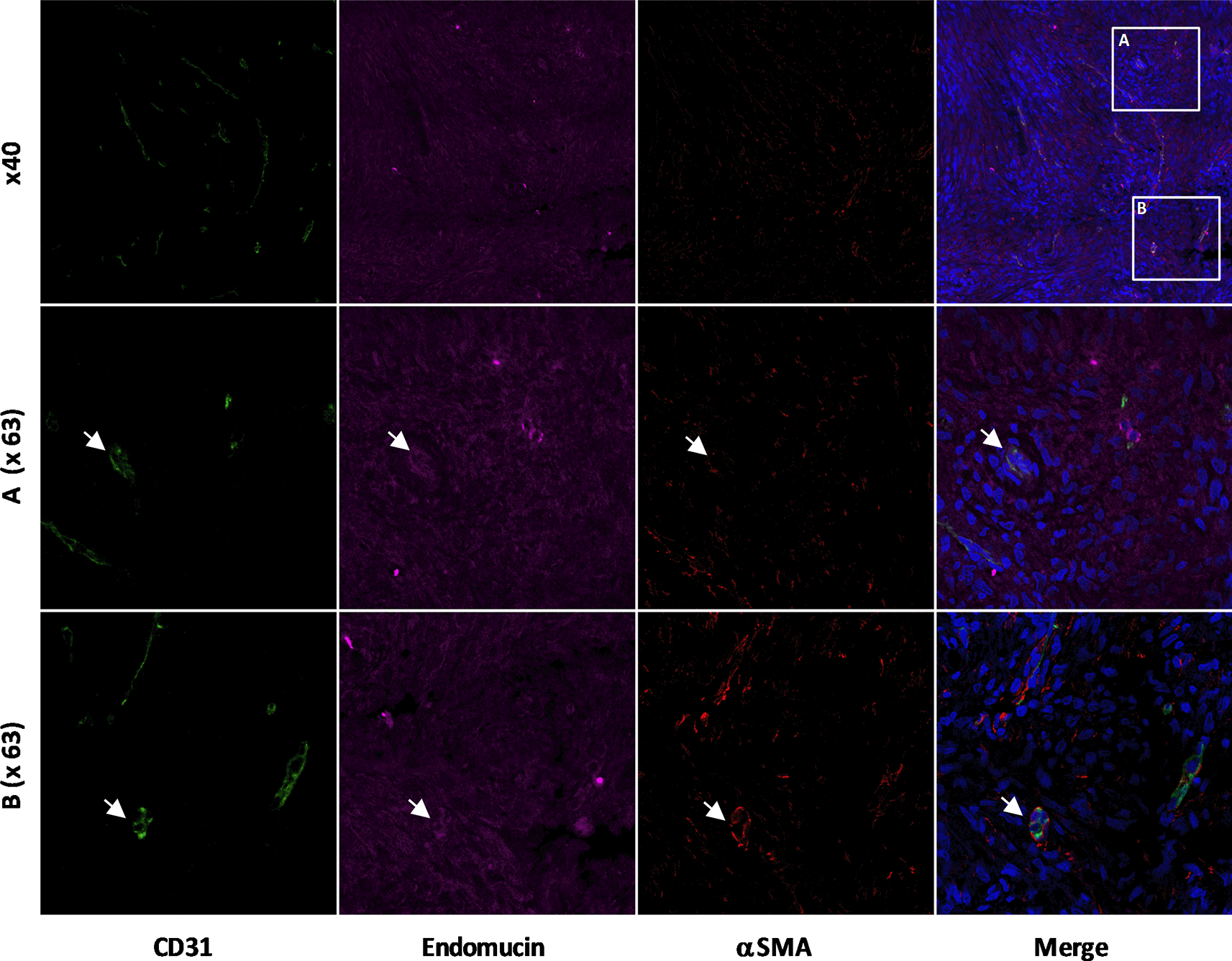
At 40× magnification, and two areas in Z-stack projections at 63× magnification. DAPI: nuclear staining.
Co-staining of CD31+/endomucin vessels seemed to show smaller vascular structures diameter-wise, unfortunately we could not distinguish any elements of architecture other than size and neither localization part of the tumor site as the biopsy was realized without targeting any specific areas of the tumor.
The Capasquelet technique result on this patient are promising, with bone healing being achieved within a short delay (12 weeks after the second stage), therefore allowing an early weight-bearing. We obtained a short delay for radiological bone healing with radiological follow up. The bone healing delay in our case (7 months) and in previous publication4 seems to be in the low range of an isolated Capanna technique with a 6-12 months delay or a Masquelet technique, which obtains complete bone union in 4–18 months, usually on less massive tibia defects.6,17 Despite the oncological disease evolution of the patient, the capasquelet surgery outcome was satisfactory, with early full weight-bearing, good function, and neither revisions nor complications.
A larger cohort, and a longer follow-up is needed to confirm these interesting results, especially in a limb length discrepancy case, for sequential restoration of femur length in pathological fractures. With our method, the osteoinductive action played by the induced membrane may exert a positive impact on the graft bone healing, with fast fibula union and allograft, early full weight-bearing, and a satisfactory functional score.
However, this two-stage technique got some drawbacks, the fibula harvest might results in morbidity,18 and it requires microsurgical expertise and access to a bone bank facility. The operating times involved in the two stages must also be noted, compared to a simpler technique.
The pejorative oncological evolution of the patient, despite an initial good pathology response (1.5% residual viable cells) and R0 margins was unusual, despite the controversial potential pejorative roles of the pathological fracture19,20 and the Li-Fraumeni Syndrome.21 In this context, it seems relevant to find new prognostic markers, to better adapt adjuvant chemotherapy in osteosarcoma patients, especially focusing on tumor microenvironment.11 The complementary histologic analysis on bone biopsy in this patient was performed aiming to explore vascularization with original stainings. Previous preclinical report investigated the implication of Endomucin expression in endothelial cells in a mouse model.12 We have shown that low expression in endothelial cells associated with both CD31 and α-SMA high expression could be associated as a better response to doxorubicin chemotherapy.
We obtained interpretable multiplexing analysis focusing on the human bone biopsy of our patient, validating the IHC methodological process. We identified CD31+ and α-SMA+ elements which looked like vascular mature structures, as well as thinner endomucin+ and CD31+, as in our preclinical mouse model.12
Concerning endomucin, cells seem to express endomucin with a diffuse staining along a rare expression of endothelial endomucin. Interestingly, endomucin seems to be also expressed in tumoral cells when it comes to human tumors, opposite to our previous mouse model. Other reports show endomucin expressed in HUVECs cells and related a role of endomucin in the vascular function.22 A recent report on digestives carcinomas23 highlight a link between survival in stomach adenocarinoma and EMCN gene, with high expression on gene datasets correlated with poorer overall survival and disease-free survival. However, our case report focused on vascular expression of Endomucin and α-SMA in a mesenchymal bone cancer and did not explore precisely the expression of endomucin in osteosarcoma tumor cells themselves. Further investigation with quantitative analysis including mass spectrometry to segregate endothelial cells and tumor cells might be needed.24
To our knowledge, it is the first description of endomucin expression via immunofluorescence staining in human osteosarcoma tumor, its high expression might be a pejorative prognosis factor as suggested in other oncological disease.23 Nonetheless, further works are needed to understand its relevance in an osteosarcoma prognosis context, as literature shown that endomucin could be highly expressed among cancerous cells, as well as in other human tissues (proteinatlas.org) with various expressions. Other experiments focusing on endomucin expression after decalcification process are also mandatory as it is well known that decalcification, and method of decalcification have an impact on antigen retrieving.25,26
In our pre-clinical model of long bone osteosarcoma, endomucin expression seems not to be affected by administration of doxorubicin.12 We could not find any clear evidence of differentiated expression of endomucin after the treatment, but model of good response seems to show a different ratio of immature/mature vessels with maturity of the vasculature associated with good response to chemotherapy.12 In this patient case, moderate to high intensity staining of endomucin among cancerous cell cause us to hypothesize whether its expression is related to the patient’s early metastatic relapse despite histologic good response.
Checking on RNA-seq of EMCN and ACTA1 publicly available on TCGA (https://portal.gdc.cancer.gov/) did not seem to show significant difference in survival related to the expression of endomucin and α-SMA in OS cells (supp Figures 1 and 2). Nevertheless, the latter lacks qualitative analysis on the vessel counts in the mesenchymal bone cancer and this type of analysis may be not suited to analyze the tumor and its microenvironment as we hypothesize the vessel maturity and architecture may have an influence on the chemotherapy response in OS. This hypothesis needs further exploration with simple techniques, such as tissue micro array, and survival cohort studies.
This case report of a Li-Fraumeni osteosarcoma patient illustrates two innovative specialized approaches, on one hand an original surgical reconstruction technique, with the Capasquelet reconstruction, allowing a good functional results and early weight-bearing in an intercalary large defect tumor resection. In the other hand, it shows promising data focusing on vascular tumor analysis with multiplexing immunofluorescent technique on tumor biopsy, aiming to better understand why patients with good tumor response will nonetheless still relapse. The Capasquelet technique needs larger cohort and longer follow-up to better asses its place in the therapeutic arsenal. Considering the immunofluorescent multiplexing analysis, the technique seems feasible and it needs to be analyzed on a larger human cohort in order to assess quantitatively if the CD31+/α-SMA+, the CD31+/endomucin+ co-labelling, or Endomucin alone might be considered as potentials predictive factor of recurrence or survival in osteosarcoma.
Conceptualization, V.C.; formal analysis, G.T. and J.A.; investigation, G.T. and J.A.; data curation, G.T., J.A. F.D. and A.C.; writing—original draft preparation, G.T., J.A. and V.C.; writing—review and editing, G.T., J.A., A.C., F.D., F.R., F.V. and V.C.; supervision, V.C.; All authors have read and agreed to the published version of the manuscript.
The data sets generated and/or analyzed during the current study are available from the corresponding author upon reasonable request.
Zenodo, 5 year survival in osteosarcoma, https://doi.org/10.12688/f1000research.124846.1. 27
This project contains the following underlying data:
Data are available under the terms of the Creative Commons Attribution 4.0 International license (CC BY 4.0).
We wish to thank the MicroPIcell platform (Stéphanie Blandin, Steven Nedellec, and Philippe Hulin). This work was supported by the Fondation pour la Recherche Médicale, FRM grant number DEA20150633177 awarded to Vincent Crenn. Experimental Therapeutic unit (Guylène Hamery, UTE Phan, IRS-UN, 8 quai Moncousu, BP70721, 44007 Nantes Cedex 1).
| Views | Downloads | |
|---|---|---|
| F1000Research | - | - |
|
PubMed Central
Data from PMC are received and updated monthly.
|
- | - |
Competing Interests: No competing interests were disclosed.
Reviewer Expertise: Osteosarcoma genomics, molecular biology, system biology
Is the background of the case’s history and progression described in sufficient detail?
Yes
Are enough details provided of any physical examination and diagnostic tests, treatment given and outcomes?
Yes
Is sufficient discussion included of the importance of the findings and their relevance to future understanding of disease processes, diagnosis or treatment?
Partly
Is the case presented with sufficient detail to be useful for other practitioners?
Yes
Competing Interests: No competing interests were disclosed.
Reviewer Expertise: Osteosarcoma genomics, molecular biology, system biology
Alongside their report, reviewers assign a status to the article:
| Invited Reviewers | |
|---|---|
| 1 | |
|
Version 2 (revision) 30 Sep 24 |
read |
|
Version 1 20 Sep 22 |
read |
Provide sufficient details of any financial or non-financial competing interests to enable users to assess whether your comments might lead a reasonable person to question your impartiality. Consider the following examples, but note that this is not an exhaustive list:
Sign up for content alerts and receive a weekly or monthly email with all newly published articles
Already registered? Sign in
The email address should be the one you originally registered with F1000.
You registered with F1000 via Google, so we cannot reset your password.
To sign in, please click here.
If you still need help with your Google account password, please click here.
You registered with F1000 via Facebook, so we cannot reset your password.
To sign in, please click here.
If you still need help with your Facebook account password, please click here.
If your email address is registered with us, we will email you instructions to reset your password.
If you think you should have received this email but it has not arrived, please check your spam filters and/or contact for further assistance.
Comments on this article Comments (0)