ALL Metrics
-
Views
-
Downloads
Get PDF
Get XML
Cite
Export
Track
Research Article

Inflammatory cytokines affecting cardiovascular function: a scoping review

[version 1; peer review: 2 approved with reservations, 1 not approved]
PUBLISHED 21 Sep 2022
Author details Author details
OPEN PEER REVIEW
REVIEWER STATUS

This article is included in the Cell & Molecular Biology gateway.

Abstract

Background: A chronic inflammatory process can cause disorders on the cardiovascular system. It is caused by an enhancement of inflammatory cytokines that can decrease the heart working-function. This article aims to review inflammatory cytokines that can cause the cardiovascular system disease.
Methods: The review process began by taking articles from two databases, namely PubMed and SpringerLink without using the publication year limit. The reviewed article was a research article using human samples, which analyzed the impact of inflammatory cytokines on cardiovascular disease or the risk level of cardiovascular disease.
Result: From a total of 3926 articles originating from two databases, 21 obtained articles have matched with the inclusion criteria for the review process. Of those 21 articles, 17 reported an increased effect of inflammatory cytokines on cardiovascular disease, while four articles showed no association between increased inflammatory cytokines and cardiovascular disease. There were six inflammatory cytokines that could affect cardiovascular disease, namely: TNF-α, IL-1β, IL-2, IL-6, IL-8, and IL-17 where cytokine IL-6 is confirmed by as many as 14 articles, TNF-α as many as six articles, and IL-1 β as many as three articles as inflammatory cytokines that could affect cardiovascular function.
Conclusion: The increased level of inflammatory cytokines can be a trigger for decreased function and cause the disease in the cardiovascular system.

Keywords

interleukin 6, tumor necrosis factor-α, medicine, cardiovascular disease, dentistry, non-communicable disease.

Introduction

One of the diseases that currently can cause health and socio-economic losses, both in developing and developed countries is cardiovascular disease.1 This is due to the increase in the number of cardiovascular diseases. Cardiovascular diseases that cause mortality and disability in the world include heart disease, vascular disease, and atherosclerosis.2,3

The major risk factor for the development of cardiovascular disease is inflammation.4,5 The occurrence of the inflammatory response is regulated by cytokines.6 Several studies have shown that cardiovascular disease can be triggered by some inflammatory cytokines and their receptors at a time when the inflammatory process occurs on an ongoing basis.7,8 Cardiovascular diseases such as atherosclerotic heart disease, coronary heart disease, and congestive heart failure are associated with an increase in inflammatory cytokines such as Interleukin 1 Beta (IL-1β), Interferon-γ (IFN-γ), and Tumor Necrosis Factor - Alpha (TNF-α), these cytokines are known to induce atherosclerotic plaque formation.9 Additionally, higher quantities of the cytokines TNF-α, IL-1, and IL-6 have been demonstrated to induce the expression of proatherogenic genes in new findings.10

Infection, nutritional imbalance, genetic factors, hypoxia, UV, allergic irritant, drugs, and physical stress can cause inflammation resulting in increased level of inflammatory cytokines. Enhancement of inflammatory cytokines induce cardiovascular diseases by triggering the expression of proatherogenic gen, induce the formation of atherosclerotic lesion, causing myocardial disfunction and cardiac hypertrophy.11

This scoping review will map cytokines that have elevated in some inflammatory conditions and their effects on cardiovascular function. Exposure factor mapping will show various inflammatory conditions that can increase cytokines. The results of the outcome mapping will provide an overview of the effect of increasing cytokines on cardiovascular function while from the results of each study that we present will inform cytokine levels that can interfere with cardiovascular function.

Methods

Scoping review was chosen because the review does not attempt to answer very specific study questions, but rather discusses a broader topic so that it is possible to apply more various study designs. We used guidance from Hilary Arksey & Lisa O’Malley framework in implementing the scoping review protocol which consisted of the following stages.

Stage 1: Identifying the research question

Discussions are carried out with the team to determine research questions by considering several key aspects such as study population, exposure, and outcomes. The research questions we will try to answer are : (1) What cytokines can affect cardiovascular function? (2) What inflammatory conditions can increase cytokine levels and interfere with cardiovascular function? (3) What types of disorders in cardiovascular function can appear due to increased cytokine levels? (4) What are the levels of cytokines that can cause impaired cardiovascular function?

Stage 2 : Identifying relevant studies

The articles were taken from two database namely Pubmed (https://pubmed.ncbi.nlm.nih.gov) and SpringerLink(https://link.springer.com). Articles were searched using the terms related to cytokine, interleukin, inflammation, infection, cardiovascular. The search keywords for each database presented below: Search keywords for PUBMED:((cytokine OR interleukin[MeSH Terms] AND ((fft[Filter]) AND (english[Filter]))) AND (inflammation OR infection[MeSH Terms] AND ((fft[Filter]) AND (english[Filter])))) AND (cardiovascular AND coronary artery[MeSH Terms] AND ((fft[Filter]) AND (english[Filter]))) Search keywords for SPRINGER LINK:(cytokine OR interleukin) AND inflammation AND cardiovascular AND “coronary artery” AND human NOT extract NOT “a review” NOT “systematic review” Selected articles are restricted to English language and research articles only without using the publication year limit. The most recent search was executed on September 15, 2021 and duplicate articles will be filtered immediately.

Stage 3: Study selection

Two reviewers selected articles that were free from duplication. The selection of articles that suitable with the inclusion criteria is carried out based on reading the tittle and abstract. Studies that included in this step are: (1) Research articles both observational and experimental with human subjects that analyze changes in cytokines concentrations due to inflammatory condition and their effects on heart function, (2) The outcome of the research is the type of the cytokine that has increased and the type of disturbance in cardiovascular system, (3) no review articles. Furthermore, selection is carried out according to the eligibility criteria through full text reading to reselect articles that will be included in the scoping review process. The selected articles were articles that used the parameters of increasing cytokines in the population experiencing inflammatory conditions in their studies. Inflammatory conditions in the cardiovascular system that can elevate cytokines systemically and weaken cardiovascular function will be included. Any disagreement resolved through a third reviewer.

Stage 4: Charting the data

Two reviewer indenpendently charted the data from final articles used MS Word 2019 form. The key data entered are: tittle, author, study design, subject, types of inflammatory conditions that cause an increase in cytokine levels and types of cytokines that have increased (cytokines exposure), outcomes on cardiovascular function, study results including cytokine levels that can interfere cardiovascular function, and research conclusion.

Results

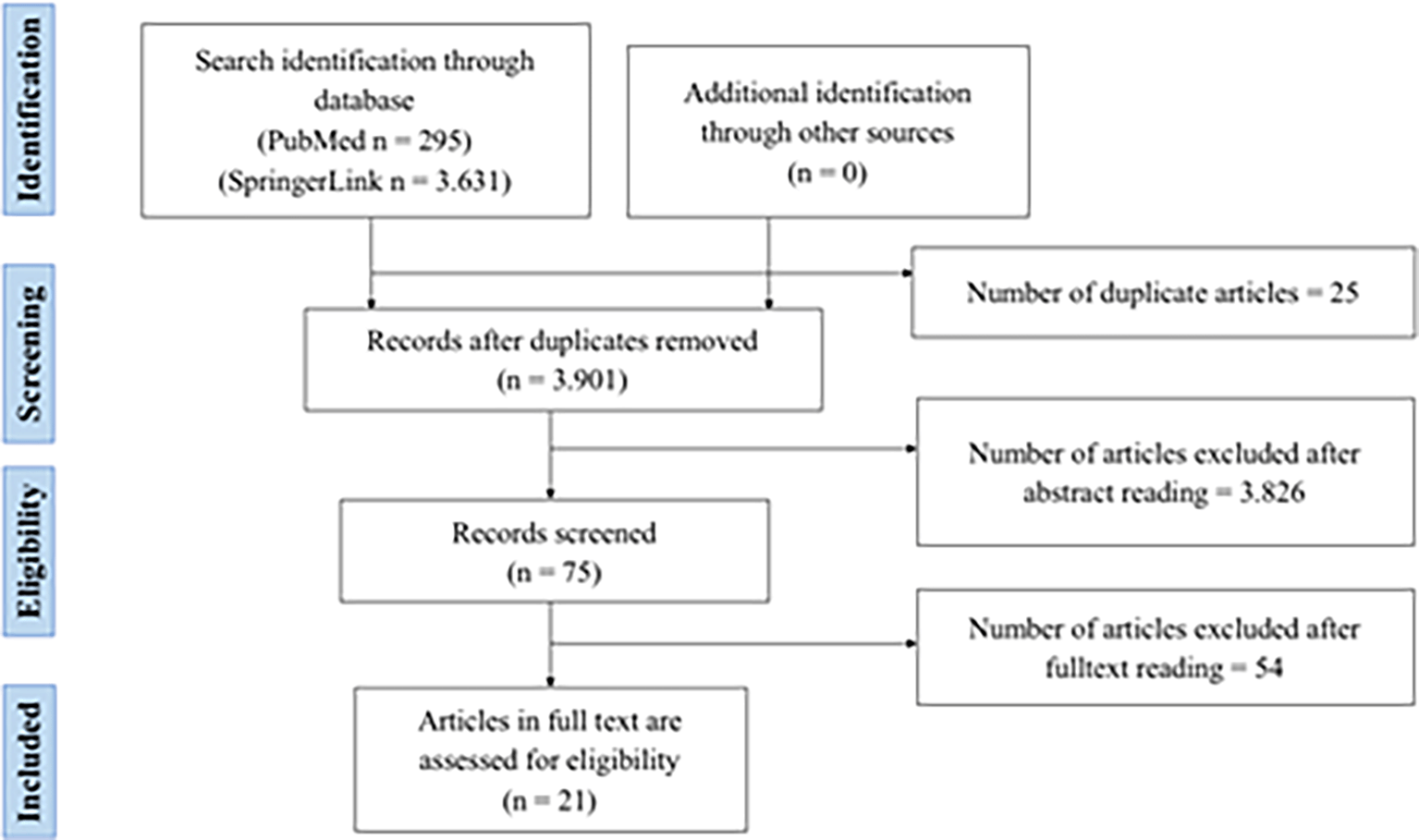
Firstly, the researcherr collected the articles from two databases and found 3,926 articles, 295 of those were taken from PubMed and 3,631 articles were taken from Springerlink. Those articles were selected first to find the duplicated articles from both databases. 24 duplicated articles were found and removed, so there were 3901 remaining articles. Then, from the remaining articles, there were 75 selected articles that met the criteria based on abstract readings. Furthermore, the researcher selected articles by reading the entire content and obtained as many as 21 articles for review (Figure 1). More information about the final selected articles has been listed in Table 1.

49e90482-10db-4672-85b5-abab375ba140_figure1.gif

Figure 1. Flow chart of PRISMA-ScR literature searching.48

Table 1. Conclusions of articles that meet inclusion criteria.49

NOHeadingWriterStudy designSubjectExposure/cytokine parametersOutcome on cardiovascular functionResultConclusion
1Coronary artery ectasia is related to coronary slow flow and inflammatory activationNatale Daniele Brunetti, Giuseppe Salvemini, Andrea Cuculo, Antonio Ruggiero, Luisa De Gennaro, Antonio Gaglione, Matteo Di BiaseCross sectional

  • 14 Coronary artery ectasia (CAE) patients

  • 17 coronary atherosclerosis patients

  • 15 people as controlled patients

  • Coronary artery ectasia, coronary atherosclerosis

  • TNF-α, IL-1β, IL-10, IL-8, IL-2

  • Coronary flow

  • Inflammantory activity

  • CAE patients had higher TNF-a, IL-1β, IL-10 levels compared to coronary atherosclerosis and the control (p < 0.05)

  • Coronary segments experiencing CAE correlated with levels of IL-10 (p < 0.01), IL-1β (p < 0.01)

  • Coroner flow in the left anterior descending artery coroner (p<0.05), at right coronary artery (p<0.001), IL-1 β (p<0.01), IL-8(p<0.05), IL-10 (p<0.01) in CAE sufferers increased, compared to control and subject with atherosclerosis without CAE

  • Coroner flow in the left anterior descending segment is associated with TNF-α (r=0.92, p<0.05) and IL-8 (r=0.88, p<0.05) in CAE sufferers

  • Subjects with above-average IL-1β showed higher coroner flow values at left anterior descending (p<0.01)

  • In patients with CAE, the disease progression was associated with inflammatory activation and impaired coronary circulation.

  • Inflammatory responses were also associated with impaired coronary circulation.

2Association between Adipokines and Coronary Artery Lesions in Children with Kawasaki DiseaseHyun Jung Kim, Eun Hye Choi, Hong Ryang KilCross sectional

  • 15 afebrile controls patients

  • 32 febrile controls patients

  • 40 Kawasaki disease patients

  • Kawasaki disease (KD), resistin

  • IL-6, TNF-α

Coronary artery lesions

  • Serum resistin levels in subjects with KD were higher than in the control group (KD had a level of 177.56 ng/mL, whereas febrile controls had a level of 76.48 ng/mL and afebrile controls had a level of 17.95 ng/mL)

  • Kawasaki disease patients experienced an increase in IL-6 (p =0.014)

  • Levels of serum IL-6 were statistically higher, while BMI was statistically lower in the KD group with coronary artery lesions than in the KD group without coronary artery lesions (p=0.016)

  • Levels of serum IL-6 in KD with coronary artery lesions = (228.26±303.97) pg/ml and in KD without coronary artery lesions group = (39.18±107.20) pg/ml

  • Levels of serum TNF-α in KD with coronary artery lesions = (9.21±31.1) pg/ml tand in KD without coronary artery lesions group = (0.21±0.22) pg/ml

Resistin levels are higher in Kawasaki disease patients, although this hasn't been used as a predictor of coronary artery lesions in the acute phase.
3Myocardial Microvascular Dysfunction in Rheumatoid Arthritis (Quantitation by (13)N-Ammonia Positron Emission Tomography/Computed Tomography)Isabelle Amigues, Cesare Russo, Jon T Giles, Aylin Tugcu, Richard Weinberg, Sabahat Bokhari, Joan M. BathonCohort

  • 76 rheumatoid arthtitis (RA) patients

  • 34 Non RA symptomatic controls patients

  • 14 people for Non RA asymptomatic controls

  • Rheumatoid arthtitis

  • IL-6

  • Myocardial flow reserve (MFR),

  • Myocardial blood flow

  • RA patients experienced a 29% decrease in MFR (<2.5) and an increase in IL-6.

  • High IL-6 levels associated with lower MFR values (p=0.024)

One-third of RA patients without cardiovascular illness had lower MFR, which is related with inflammation and increased mass and volume of the left ventricle.
4Systemic inflammation is related to coronary microvascular dysfunction in obese patients without obstructive coronary diseaseF. Tona, R. Serra, L. Di Ascenzo, E. Osto, A. Scarda, R. Fabris, R. Montisci, G. Famoso, S. Tellatin, M. Foletto, A. Giovagnoni, S. Iliceto, R. VettorCross sectional

  • 86 obese patients

  • 48 healthy people as controls

  • Obesity without obstructive coronary disease

  • IL-6, TNF-a, adiponectin

  • Coronary Flow Reserve (CFR)

  • Coronary microvaskular dysfunction

  • CFR was higher in non-obese patients (p=0.02), compared to obese patients

  • Abnormal CFR occurs in 27 (31%) obese patients and in 1 (2%) control subjects (p=0.0001)

  • Multivariate analysis results show TNF-a and IL-6 are determinants of CFR (p<0.02)

  • The results of a multivariable logistic regression analysis show IL-6 (p=0.03) and TNF-α (p=0.03) are determinants of CFR ≤ 2.5

  • IL-6 levels were higher in the group with decreased CFR (4.6 [3.6-6.1]) ng/l than the group with normal CFR (3 [2-3.8]) ng/l

  • TNF-α levels were higher in the group with decreased CFR (12 [10.5-15.0]) ng/l than the group with normal CFR (10 [8.8-11.5]) ng/l

  • Obese people with no clinical indications of cardiac disease had lower CFR.

  • Microvascular dysfunction is associated with chronic inflammation mediated by adipocytokines.

5Role of interleukin-17 and interleukin-17-induced cytokines
interleukin-6 and interleukin-8 in unstable coronary artery disease
Satwat Hashmi and Qiu Tang ZengCross sectional58 patients consisting of:

  • 14 stable angina patients

  • 24 unstable angina patients

  • 20 acute myocardial infarction subjects

  • 20 healthy people as a control

The 58 subjects were divided into two groups according to plaque morphology in the coronary arteries: the group with simple lesions (n=22) and the group with complex lesions (n=36).

  • Culprit lesion,

  • IL-17, IL-10, IL-8, IL-6, and hs-CRP

  • Acute Myocardial Infarction (AMI)

  • Unstable angina

  • Stable angina

  • Increased concentrations of proinflammatory cytokines IL-6 (p<0.05), IL-17 (p<0.05), IL-8 (p=0.07 for unstable angina and p<0.05 for AMI), hs-CRP p<0.05 for unstable angina) and decreased IL-10 concentration (p<0.01) in acute myocardial infarction and unstable angina compared with the control group.

  • Plasma IL-17 concentrations were positively correlated with hs-CRP (r=0.655, p<0.001) and plasma IL-6 concentrations (r=0.65, p<0.01)

  • IL-17 levels were greater (326.47±126.10 pg/ml) in patients with more lesions, but no significant difference were compared to the group with a simple picture of lesions (299.47±123.14 pg/ml)

  • IL-6 levels in the group with complex lesions = (53.24±33.30 pg/ml) and in the group with simple lesions = (46.62±16.33 pg/ml)

  • IL-6 levels in the unstable angina group = (53.63±31.92 pg/ml) and in the acute myocardial infarction group = (46.91±16.11 pg/ml)

Inflammation appears to play a role in boosting the activity of IL-6, IL-17, and IL-8 in people with unstable angina and acute myocardial infarction. Increased IL-17 triggered by inflammation has a role in causing instability in people with artery coronary disease.
6IL 6 but not TNF is linked to coronary artery calcification in patients with chronic kidney diseaseJoanna Kaminska, Marek Stopinski, Krzysztof Mucha, Anna Jędrzejczak, Marek Gołębiowski, Monika A. Niewczas, Leszek Pączek, Bartosz ForoncewiczCohort57 patients with chronic kidney disease (CKD):

  • 38 people stages 5

  • 19 people stages 3 or 4

  • 19 controlled people

  • Chronic kidney disease (CKD)

  • Fetuin A, adiponectin, leptin,

  • TNF, IL-6

  • MMP9, ICAM1, VCAM1

Artery calcification

  • VCAM1, TNF, IL-6 have higher levels of CKD with higher stages (p<0.001)

  • VCAM 1 is related to ICAM1, TNF, IL-6 (p<0.05)

  • TNF and IL-6 are related to each other (p<0.05)

  • IL-6 has significant association with CRP, kidney function, age, and calcium score (p<0.05)

  • Increase in IL-6 and calcium score occurred up to more than 3 times over 5 years as a cause of risk of death due to CKD (p<0.0001)

  • IL-6 is related to calcium score

  • Chronic inflammation such as coronary artery calcification contributes to the mortality rate of cardiovascular patients with impaired kidney function

7Association between interleukin-6 and the risk of cardiac events measured by coronary computed tomography angiographyLei Zhao, Xilin Wang, Yuhai YangCohort303 patients:

  • 101 people with Low IL-6

  • 101 people with intermediate IL-6

  • 101 people with High IL-6

  • Coronary Artery Disease

  • IL-6

Atheroscleroticoronary plaques

  • Subjects with high IL-6 levels showed higher atherosclerotic load than subjects with lower IL-6 levels (p<0.001)

  • Greater IL-6 levels were associated with a higher incidence of MACE and mortality than lower IL-6 levels (p<0.0001).

Subjects with elevated levels of IL-6 have a higher risk of atherosclerosis and MACE
8Associations of inflammatory markers with coronary artery calcification: results from the Multi-Ethnic Study of AtherosclerosisNancy Swords Jennya, Elizabeth R Brown, Robert Detrano, Aaron R Folsom, Mohammed F Saad, Steven Shea, Moyses Szklo, David M. Herrington, David R Jacobs Jr.Cross sectional6783 multi-etnic participants

  • coronary artery calcification (CAC), C-reactive protein, fibrinogen

  • IL-6

  • artery calcification

  • atherosclerotic

  • CRP has a relative risk of 1.13 for CAC based on age, gender and ethnicity

  • IL-6 has a relative risk of 1.22 and fibrinogen 1.18 based on gender and ethnicity.

  • Inflammatory markers have a weak relationship with CAC.

  • Inflammatory markers and CAC have different pathophysiologies.

9Role of superoxide dismutase in vascular inflammation and in coronary artery diseaseV. Lubrano, P. Di Cecco, G.C. ZucchelliCross sectional

  • 36 coronary artery disease (CAD) patients (33 men and 3 women)

  • 19 normal subjects (16 men and 3 women)

  • Measurement of extracellular superoxide (EC-SOD) levels, CRP

  • Measurement of IL-6 and TNF-α levels mm

Impairment of coroner artery function

  • The levels of EC-SOD in the control and CAD groups were the same.

  • There was a significant difference in levels of radical peroxide (p<0.01), hs-CRP (p<0.01), and hs-IL-6 (p<0.001) between subjects with CAD and control groups.

  • There was an increase in hs-IL-6 levels in subjects with more impaired coroner's arteries (p<0.05).

  • Hs-IL-6 concentrations are associated with CRP p<0.05, r=0.14) and peroxide radicals (p<0.05, r=0.1)

  • Average levels of IL-6:

    control = (1.05±0.2 pg/ml)

    1 artery involved = (2.7±0.7 pg/ml)

    2 artery involved = (2.6±1.7 pg/ml)

    3 artery involved = (3.3±1.0 pg/ml)

    4 artery involved = (3.5±0.6 pg/ml)

  • Average levels of TNF-α:

    control = (2.50±1.1 pg/ml)

    1 artery involved = (1.05±0.3 pg/ml)

    2 artery involved = (1.34±0.2 pg/ml)

    3 artery involved = (0.98±0.4 pg/ml)

    4 artery involved = (3.90±1.5 pg/ml)

  • EC-SOD has a small role to play in CAD prevention.

  • hs-IL-6 can be used as a marker in efforts to prevent and characterize artery coronary disease.

10Biomarkers of endothelial dysfunction, cardiovascular risk factors and atherosclerosis in rheumatoid arthritisPatrick H Dessein, Barry I Joffe, Sham SinghCross sectional

  • 74 people with rheumatoid arthritis

  • 80 healthy individuals (control)

  • Rheumatoid arthritis

  • hs-CRP, IL-1, IL-6, and TNF-α

Marker levels of endothelial function impairment (ICAM-1, VCAM-1, ELAM-1)

  • TNF-, IL-6, IL-1, and hs-CRP levels were higher in patients with rheumatoid arthritis than in healthy people (p<0.0001).

  • Endothelial function does not differ significantly between control and people with rheumatoid arthritis (p=0.08)

  • Levels of IL-6 in rheumatoid arthritis subjects are highly related to endothelial dysfunction marker levels. (ICAM-1, VCAM-1, and ELAM-1) (p≤0.03)

  • Rheumatoid titer and GFR levels have relationships with levels of VCAM-1 and ICAM-1 (p≤0.02)

  • In rheumatoid arthritis survivors, VCAM-1 is linked to intima-media carotid artery thickness (p=0.02) and plaque thickness (p=0.04).

  • People with rheumatoid arthritis have worse endothelial function than controls, but also have higher levels of hs-CRP and cytokines.

  • In persons with rheumatoid arthritis, IL-6, rheumatoid tititer levels, and GFR may predict the probability of impaired endothelial function.

11Markers of inflammation are cross-sectionally associated with microvascular complications and cardiovascular disease in type 1 diabetes”the EURODIAB Prospective Complications Study”MT Schram, N Chaturvedi, CG Schalkwijk, JH Fuller, CDA StehouwerCross sectional543 subjects with type 1 diabetes were divided into 2 groups:

  • 348 diabetics with complications

  • 195 people with diabetes without complications

  • Type 1 Diabetes mellitus

  • IL-6, CRP, and TNF-α

  • Cardiovascular disease

  • Albuminuria

  • Retinopathy

  • IL-6, CRP, and TNF- levels aggregated in one inflammatory marker Z-score were significantly linked with retinopathy, albuminuria, and cardiovascular disease, and negatively with GFR.

  • Albuminuria, retinopathy, and cardiovascular disease were all substantially linked with levels of IL-6, CRP, and TNF- combined in one Z-score inflammatory measure, which were inversely proportional to GFR.

  • In subjects normo-, micro-, and microalbuminaria Z score = -015; 0,10; 0.28 with CI = 95%, value p<0.0001

  • On subject no, non-proliferative and proliferative retinopathy Z score for -023; 0.14; 0.20 with CI = 95%, p<0.0001

  • In subjects with cardiovascular disease and not suffering from cardiovascular disease Z score -028, 0.06, with CI = 95% p<0.001

  • Each increase of 1 SD from Z inflammatory score marker will decrease GFR by 4.6 ml per min per 1.73 m2 (p<0.001)

  • IL-6 levels were higher in the diabetes group with cardiovascular disease (≥2 pg/ml) than the diabetes group without cardiovascular disease (≥1.5 pg/ml)

  • TNF-α levels were higher in the diabetes group with cardiovascular disease (≥3 pg/ml) than the diabetes group without cardiovascular disease (≥2 pg/ml)

  • In subjects with type 1 diabetes, elevated inflammatory markers such as IL-6, CRP, and TNF-α will increase microvascular problems and the risk of cardiovascular disease.

  • Reduced inflammatory activity can help to reduce vascular problems in people with type 1 diabetes.

12Associations between serum uric acid and adipokines, markers of inflammation, and endothelial dysfunctionS Bo, R Gambino, M Durazzo, F Ghione1, G Musso, L Gentile, M Cassader, P Cavallo-Perin, and G PaganoCross sectional100 healthy men

  • Uric acid, resistin, hs CRP, leptin,nitrotyrosine

  • Adiponectin, IL-6, TNF-α

Endothelial dysfunction marker levels:

  • E-selectin

  • intercellular adhesion molecule-1 (ICAM-1)

  • vascular adhesion molecule-1 (VCAM-1)

  • Total Antioxidant Status (TAS)

  • Subjects with higher uric acid levels had worse metabolic patterns and higher prevalence rates suffering from metabolic syndrome (p<0.001; 95%CI 1.6-8.2; OR=3.6) for a 50 μmol/l increase in uric acid levels.

  • Nitrotyrosine (p 0.001) and adiponectin (p=0.02) levels are inversely proportional to uric acid levels, which are directly related to E-selectin (p=0.006) and TAS (p 0.001) levels

  • Il-6 (p=0.55) and TNF-α (p=0.55) levels do not differ significantly between groups

  • Uric acid levels are inversely proportional to nitrotyrosine levels and directly proportional to TAS

  • The levels of uric acid are inversely proportional to adiponectin and are directly proportional to E-selectin

  • There is no linear relationship between cardiovascular disease risk and uric acid levels

13Serum levels of interleukin-22, cardiometabolic risk factors and incident type 2 diabetes: KORA F4/FF4 studyChristian Herder, Julia M Kannenberg, Maren Carstensen-Kirberg, Cornelia Huth, Christa Meisinger, Wolfgang Koenig, Annette Peters, Wolfgang Rathmann, Michael Roden, Barbara ThorandCross sectional and CohortCross sectional = 1107 people
Cohort = 504 people

  • Type 2 Diabetes mllitus, glucose statuse, HDL, GFR

  • IL-22 levels, IL-1R

Risk factors for:

  • Cardiometabolic disease

  • Diabetes mellitus

  • Male sex, smoking, low HDL levels, low GFR, and low IL-1RA levels are associated with high IL-22 levels (p<0.05)

  • The average IL-22 levels in type 2 diabetics was 6.28 (1.95; 12.35) pg/ml and in the control group was 6.45 (1.95; 11.80) pg/ml.

  • OR=1.02 (0.85; 1.23) CI 95% for the incidence of type 2 diabetes based on age and gender factors affected by IL-22

  • Increased IL-22 levels are inversely related to various parameters linked with cardiometabolic disease risk, but not with the risk of type 2 diabetes.

  • Although IL-22 cannot be utilized as a biomarker for type 2 diabetes in humans, it has the potential to be used as a marker for cardiovascular disease if more study is done on it.

14Relationship between IL-27 and coronary arterial lesions in children with Kawasaki diseaseFeifei Si, Yao Wu, Fang Gao, Siqi Feng, Ruixi Liu, Qijian YiCross sectional81 children with Kawasaki disease (48 men, 33 women)

  • Kawasaki disease

  • Levels of IL-27, IL17A, IL-10, IL-6, IL-1 β, TNF-a in serum

Coronary artery lesions

  • Serum levels IL-27 (p=0.004), IL-6 (p<0.05), IL-1 β (p<0.05), and TNF-α (p<0.05) were higher in the Kawasaki Disease patient group accompanied by coroner artery lesions than the Kawasaki Disease patient group without coroner artery lesions.

  • There is a relationship between WBC (r=0.185), CRP (r=0.291), PCT (r=0.400), IL-10 (r=0.454), IL-17A (r=0.618), IL-6 (r=0.263), IL-1 β (r=-0.13) and TNF-α (r=0.372) with IL-27 in subjects with KD

  • Levels of serum IL-6 in KD with coronary artery lesions = (234.20±133.10) pg/ml and in KD without coronary artery lesions group = (110.50±73.70) pg/ml

  • Levels of serum TNF-α in KD with coronary artery lesions = (56.00±15.40) pg/ml and in KD without coronary artery lesions group = (21.60±4.60) pg/ml

  • Levels of serum IL-1β in KD with coronary artery lesions = (33.80±18.70) pg/ml and in KD without coronary artery lesions group = (3.60±0.60) pg/ml

  • Increased IL-27 may be the cause of increased pro-inflammatory cytokines in acute KD

  • IL-27 may have a key role in the progression of coronary artery lesions in persons with acute KD.

15Effect of ABCA1 promoter methylation on premature coronary artery disease and its relationship with inflammationFang An, Chao Liu, Xiujuan Wang, Tan Li, Hao Fu, Buhe Bao, Hongliang Cong, Jihong ZhaoCross sectional

  • 90 patients with premature coroner artery disease

  • 90 healthy people

  • ABCA1 rates promoter methylation,

  • CRP, cfDNA/NETs HDL,

  • IL-1 β

Premature coroner artery disease (pCAD)

  • People with premature coronary artery disease had a higher ABCA1 transcriptional activation rate than the healthy group (44.24%±3.66 vs. 36.05%±2.99, p<0.001)

  • The methylation of the ABCA1 promoter is a risk factor for coronary artery disease (OR=2,878.95% CI: 1,802–4,594, p<0,001)

  • ABCA1 promoter methylation levels were positively correlated with IL-1 β, cfDNA/NETs, and CRP (r=0.385; 0.404; 0.389; p<0.001) and negatively correlated with HDL (r=0.488, p<0.001)

  • The levels of ABCA1 promoter methylation are affected by IL-1 β (p=0.001), CRP (p=0.002), cfDNA/NETs (p=0.001).

  • IL-1β levels were higher in the pCAD group (57.74±12.55 pg/ml) than the control group (47.66±14.77 pg/ml)

  • The higher the level of ABCA1 promoter methylation, the lower HDL levels and high levels of ABCA1 promoter methylation are risk factors for the development of premature coroner artery disease.

  • The methylation of the ABCA1 promoter is influenced by inflammatory cytokines and cfDNA/NETs, which leads to the development of premature coronary artery disease.

16Epicardial adipose tissue is a predictor of decreased kidney function and coronary artery calcification in youth- and early adult onset type 2 diabetes mellitusM Reinhardt, T R Cushman, MS Thearle, J KrakoffCross sectional149 Americans (92 women, 57 men of whom 95 were diagnosed with type 2 diabetes)

  • Type 2 diabetes mellitus

  • Epicardial fat

  • Pericardial fat

  • IL-6 levels,

  • Calcification of coroner's arteries

  • Kidney function

  • BMI was associated with pericardial fat volume and epicardial fat volume (r=0.26, p=0.001; r=0.37, p 0.0001).

  • In the control group, the volume of epicardial fat associated with coronary artery mass (r=0.51, p<0.0001)

  • The presence of IL-6 levels and coronary artery calcification were associated with epicardial fat volume (β=0.05±0.02 cm3, p=0.03; β=005±0.01 pg/ml/cm3, p=0.002).

  • In the group of young type 2 diabetes mellitus subjects, pericardial fat volume and epicardial fat volume can be used as predictors of decreased eGFRcr (CKD-EPI) (β=−0.25 ± 0.05 ml/min/cm3, p<0.0001; β=−0.3 ± 0.08 ml/min/cm3, p=0.001)

Epicardial fat volume is a risk factor for heart disease and can be a predictor of poor kidney function in young type 2 diabetes mellitus patients.
17Is serum Interleukin-17 associated with early atherosclerosis in obese patients?Giovanni Tarantino, Susan Costantini, Carmine Finelli, Francesca Capone, Eliana Guerriero, Nicolina La Sala, Saverio Gioia, Giuseppe Castello2Cross sectional80 obese people with normal liver enzyme levels

  • Obesity

  • C-reactive protein

  • Eotaxin-3 CCL4/MIP1β, fibrinogen, ferritin

  • IL-8, IL-17, and TNF-α

Thickness of carotid intima-media

  • There was an association between inflammatory cytokine IL-6 levels and serum IL-17 levels (p<0.0001), TNF- α (p<0.0001), IFN-γ (p < 0.0001)

  • IL-17 levels are associated with IL-8 (95%CI: p<0.0001), eotaxin (95%CI: p<0.0001), and CCL4/MIP1β (95%CI: p<0.0001)

  • Examination of serum eotaxin levels (p=0.0059) and visceral fat (p=0.0010) can predict carotid intima media thickness.

  • The thickness of the carotid intima media and the Il-17-related eotaxin have a strong association.

  • The link between visceral fat, serum eotaxin levels, and carotid intima media thickness supports the idea that IL-17 produced by visceral fat tissue can cause atheromatous vascular smooth muscle cells to secrete eotaxin.

18Association of systemic inflammation with epicardial fat and coronary artery calcificationSoren Gauss, Lutz Klinghammer, Alina Steinhoff1, Dorette Raaz-Schrauder, Mohamed Marwan, Stephan Achenbach, Christoph D. GarlichsCross sectional391 people at high risk of coronary artery disease

  • Epicardial fat volume and inflammatory markers

  • (IL-17, IL-15, IL-13, IL-12, IL-10, IL-8, IL-7, IL-6, IL-4, IL-2, IL-1α, IP-10, Eotaxin, MIP-1, MCP-1, G-CSF, GM-CS, hs-CRP, TNF-α, IFN-γ)

Calcification of coronary arteries

  • Epicardial fat volume and CAC have a significant correlation (p<00.001; p=0.37)

  • Coronary artery calcification and epicardial fat volume were significantly related to age (p=0.032), gender (p<0.001), diabetes (p=0.036), smoking (p<0.001), and hypertension (p<0.001).

  • Calcification of coroner arteries is negatively correlated with IL-13 (r=-0.10) and G-CSF (r=-0.12)

  • Epicardial fat volume is positively correlated with IP-10 (p=0.002), MCP-1 (p=0.037)

  • The development of coroner artery calcification is significantly related to the expansion of epicardial fat (p<0.001)

  • Results showed no association between increased coronary artery calcification, epicardial fat volume, cytokines indicating systemic inflammation

  • Epicardial fat volume indicates the development of atherosclerosis as measured through coronary artery calcification scores

  • Systemic inflammatory marker levels do not predict increased coronary artery calcification and epicardial fat volume.

19Comparison of inflammation, arterial stiffness and traditional cardiovascular risk factors between rheumatoid arthritis and inflammatory bowel diseaseFenling Fan, Abby Galvin, Lu Fang, David Andrew White, Xiao-lei Moore, Miles Sparrow, Flavia Cicuttini, Anthony Michael DartCross sectional

  • 43 rheumatoid arthritis patients

  • 42 inflammatory bowel disease (IBD) patients

  • 73 healthy people

  • Rheumatoid Arthtritis

  • Inflammatory Bowel Disease

  • CRP, TNF-α, IL-1 β, IL-10, IL-6

  • von Willebrand factor (vWF)

  • Endothelial dysfunction,

  • Arterial stiffness

  • TNF-, IL-10, IL-1β, IL-6, and von Willebrand factor (vWF) levels were greater in patients with IBD and rheumatoid arthritis compared to controls.

  • When compared to the healthy group, FRS, waist circumference, triglycerides, and BMI increased in rheumatoid arthritis but not in IBD.

  • baPWW did not differ substantially in the three groups, while the IBD group showed a much lower ABI than the control group

  • Marker macrophage migration inhibitory factor, vWF, CRP, and tumour necrosis factor-α were all statistically associated with baPWW.

  • vWF, tumour necrosis factor-α, marker macrophage migration inhibitory factor and CRP significantly associated with baPWW

  • In rheumatoid arthritis CRP levels are higher than IBD.

  • There is no link between inflammatory markers and arterial stiffness

20Association of influenza virus infection and inflammatory cytokines with acute myocardial infarctionXiuru Guan, Wei Yang, Xijuan Sun, Lanfeng Wang, Benjiang Ma, Hongyuan Li, Jin ZhouCase control

  • 120 acute myocardial infarction (AMI) patients

  • 160 controls

  • Influenza virus

  • Titer IgG,

  • levels of TNF-α, IL-18, IL-10, IL-6, IL-2, ET-1, IFN-γ, sVCAM-1, sICAM-1

Acute Myocardial Infarction (AMI)

  • The AMI group has positive IgG antibody titers for IV-B and IV-A (IV-B, OR: 10.2, 95% CI: 5.7–20.0; IV-A: OR: 3.1, 95% CI:1.5–6.4)

  • The risk of AMI is related to the presence of IgG antibodies.

  • For IV-B (OR: 20.3, 95% CI: 5.6–40.8) and IV-A (OR: 5.5, 95% CI: 1.3–23.0)

  • Levels of TNF-α, IL-18, IL-10, IL-6, IL-2, ET-1, IFN-γ, sVCAM-1, and sICAM-1 in AMI patients are significantly higher (P=0.01)

  • AMI is associated to influenza virus infection.

  • The development of atherosclerosis in inflammatory cytokines plays a role in triggering AMI

21Plasma concentration and expression of adipokines in epicardial and subcutaneous adipose tissue are associated with impaired left ventricular filling patternKacper Toczylowski, Tomasz Hirnle, Dorota Harasiuk, Piotr Zabielski, Anna Lewczuk, Iwona Dmitruk, Monika Ksiazek, Artur Sulik, Jan Gorski, Adrian Chabowski, Marcin BaranowskiCross sectional55 male patients with Coronary Artery Disease (CAD)

  • Fat tissue

  • TNF-α, IL-6, apelin, visfatin, resistin, adiponectin lvel, leptin in plasma

  • Expression of mRNA and protein in epicardial fat tissue and subcutaneous fat tissue

Decreased left ventricular filling function

  • Diabetes and obesity are linked to lower levels of plasma adiponectin, higher levels of leptin, higher levels of leptin and IL-6 expression, and higher levels of visfatin expression in epicardial fat tissue (p<0.001).

  • Decreased diastole function in the left ventricle is inversely proportional to the increase in plasma IL-6 concentration (β=-0.28±0.13; p=0.03) for the initial diastole velocity and (β=-0.34±0.13; p=0.014) for the ratio of blood flow speed at the peak of the initial diastole versus the speed of blood flow at the end of diastole

  • Adipokin expression in subcutaneous fat tissue and epicardial fat tissue is associated with heart function

  • Diastole dysfunction in the left ventricle is strongly associated with systemic adipokin levels

Characteristics of the articles

The articles reviewed based on research from eight countries, include: five articles from China, five from Italy, three from America, two each from Poland and Germany, and one each from Korea, Johannesburg and Australia. One of the studies took the samples from 31 health centers in Europe. The number of the sample from each study varied from the smallest amount 55 to the largest amount 6783with the sample classification consisting of two articles using samples from children and 19 articles using sampes from adults. A total of 16 studies used cross sectional research design, three studies used cohort design, one study used case control, and one study used a combination of cross sectional and cohort research designs. Article selection was conducted without limitation of the year of publication.

Cytokines that affect cardiovascular function

There are 6 cytokines that can affect cardiovascular function, namely:

IL-6

A total of 14 studies reported that an increased in IL-6 can lead the cardiovascular function disorders, in which two studies used samples of children suffering from Kawasaki Disease. A research found that IL-6 concentration were higher in patient with Kawasaki Disease with coronary artery lesion compared to people Kawasaki Disease without coronary artery lesion (p=0.016).12 The study Si, et al., 2017, showed serum IL-6 concentration, higher in the group of Kawasaki Disease patients accompanied by coroner artery lesions than the Kawasaki Disease patient group without coroner artery lesions (p<0.05).13

Two studies confirmed the presence of impaired cardiovascular function in rheumatoid arthritis patients. One study found that rheumatoid arthritis sufferers had an increase in IL-6 (p=0.024) levels as well as a 29%14 decrease in myocardial flow reserve. This study was supported by other studies that showed an increased in IL-6 in people with rheumatoid arthritis can lead to impaired function in the endothelial (p≤0.03).15

According to a study, greater IL-6 levels were linked to a higher calcium level (p<0.05) and the development of calcification of the coroner’s arteries in individuals with chronic kidney disease (p<0.05).16 There were 2 other studies with samples of people with diabetes mellitus that showed an increased association of IL-6 and the risk of cardiovascular disease. The first researcher proved that the elevated risk of cardiovascular disease was caused by an increase in IL-6 in patients with type 1 diabetes mellitus (Z score=-0.28, 0.06 with CI=95% and a value of p<0.001).17 Another study found that the volume of epicardial fat in young early-adult-onset diabetes mellitus patients is linked to the presence of coronary artery calcification and IL-6 levels (β=0.05±0.02 pg/ml/cm3, p=0.03; β= 005 ± 0.01 pg/ml/cm3, p=0.002).18

There was one study that reported an increase in IL-6 in obesity conditions at risk of cardiovascular disease, where coronary flow reserve in obese people was lower than that of people without obesity (p=0.02) and a multivariate analysis revealed that IL-6 was a determinant of coronary flow reserve (p<0.02).19 In addition, one study showed that if a person has a high level of IL-6, he will have a higher atherosclerotic load (p<0.001) and increase the risk to develop a major adverse cardiac events (MACE) (p<0.0001).20

Three studies reported that cardiovascular patients may experience an increased in IL-6 which ultimately increases the risk of developing the disease. Research Lubrano et al. (2006), found that subjects with coronary artery disease who had higher levels of IL-6 had more impaired coronary arteries than those who had lower levels of IL-6 (p<0.05).21 The study was supported by other studies proving that people suffering from coronary artery disease with higher levels of IL-6 are at risk of developing impaired diastole function in the left ventricle (β=-0.28±0.13; p=0.03) for the initial diastole velocity and (β=-0.34±0.13; p=0.014) for the ratio of blood flow speed at the peak of the initial diastole versus the speed of blood flow at the end of diastole.22 One study reported that people with acute myocardial infarction infected with influenza virus had markers of atherosclerosis and higher levels of IL-6 than the control group (p=0.01).23 Other studies also confirmed that IL-6 can be an independent predictor for acute myocardial infarction for someone who has suffered from cardiovascular disease (p=0.03).24

Anoher study proving that an increased cytokine IL-6 can also increase the risk of cardiovascular disease was also conducted by Jenny et al. (2010), against a multiethnic sample in America. According to the findings, increased levels of IL-6 had a relative risk of 1.22 based on gender and ethnicity for causing coronary artery calcification.25

TNF-α

Six studies found a link between an increase in TNF-α, which occurs in an elevated risk of cardiovascular disease and inflammatory diseases. One study in people with chronic kidney failure showed that elevated concentrations of TNF-α (p<0.01) were associated with vascular cellular adhesion molecule-1 (VCAM-1) (p<0.05) where an increased in VCAM-1 will increase the risk of calcification of the coroner’s arteries.16

This study is also supported by other studies where the increased risk of cardiovascular disease is caused by an increase in TNF-α in patients with type 1 diabetes mellitus (Z score=-0.28, 0.06 with CI=95% and a value of p<0.001.17 The study Si F et al. (2017), showed serum TNF-α levels, higher in the Kawasaki Disease patient group accompanied by coroner’s artery lesions than the Kawasaki Disease patient group without coroner artery lesions (p<0.05).13

Two studies showed that increased levels of TNF-α in someone suffering from cardiovascular disease will aggravate the progression of the disease. One study proved an increase in TNF-α in people with coronary artery ectasia which resulted in an increase in flow in the coroner’s arteries (r=0.92, p<0.05).26 One study reported that people with acute myocardial infarction infected with influenza virus had higher levels of TNF-α and markers of atherosclerosis than the control group (p=0.01).23

Another condition that can increase TNF-α levels is obesity. The study showed that, from the results of multivariate analysis in a sample of people with obesity, TNF-α levels are a determinant of coronary flow reserve (p<0.02) where with increasing levels of TNF-α there will be a decrease in coronary flow reserve.19

IL-1β

Three studies reported a level that increases the risk of cardiovascular disease is IL-1β, whereas one study showed an increased association of IL-1β and cardiovascular disease risk in inflammatory diseases. The study Si F et al. (2017), showed serum IL-1β levels is higher in Kawasaki Disease patient group accompanied by coroner artery lesions than the Kawasaki Disease patient group without coroner artery lesions (p<0.05).13

Another study confirmed that people with cardiovascular disease itself were at risk of increased levels of IL-1β which worsened the disease. An increase in IL-1β in coronary artery ectasia compared to atherosclerosis and control (p<0.05) can result in increased flow in right coronary artery and left anterior descending coronary artery (p<0.01).26 Other studies reported an increased in IL-1β (p<001) as a risk factor for increased levels of ABCA1 promoter methylation (r=0.385, p<0.01) where the higher the level of ABCA1 promote methylation, the higher the risk of premature coroner artery disease.27

IL-17

Research in the obese group showed that at higher eotaxin levels there was an increase in carotid intima thickening (p=0.0059), an increase in IL-17 was related with an increase in serum eotaxin levels (95%CI: p<0.0001), which is a risk factor for atherosclerosis.28 This study was supported by another study found an increase in the cytokine IL-17 (p<0.05) in unstable angina patients and acute myocardial infarction compared to the control group.24

IL-18

One study showed that people with acute myocardial infarction infected with influenza virus had higher levels of IL-18 and atherosclerosis marker markers (p=0.01) than the control group.23

IL-2

The study from Guan et al. (2012), proved that there was an increased in IL-2 and marker markers of atherosclerosis in acute myocardial infarction sufferers infected with influenza virus (p=0.01).23

Cytokine levels that affect cardiovascular function

The summary for cytokines that have increased in inflammatory conditions and their effect on the cardiovascular system can be seen in Table 2. One study showed that compared to type 1 diabetes mellitus without cardiovascular disease, type 1 diabetes mellitus patients with cardiovascular disease had higher levels of inflammatory cytokines (IL-6: 2 pg/ml and 1.5 pg/ml; TNF-α: 3 pg/ml and 2 pg/ml for).17

Table 2. Summary for cytokines that have increased in inflammatory conditions and their effect on the cardiovascular system.

Inflammatory conditionsCytokineCardiovascular outcomeNumber of studies
Kawasaki diseaseIL-6Coronary artery lesion2 (9.5%)
TNF-αCoronary artery lesion1 (4.8%)
IL-1βCoronary artery lesion1 (4.8%)
Rheumatoid arthtitisIL-6

  • Decrease in myocardial flow reserve

  • Impaired function in the endothelial

2 (9.5%)
Chronic kidney diseaseIL-6Artery coronary calcification2 (9.5%)
TNF-αArtery coronary calcification1 (4.8%)
Diabetes mellitus type 1IL-6Artery coronary calcification1(4.8%)
TNF-αArtery coronary calcification1 (4.8%)
ObesityIL-6Decrease in coronary flow reserve1 (4.8%)
TNF-αDecrease in coronary flow reserve1 (4.8%)
IL-17

  • Atherosclerosis

  • Unstable angina

  • Acute myocardial infarction

2 (9.5%)
Coronary artery disease cause by inflammationIL-6Atherosclerosis
Impaired heart function
3 (14.3%)
TNF-α

  • Atherosclerosis

  • Impaired heart function

2 (9.5%)
IL-1β

  • Atherosclerosis

  • Impaired heart function

2 (9.5%)
Acute myocardial infarction invected with Influenza virusIL-18Atherosclerosis1 (4.8%)
IL-2Atherosclerosis1 (4.8%)

In the Kawasaki Disease with coronary artery lesions group, IL-6 levels were 228.26 ± 303.97 pg/ml and TNF-α levels were 9.21 ± 31.10 pg/ml, while the Kawasaki Disease group without coronary artery lesions had IL-6 levels was 39.18 ± 107.20 pg/ml and TNF-α levels were 0.21 ± 0.22 pg/ml.12 Another similar study showed that the Kawasaki Disease with coronary artery lesions group, the levels of IL-6, IL-1β, and TNF-α were 234.1 ± 133.1 pg/ml, 33.8 ± 18.7 pg/ml, 56.0 ± 15.4 pg/ml, while the Kawasaki Disease group without coronary artery lesions the levels of IL-6, IL-1β, and TNF-α were 110.5 ± 73.7 pg/ml, 3.6 ± 0.6 pg/ml, and 21.6 ± 4.6 pg/ml.13

One study in the obese group showed that the group with decreased coronary flow reserve, the average levels of IL-6 and TNF-α were 4.6 (3.6 – 6.1) ng/l and 12 (10.5 – 15.0) ng/l, higher than the group without a normal coronary flow reserve rates where IL-6 ang TNF-α levels were three (2 – 3.8) ng/l and ten (8.8 – 11.5) ng/l.19

Another study using samples of patients with cardiovascular disease proved that the group with simple lesions on the coronary arteries, the IL-6 levels were 46.62 ± 16.33 pg/ml and the group with complex lesions, the levels of IL-6 were 53.24 ± 33.30 pg/ml.24 One study showed that the average IL-6 concentration in the group with narrowed lumen diameter in 1 coronary artery was 2.7 ± 0.7 pg/ml, 2 arteries was 2.6 ± 1.7 pg/ml, 3 arteries was 3.3 ± 1.0 pg/ml, and four arteries was 3.5 ± 0.6 pg/ml and also the average concentration of TNF-α in the group with narrowed lumen diameter in one coronary artery was 1.05 ± 0.3 pg/ml, two arteries was 1.34 ± 0.2 pg/ml, three arteries was 0.98 ± 0.14 pg/ml, and four arteries was 3.9 ± 1.5 pg/ml.21 The group with premature coronary artery disease also showed a higher average IL-1β level of 57.74 ± 12.55 pg/ml compared to the healthy group of 47.66 ± 14.77 pg/ml.27

Discussion

In this present study, the investigation of inflammatory cytokines that associated to cardiovascular system disease was done. According to the review that has been conducted, it was found that the increased in some cytokines can cause impaired function in cardiovascular system, especially in the coroner’s arteries. There were three most widely reported inflammatory cytokines: IL-1β, IL-6 and TNF-α. Elevated IL-6 may be at risk of causing lesions of artery coronaria.12,13,16,18,20,24,25 IL-6 increased the angiotensin II receptors activity and expression of adhesion molecules in endothelial cells, triggering inflammation of blood vessels.29 The development of atherosclerosis and vascular diseases is complicated by blood vessel inflammation.30,31 Pro-inflammatory cytokine may enhance the risk factor of cardiovascular system through narrowing blood vessel resulted to high blood pressure. The increased level of pro-inflammatory cytokines can be a trigger for decreased function and cause the disease in the cardiovascular system.

Cell adhesion molecules (VCAM1 and ICAM-1) promote leukocyte adhesion and migration to the subendothelial space.32 In addition, increased levels of IL-6 can also cause disruptions in the flow in the coronary arteries.8,19,22 The complex interaction between TF and Von Willebrand Factor (vWF), vascular dysfunction and endothelial damage is triggered by inflammation of various cells in the walls of blood vessels.33,34 IL-6 can trigger the expression of tissue factor (TF) (index for coagulation), chemokines, and adhesion molecules on endothelial cell surfaces.33,35,36 Increased damage to endothelial cells can lead to a decrease in nitric oxide levels released into blood vessels resulting in impaired vascular function and decreased blood flow.37,38 In addition, the decreased in coronary flow reserve (CFR) can be caused by narrowing of the coroner’s arteries or it could be due to endothelial dysfunction that occurs before the narrowing of the arteries. Multivariate analysis shows that microvascular dysfunction is not a risk factor for the early phase of atherosclerosis in asymptomatic obese individuals, but rather the role of inflammation.39

The increased in IL-6 not only occurs in infectious disease conditions, but there are also other conditions that can cause an increased in IL-6, namely obesity and increased volume of epicardial fat.18,19 Cytokines such as TNF-α and IL-6 can be produced by fat cells which are mediators in the occurrence of inflammatory responses in subjects with obesity.40

An increased level of TNF-α can trigger the formation of lesions on the coroner’s arteries that can lead to atherosclerosis and acute myocardial infarction.13,16,23 TNF-α can accelerate the occurrence of aphrgenesis by triggering increased expression of Monocyte Chemotactic Protein-1 (MCP-1), E-selectin, VCAM-1, and ICAM-1.41 In addition, TNF-α can increase the expression of NF-κβ, so that Fractalkine expression can also increase which triggers the formation of atherosclerosis lesions.42

TNF-α can cause impaired function in endothelial and microvascular cells and potentially decrease flow in the coronary artery.19,26 Decreased in artery coronaria due to the increased of TNF-α caused by TNF-α can decrease the availability of nitric oxide in endothelial cells so that the ability of vasodilation of blood vessels is also impaired. In addition, TNF-α causes an increased in apoptosis of endothelial cells that result in injury to blood vessels.41

IL-1β triggers inflammation in endothelial cells.43 IL-1 induces the attachment of molecules such as VCAM-1, ICAM-1 which can lead to recruitment of leucocytes. IL-1 also stimulates chemokines such as monocyte chemoattractant protein (MCP)-1 (C-C motif chemokine ligand [CCL]-2) which is a chemoattractant for mononuclear phagocytes, implying a role for IL-1 in cardiovascular disease. When exposed to inflammatory stimuli, atheroma cells secrete IL-1.44,45 Moreover, IL-1β may increase the risk of lesions in the coroner’s arteries13 because IL-1 β induces IL-6 production. The increased of IL-6 production will trigger the liver to produce fibrinogen and plasminogen activator inhibitors that stimulate in the formation of thrombus accumulation as a precursor to atherosclerosis.46 In addition, increased levels of IL-6 triggered by IL-1 β also cause impaired vascular function and decreased blood flow.37,38

Elevated levels of inflammatory cytokines can also be caused by cardiovascular disease itself and lead to increased progression of more severe disease, such as calcified coronary artery lesions, increased coronary artery luminal narrowing, and coronary artery flow disturbances.21,22,23,24,26

There are several inflammatory cytokine that associated with cardiovascular disease, high risk patient should modify their lifestyle such as healthy diet and light continuous exercise to minimize the cardiovascular disease risk. Health general checkup frequently also play important role to control the risk of cardiovascular disease.

However, in the process, the scoping review that has been carried out has several limitations, the articles included do not all have the determination of cytokine levels that can cause impaired cardiovascular function and the determination of the type of disorders in cardiovascular function is not done specifically. Further study is urgently needed such as systematic review with meta-analysis to provide the higher evidence level about this topic.

Future research

Research that has been done shows that levels of inflammatory cytokines that can trigger disorders of the cardiovascular system vary widely, this seems to be strongly influenced by many factors. Further research is needed to determine the levels of inflammatory cytokines that can trigger disorders of the cardiovascular system by considering confounding factors such as type of inflammatory disease, age, BMI, and lifestyle so that more appropriate data is obtained for determining inflammatory cytokine levels.

Conclusion

Based on the results of this review, it is known that inflammatory cytokines can cause impaired function in cardiovascular system. This means that cardiovascular disease prevention approaches can be taken through controlling factors that can trigger an increase in inflammatory cytokines, especially inflammatory diseases and infections.

Data availability

All data underlying the results are available as part of the article and no additional source data are required.

Reporting guidelines

Figshare: PRISMA-ScR checklist for Inflammatory cytokines affecting cardiovascular function: a scoping review. https://doi.org/10.6084/m9.figshare.20277921.v1.47

Figshare: PRISMA flowchart Inflammatory cytokines affecting cardiovascular function: a scoping review. https://doi.org/10.6084/m9.figshare.20277972.v1.48

Figshare: Table Review Inflammatory cytokines affecting cardiovascular function: a scoping review. https://doi.org/10.6084/m9.figshare.20277975.v1.49

Data are available under the terms of the Creative Commons Attribution 4.0 International license (CC-BY 4.0).

Comments on this article Comments (0)

Version 1
VERSION 1 PUBLISHED 21 Sep 2022
Comment
Author details Author details
Competing interests
Grant information
Copyright
Download
 
Export To
metrics
Views Downloads
F1000Research - -
PubMed Central
Data from PMC are received and updated monthly.
- -
Citations
CITE
how to cite this article
Kristanti RA, Bramantoro T, Soesilawati P et al. Inflammatory cytokines affecting cardiovascular function: a scoping review [version 1; peer review: 2 approved with reservations, 1 not approved]. F1000Research 2022, 11:1078 (https://doi.org/10.12688/f1000research.122390.1)
NOTE: If applicable, it is important to ensure the information in square brackets after the title is included in all citations of this article.
track
receive updates on this article
Track an article to receive email alerts on any updates to this article.

Open Peer Review

Current Reviewer Status: ?
Key to Reviewer Statuses VIEW
ApprovedThe paper is scientifically sound in its current form and only minor, if any, improvements are suggested
Approved with reservations A number of small changes, sometimes more significant revisions are required to address specific details and improve the papers academic merit.
Not approvedFundamental flaws in the paper seriously undermine the findings and conclusions
Version 1
VERSION 1
PUBLISHED 21 Sep 2022
Views
1
Cite
Reviewer Report 31 Jan 2024
Zhenguo Liu, University of Missouri School of Medicine, Columbia, Missouri, USA 
Approved with Reservations
VIEWS 1
This is an interesting review article that was intended to summarize the data on inflammatory cytokines and cardiovascular diseases. The concept was interesting and important. Although the relationship between inflammation and cardiovascular diseases has been known, the relationship between individual ... Continue reading
CITE
CITE
HOW TO CITE THIS REPORT
Liu Z. Reviewer Report For: Inflammatory cytokines affecting cardiovascular function: a scoping review [version 1; peer review: 2 approved with reservations, 1 not approved]. F1000Research 2022, 11:1078 (https://doi.org/10.5256/f1000research.134372.r208850)
NOTE: it is important to ensure the information in square brackets after the title is included in all citations of this article.
Views
2
Cite
Reviewer Report 24 Nov 2023
Nikolaos Chrysanthakopoulos, Medical School University of Athens, Athens, Greece 
Not Approved
VIEWS 2
First of all the Authors should carry out a Systematic Review and a Meta- Analysis. They stated the outcomes of other similar studies. Therefore the
possible Conclusions/Comparisons, etc. could be unreliable.

Regarding the methodology, they did ... Continue reading
CITE
CITE
HOW TO CITE THIS REPORT
Chrysanthakopoulos N. Reviewer Report For: Inflammatory cytokines affecting cardiovascular function: a scoping review [version 1; peer review: 2 approved with reservations, 1 not approved]. F1000Research 2022, 11:1078 (https://doi.org/10.5256/f1000research.134372.r220700)
NOTE: it is important to ensure the information in square brackets after the title is included in all citations of this article.
Views
16
Cite
Reviewer Report 06 Oct 2023
Thiago Bruder-Nascimento, Department of Pediatrics, University of Pittsburgh, Pittsburgh, PA, USA 
Approved with Reservations
VIEWS 16
In this article, Kristanti et al. is describing the association between Inflammatory cytokines and cardiovascular function. The authors used two databases PubMed and SpringerLink to find six major inflammatory cytokines that could affect cardiovascular function (TNF-α, IL-1β, IL-2, IL-6, IL-8, ... Continue reading
CITE
CITE
HOW TO CITE THIS REPORT
Bruder-Nascimento T. Reviewer Report For: Inflammatory cytokines affecting cardiovascular function: a scoping review [version 1; peer review: 2 approved with reservations, 1 not approved]. F1000Research 2022, 11:1078 (https://doi.org/10.5256/f1000research.134372.r199157)
NOTE: it is important to ensure the information in square brackets after the title is included in all citations of this article.

Comments on this article Comments (0)

Version 1
VERSION 1 PUBLISHED 21 Sep 2022
Comment
Alongside their report, reviewers assign a status to the article:
Approved - the paper is scientifically sound in its current form and only minor, if any, improvements are suggested
Approved with reservations - A number of small changes, sometimes more significant revisions are required to address specific details and improve the papers academic merit.
Not approved - fundamental flaws in the paper seriously undermine the findings and conclusions
Sign In
If you've forgotten your password, please enter your email address below and we'll send you instructions on how to reset your password.

The email address should be the one you originally registered with F1000.

Email address not valid, please try again

You registered with F1000 via Google, so we cannot reset your password.

To sign in, please click here.

If you still need help with your Google account password, please click here.

You registered with F1000 via Facebook, so we cannot reset your password.

To sign in, please click here.

If you still need help with your Facebook account password, please click here.

Code not correct, please try again
Email us for further assistance.
Server error, please try again.