Keywords
Barium contrast, upper gastrointestinal study, barium level
Barium contrast, upper gastrointestinal study, barium level
Barium sulfate is an inert and insoluble salt used in radiographic contrast media for upper-gastrointestinal studies.1–4 Aspiration of barium sulfate is a rare occurrence during the procedure and eventually barium sulfate could end up deposited into either one or both lungs.1–3,5 Predisposing factors of barium sulfate aspiration in upper-gastrointestinal studies include alcoholism, advanced age, known oropharyngeal dysphagia, head and neck cancers, vomiting, low level of consciousness, swallowing disorders such as decreased esophageal motility, unopposed esophageal mass or neurological deficiencies.2,4,6–8 Barium sulfate is mostly aspirated bilaterally, followed by right and then left side. Most clinical manifestations are dyspnea, hypoxemia, and acute respiratory distress syndrome.2,4,9–12 Currently, the proper management of barium sulfate aspiration is not indicated by standardized procedures or guidelines from evidence-based medicine at present. The main treatment is supportive and symptomatic care, particularly respiratory support.1,4,6,7,9,13
We report a case of acute shortness of breathing immediately after barium sulfate aspiration with a finding of barium contrast in the pulmonary parenchyma on a chest radiograph (CXR).
An 88-year-old unemployed Thai man with known diabetic mellitus, hypertension, prior ischemic stroke and dementia had a history of dysphagia for a year. Upper-gastrointestinal study was ordered by the attending physician to investigate for the cause of dysphagia.
During the examination, while drinking the contrast medium (approximately 250 mL, 250% weight/volume), he started coughing and choking. After an hour, he developed dyspnea with oxygen saturation by pulse oximetry (SpO2) of 93% on room air. Consequently, he had fever (38°C) and tachycardia (110 beats/min). Oxygen therapy via a non-rebreather mask was given and administration of intravenous (i.v.) fluid.
An esophagogram showed barium contrast had passed the laryngeal area and also demonstrated presence of contrast in the trachea and bronchial tree. No tracheoesophageal fistula were well visualized (Figure 1).

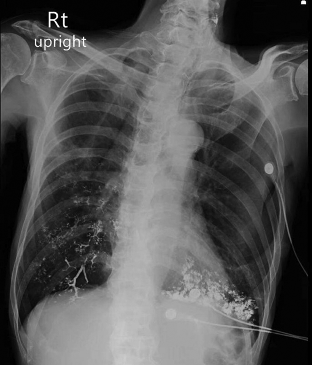
Complete blood cell counts showed leukocytosis (14,400 cells/μL) with neutrophil predominance. The serum electrolytes, kidney and liver function tests were within normal range. The CXR an hour after aspiration showed that the contrast was trapped and deposited in his tracheobronchial tree and bilateral upper lung fields (Figure 2).

He was admitted into the general ward and treated supportively with i.v. fluids, i.v. antibiotics (piperacillin/tazobactam) and supplementary oxygen. The follow-up CXR, the next day, showed progression of contrast through bilateral bronchioles and alveoli with left-side predominance.
The follow-up CXR at three days after aspiration showed subsequent resolution of contrast depositions in both lungs (Figure 3).

The lowest SpO2 during admission was 91%. His condition improved within a few days after the aspiration event. After one week of admission, he was finally discharged with no respiratory symptoms and normal SpO2.
Two years later, follow-up CXR and blood examination were done. The CXR showed considerable interval clearing of the aspirated contrast material but barium was still discovered in both sides of lungs (Figure 4). The plasma and urine barium concentration at 2 years after the aspiration were <0.5 μg/L and 13.6 μg/L (Inductively coupled plasma mass spectrometry technique), respectively.
Barium sulfate is used for examination of the upper gastrointestinal tract. Aspiration of barium contrast during radiological investigations occurs in 8% of children with gastroesophageal reflux.3,14 This complication is rare and does not commonly cause chemical pneumonitis.5,9 In cases of severe respiratory compromise after aspiration, it occurred frequently in patients with other comorbid diseases.9,15 A fatal case of barium sulfate aspiration mainly caused by the barium deposited inside both lungs with altered ventilation/perfusion (V′/Q′) ratio. The autopsy showed the diffused intra-alveolar spreading of barium throughout the lung parenchyma.2,16 Fatal consequences are considered to be related with large amounts of aspirated barium, which may reflect occlusion of the airway.1,2,12,17,18 Acute inflammation of the bronchial wall after barium aspiration has been attributed to high density preparations.1,5,18 Moreover, two fatal case reports4,19 are described as a result of the aspiration of a very high concentration (250% weight/volume) of barium sulfate. Severe chemical bronchitis and pneumonia induce acute respiratory distress syndrome which also causes death.1,4,16,17 The survival rate of barium aspiration among infants was 100% while it was 56.5% in adults.4
Here, we demonstrate the case of an 88-year-old man who aspirated barium sulfate and developed chemical pneumonitis. The predisposing factor for aspiration in this case might be the old age.
Treatment of barium aspiration depends on the severity of lung reaction. The management is mainly supportive care with oxygen therapy; however antibiotics use is not routinely recommended.1,7,11,13,18,19 One case report described successful treatment with early bronchioalveolar lavage (BAL) and positive pressure mechanical ventilation.20
Even the concentration of barium sulfate aspirated in our case was very high, our patient did not need intubation with ventilation support. Finally, he survived. Therefore, the prompt diagnosis and good supportive care might have contributed to the excellent prognosis in our case.
Two years after exposure, barium was not detected in the patient’s plasma, however barium was detectable in his urine (13.6 μg/L). His urine level was a bit higher than 95th percentile of urine level of metals in a reference United States population selected from the Third National Health and Nutrition Examination Survey which is 8.65 μg/L.21 In one study, barium was detected from blood and urine several hours after ingestion of commercial barium product for gastrointestinal examination.22 Currently, there is no reference urine barium level in the Thai population. These finding may be partially explained by our patient’s exposure of barium from food or water23 or by gradual systemic absorption of the residual barium in the lungs.
Due to the findings of the follow-up CXR, barium sulfate could be gradually cleared from the respiratory tract, however as long as 2 years, barium is still discovered in the lung. To the best of our knowledge, there is no any case report which follows up the patients until normal CXR is noted after aspiration.
In this case, we present an elderly male who aspirated a high concentration and a large amount of barium contrast, however he survived and had good prognosis.
Written informed consent for publication of the case details included accompanying images was obtained from the patient. This report was approved by the Institutional Ethic Committee Board of Ramathibodi Hospital Faculty of Medicine, Mahidol University. Ethics Approval Reference Number is COA.MURA 2022/138.
The data is not available for public access because of patient privacy concerns, but is available from the corresponding author upon reasonable request.
| Views | Downloads | |
|---|---|---|
| F1000Research | - | - |
|
PubMed Central
Data from PMC are received and updated monthly.
|
- | - |
Provide sufficient details of any financial or non-financial competing interests to enable users to assess whether your comments might lead a reasonable person to question your impartiality. Consider the following examples, but note that this is not an exhaustive list:
Sign up for content alerts and receive a weekly or monthly email with all newly published articles
Already registered? Sign in
The email address should be the one you originally registered with F1000.
You registered with F1000 via Google, so we cannot reset your password.
To sign in, please click here.
If you still need help with your Google account password, please click here.
You registered with F1000 via Facebook, so we cannot reset your password.
To sign in, please click here.
If you still need help with your Facebook account password, please click here.
If your email address is registered with us, we will email you instructions to reset your password.
If you think you should have received this email but it has not arrived, please check your spam filters and/or contact for further assistance.
Comments on this article Comments (0)