Keywords
dental implants, clinical evaluation, marginal bone loss, implant survival, zirconia, immediate loading
dental implants, clinical evaluation, marginal bone loss, implant survival, zirconia, immediate loading
Rehabilitation with dental implants has now become the mainstay of prosthetic care and greatly enhances the quality of life of patients undergoing replacement of missing teeth. For endosseous dental implants, titanium is considered the material of choice owing to its long-proven success and survival rates.1–3
The evolution of macro design and surface texture has resulted in the preferred use of rough surface implants, which contribute to increased primary stability and markedly reduced healing time.4 However, the rough surfaces of titanium are more prone to plaque accumulation and subsequent periimplantitis, as well as increased release of titanium particles and ions into the surrounding tissue.5–7
Although the precise impact of the released titanium particles needs to be further explored, there have been concerns regarding hypersensitivity to titanium in a certain minority of the population, estimated to be 0.6%.8 Another noteworthy consideration is the greyish hue of titanium that reflects through the peri-implant tissue of patients with a thin gingival biotype and poses a threat to the aesthetic outcome, especially in patients with increased aesthetic demands or in those with a high smile line.9,10
Zirconia implants have recently been recommended as an aesthetic alternative to titanium implants. The resemblance to natural tooth colour, combined with excellent biocompatibility and reduced adhesion to dental plaque, favour its use as a dental implant biomaterial.11 They also exhibit favourable physical and mechanical properties.12 However, aging or low-temperature degradation of zirconia negatively impacts the properties of this biomaterial resulting in roughness and eventual degradation of the material.13,14
Endosseous zirconia implants seem to be a preferred option in patients with a thin gingival biotype, or who express specific concern regarding the visibility of a greyish hue at the cervical margin, patients who may have sensitivity to titanium, or in those who insist on having “metal-free” dental therapy.15
Research has shown that the osseointegration of zirconia implants is similar to titanium implants.16 However, to the best of the authors’ knowledge, the clinical performance of zirconia implants has not yet been evaluated in the Indian population.16 Thus, this study was conducted to assess the short term treatment with endosseous zirconia implants in a subset of the Indian population.
The study was designed as a prospective cohort investigation in a university setup (A B Shetty Memorial Institute of Dental Sciences) and was conducted following institutional ethical committee clearance (A B Shetty Memorial Institute of Dental Sciences; Cert no: ABSM/EC66/2017). Written informed consent was obtained from all participants in this study for publication of the data and their clinical images.
All seven patients who visited the clinic between June 2018 and February 2019 were included in the current study. Follow-up duration was set to six and 12 months after the placement of the implant. Participants were selected by following stated criteria:
Sintered and yttria-stabilised, one-piece, zirconia dental implants (WhiteSky, Bredent, Germany) were used in this study. These endosseous implants have a double cylindrical thread design. The portion of the implant surface that is intended to remain embedded in bone has a sandblasted surface, whereas the transmucosal portion is smooth, with a height of 2 mm. The abutment has a height of 6.8 mm which can be modified according to the clinical situation.17
The implants were placed according to the manufacturer’s instruction in a prosthetically determined 3D position. An immediate post-operative orthopantomogram (OPG)/intraoral periapical (IOPA) was taken to evaluate the positioning of the implants and to serve as a baseline for future bone level assessment.
The one-piece zirconia implants were immediately restored with a highly cross-linked acrylic proshell (Visiolign, Bredent) relined with composite resin (Comboline, Bredent). The restoration was kept out of centric and eccentric occlusion and served as a durable, long-term prosthesis. The marginal bone levels (MBL) were assessed at six months and 12 months following placement and mean MBL was calculated. Data was analysed by using SPSS version 23 (IBM SPSS Statistics, RRID:SCR_019096) (An open-access alternative is R Stats (R Project for Statistical Computing, RRID:SCR_001905)).
Five implants were placed in the maxilla, out of which three were placed in well-healed sites, and two were placed following extraction (immediate and early placement, respectively). Five implants were placed in the mandible, among which one was used to replace two missing lower central incisors and the remaining four implants were used for an immediately loaded full-arch prosthesis. Two participants received implants in the mandible and five participants received implants in the maxilla.
Figures 1, 2, and 3 show single tooth replacement in a well-healed edentulous site. Figure 4 shows a cantilever prosthesis using one implant in the anterior region. Figure 5 shows extraction and immediate placement. Figure 6 shows full arch rehabilitation.
(a) Preoperative view; (b) Preoperative radiograph; (c) Implant insertion; (d) Immediate restoration; (e) Immediate post-op radiograph.
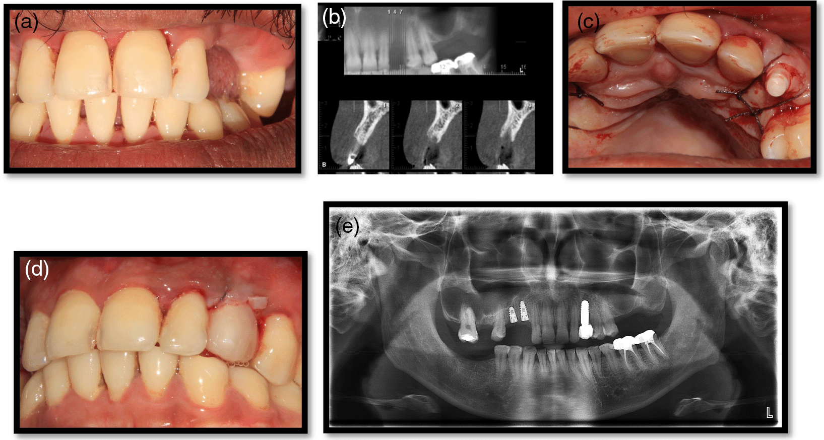
(a) Preoperative view; (b) Preoperative radiograph; (c) Initial site; (d) Implant insertion; (e&f) Immediate restoration; (g) Immediate post-op radiograph; (h) Radiograph at 1-year post-placement.
(a) Preoperative view; (b) Preoperative radiograph; (c) Implant insertion; (d) Immediate restoration; (e) Immediate post-op radiograph; (f) 1-year post-placement.
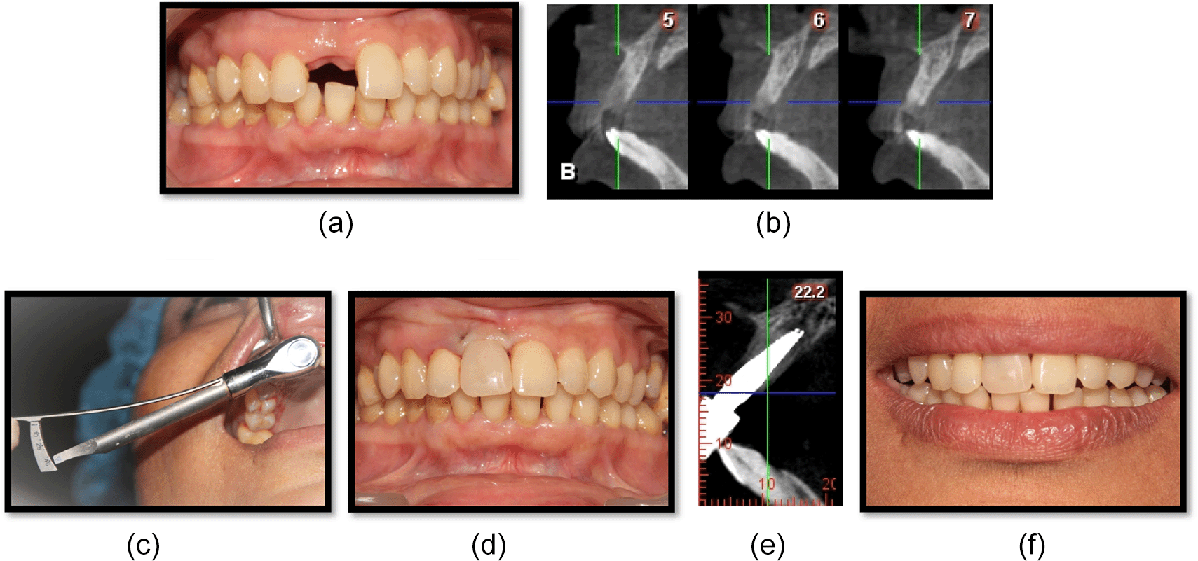
(a) Preoperative view; (b) Preoperative radiograph; (c) Paralleling place; (d) Immediate restoration; (e) Immediate post-op radiograph.
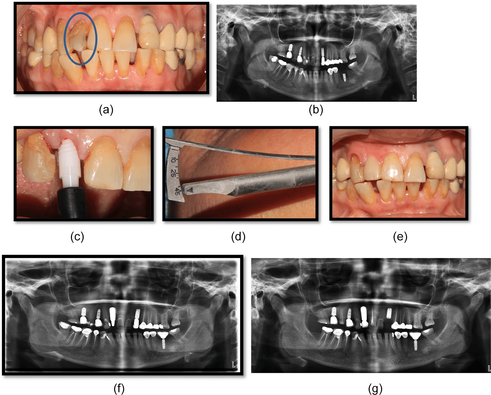
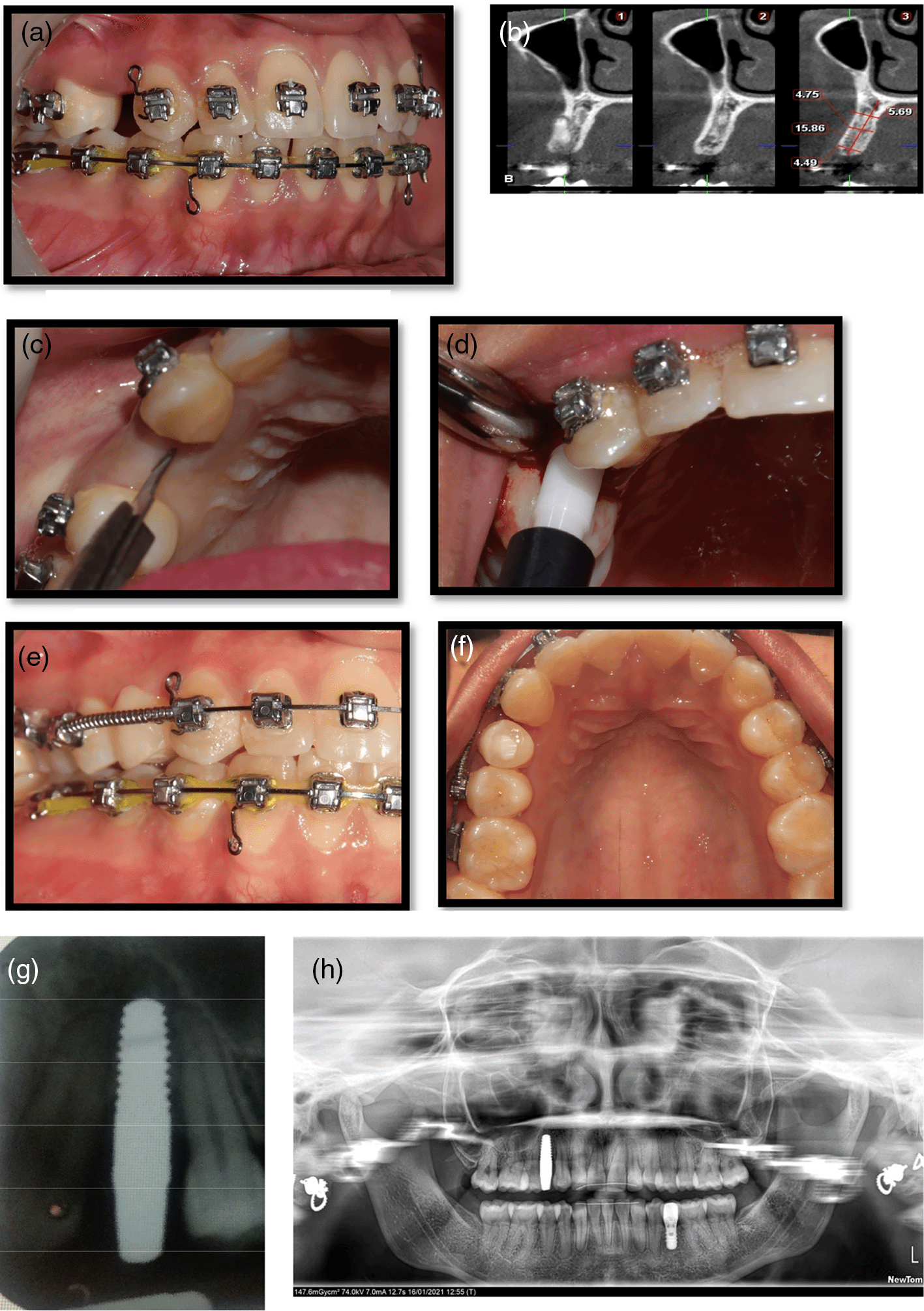
(a) Preoperative view; (b) Preoperative radiograph; (c) Implant insertion; (d) Implant insertion torque; (e) Immediate restoration; (f) Immediate post-op radiograph; (g) Radiograph 1 year post-placement.
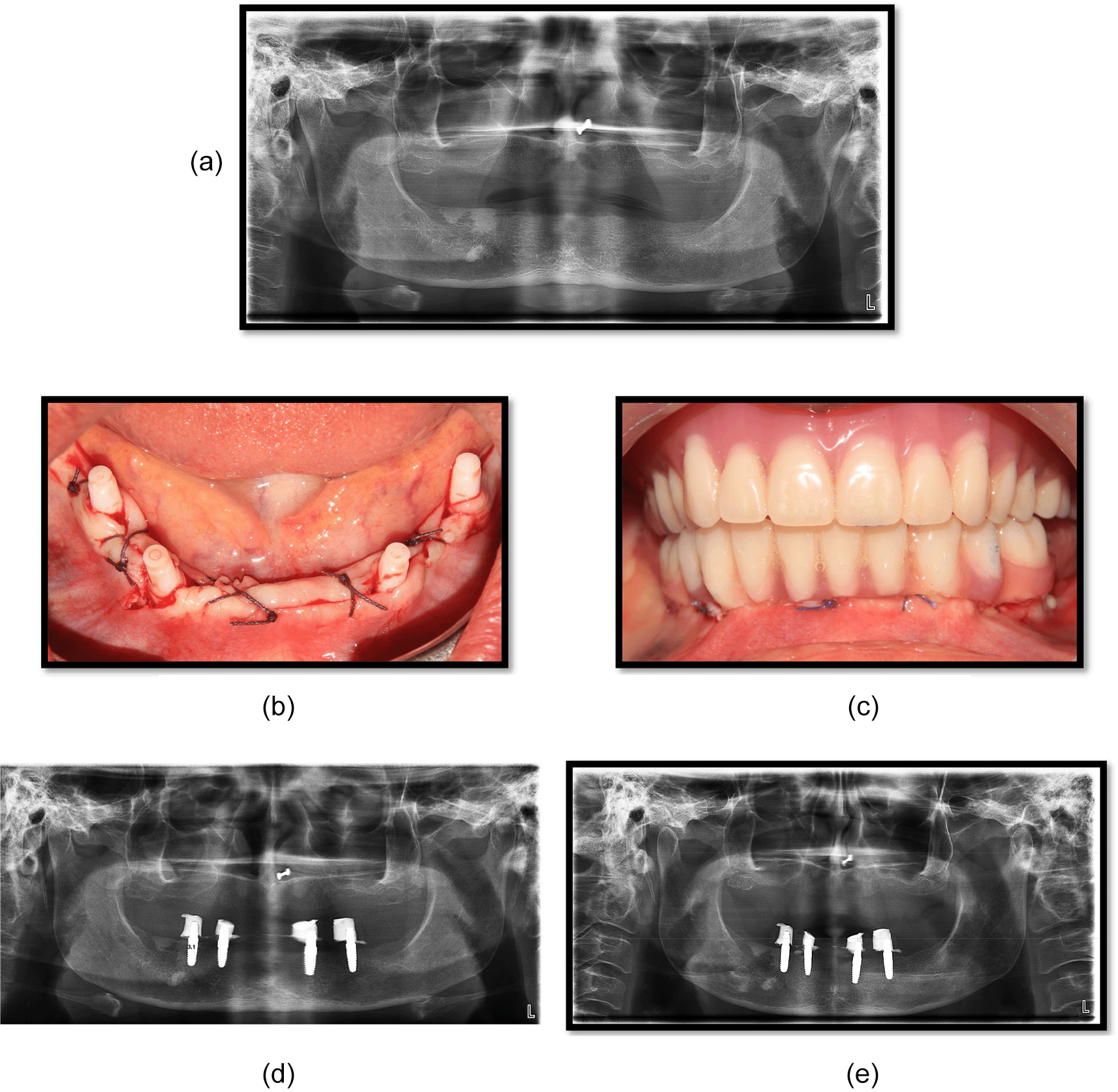
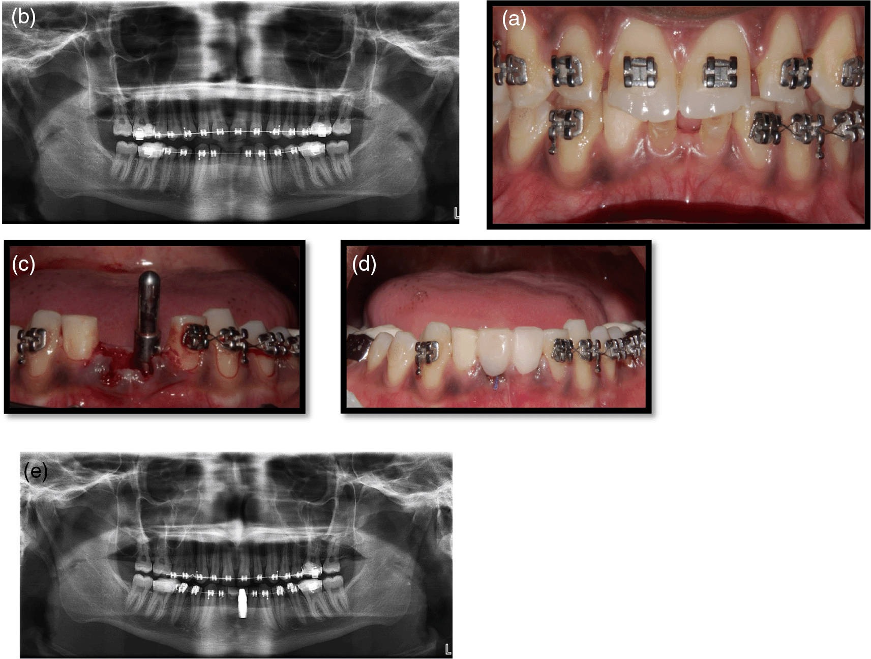
(a) Preoperative radiograph; (b) Implant insertion; (c) Immediate restoration; (d) Immediate post-op radiograph; (e) Radiograph 1-year post-placement.
Diameter: Three implants of diameter 3.5 mm were placed in the lower anterior region and the remaining implants had diameters greater than or equal to 4 mm.
Torque: A minimum torque of 25 Ncm was obtained during implant placement.
Survival rate: During the follow-up period which extended up to 12 months, two early implant failures were observed. Thus, the cumulative survival rate at the end of two years was 80%.
Marginal bone loss: The present study showed a mean marginal bone loss (MBL) of 0.84 mm at one year following implant insertion, with maximum bone loss occurring in the first six months (0.73 mm) following placement (Table 1).18
The present study evaluated the mean marginal bone loss and survival rate of 10 endosseous zirconia implants in various clinical scenarios, after 12 months of implant placement. Concern regarding the prevalence of hypersensitivity to titanium particles in peri-implant tissues, the impaired aesthetic outcome in patients with a thin biotype and increasing demand for “metal-free” therapy, have made zirconia implants a promising alternative to the conventional endosseous titanium implants.
While the mechanical and optical properties of zirconia are the major factors supporting its use as an implant material, its biocompatibility and excellent soft-tissue response also have an important contribution to make in maintaining long term peri-implant health. Since zirconia implants emerged as a potential alternative to titanium, various studies have been conducted to assess its hard and soft tissue integration.18,19
To the best of the authors’ knowledge, this is the first study conducted in the Indian population to evaluate the performance of endosseous zirconia implants in a variety of clinical situations.
Osseointegration of dental implants and long – term stability of marginal bone level remain the defining criteria for implant success.20 The present study showed a mean marginal bone level of 0.73 mm after one year, with maximum bone loss occurring in the first six months. This result is in accordance with previous studies conducted by Roehling et al., and Pieralli et al.,21,22 and following Albrektsson’s criteria of implant success.20 This may be credited to the high biocompatibility of zirconia, low plaque adhesion, and the absence of a micro gap between the fixture and abutment.23
The increased bone loss observed in the first six months is similar to the findings reported by Borgonovo et al.,23 and Balmer et al.,24 and can be attributed to the extensive bone remodelling following implant placement. Although in the present investigation, the mean MBL was 0.73 mm after one year, one implant showed MBL ≥ 2 mm. This was probably due to the greater initial implant insertion depth used in extraction sockets.
Of the 10 implants, two implants showed early failure. One of these two implants was placed in the lower anterior region to replace two missing central incisors. The provisional restoration had a cantilever which could have caused an overload and increased micromovement leading to failure. The second implant which failed was placed in the maxillary canine region in a patient who had both upper and lower posterior teeth rehabilitated with removable partial prostheses. Failure of this implant may be attributed to the patient’s preference of incising with a fixed prosthesis. Thus, the cumulative survival rate of implants in this study was 80%, lower than those reported in a recently published systematic review25 and especially with the survival rate of titanium implants.26
Of particular interest, was the reduced bone loss seen with the four implants placed in the mandible and immediately loaded with a fixed hybrid prosthesis, which can be attributed to the splinting effect of the prosthesis. However, this finding contrasted with the results of the study conducted by Borgonovo et al.,23 who observed no statistically significant difference in the marginal bone loss adjacent to free-standing vs. multiple splinted implants. Thus, while the results of this study were encouraging towards the use of endosseous zirconia implants in diverse clinical scenarios, due care and caution in case selection must be exercised. Long-term studies in this context are required.
The cantilever of an immediately loaded prosthesis can lead to implant failure. Missing posterior teeth and patients with a partial denture can lead to failure as the patients lend to bite with natural teeth. A minimum bone width of 5 mm is needed to place one-piece zirconia implants designed for immediate loading, to avoid simultaneous augmentation procedures. Splinting of zirconia implants in case of full arch reconstruction will reduce the resorption of bone.
Figshare: A short-term prospective study to evaluate the clinical performance of endosseous zirconia implants. https://doi.org/10.6084/m9.figshare.17163584.v6.18
This project contains the following underlying data:
Data are available under the terms of the Creative Commons Attribution 4.0 International license (CC-BY 4.0)
| Views | Downloads | |
|---|---|---|
| F1000Research | - | - |
|
PubMed Central
Data from PMC are received and updated monthly.
|
- | - |
Provide sufficient details of any financial or non-financial competing interests to enable users to assess whether your comments might lead a reasonable person to question your impartiality. Consider the following examples, but note that this is not an exhaustive list:
Sign up for content alerts and receive a weekly or monthly email with all newly published articles
Already registered? Sign in
The email address should be the one you originally registered with F1000.
You registered with F1000 via Google, so we cannot reset your password.
To sign in, please click here.
If you still need help with your Google account password, please click here.
You registered with F1000 via Facebook, so we cannot reset your password.
To sign in, please click here.
If you still need help with your Facebook account password, please click here.
If your email address is registered with us, we will email you instructions to reset your password.
If you think you should have received this email but it has not arrived, please check your spam filters and/or contact for further assistance.
Comments on this article Comments (0)