Keywords
gallbladder carcinoma, ovarian metastasis, radiology, pathology
This article is included in the Oncology gateway.
gallbladder carcinoma, ovarian metastasis, radiology, pathology
Ovaries can be an elective metastatic site for many cancers mostly in gastrointestinal tract, breast and endometrial cancers. Krukenberg tumor also known as signet ring adenocarcinoma, mostly from gastric origin, is the most described.1 Only a few cases of ovarian gallbladder cancer metastasis are reported in the literature.1,2 The chance of finding ovarian spread among patients with gallbladder carcinoma are up to six percent.1,2 Here we describe a case of bilateral ovarian metastasis of gallbladder carcinoma in a 44-year-old woman, suffering from pelvic pain, anorexia, weight loss and decreased visual acuity. She declined adjuvant therapy and passed away six months after surgery.
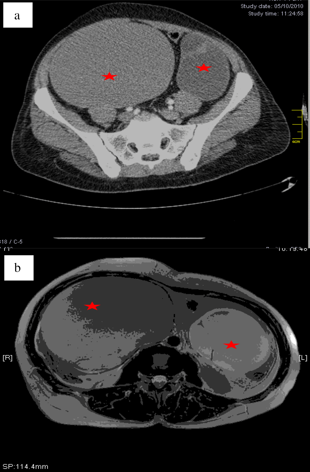
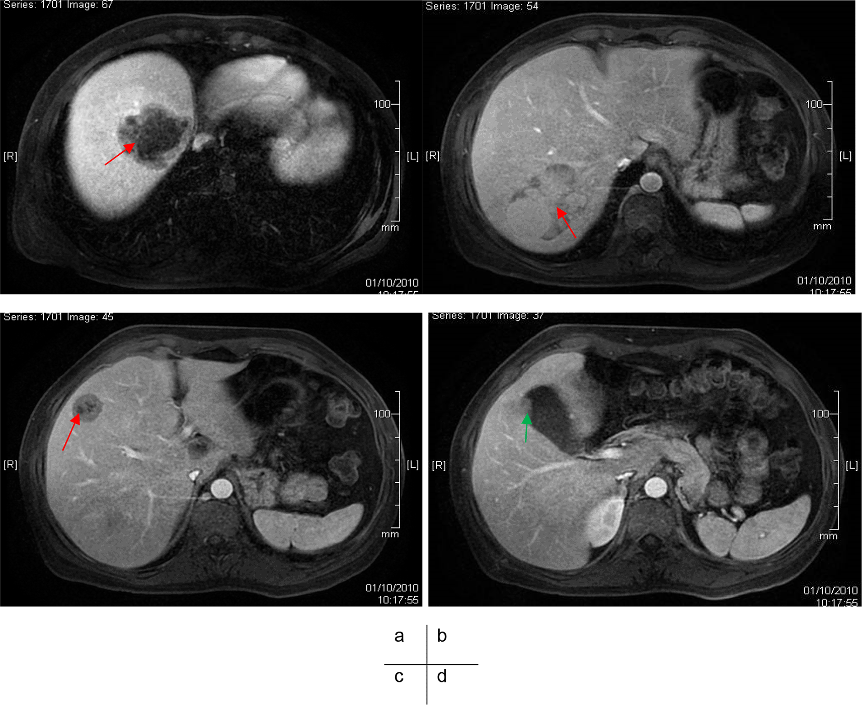
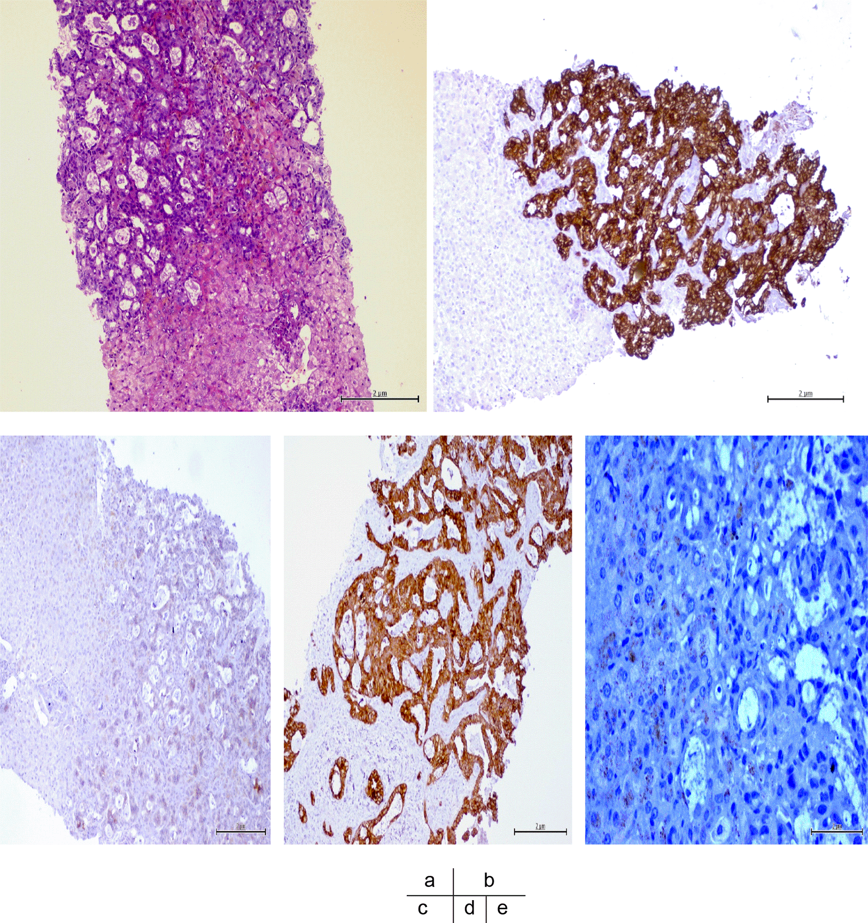
A 44-year-old Arabic woman (gravida 4, para 3) was admitted to our department of gynecology suffering from chronic pelvic pain associated with weight loss and anorexia during the last three months and had been experiencing reduced visual acuity since a week. She had no relevant medical history and did not undergo any surgical procedure. No family history of inherited gynecologic cancer was found. A physical examination found bilateral pelvic masses. An ultrasound exam showed bilateral and complex ovarian cysts with septa. Serum tumor markers (CA-125, CEA and CA19-9) were normal. A CT scan and pelvic abdominal MRI were performed. They showed bilateral ovarian masses with large cysts and enhanced septa. The tumors were isodense on the CT scan and hyper intense on T2 MRI sequences (Figure 1). We additionally found several necrotic metastatic lesions in the liver (segments I, IV, VI and VII) associated to a polyp of the gallbladder (Figure 2). Furthermore, there were metastatic microndular miliary in lungs, left pulmonary arterial embolism and bilateral choroid metastasis (Figure 3). Liver biopsy showed abnormal cell proliferation arranged in glandular structures, backed against each other. Tumor cells were cytokeratin 7 and 19 positive, cytokeratin 20 and hepatocyte antigen negative, allowing the diagnosis of a moderately differentiated adenocarcinoma arising from the gallbladder.


Multidisciplinary consultation meeting decided to perform primary staging surgery (total abdominal hysterectomy, bilateral adnexectomy, omentectomy, peritoneal biopsy, cholecystectomy and partial hepatectomy). Per-operative exploration showed suspicious bilateral mi part solid mi part cyst ovarian tumors, with a focally roughened surface. Uterus was normal in shape and size. Gallbladder had thick walls. Pathologic examination of gallbladder showed moderately differentiated adenocarcinoma. However, it showed benign ovarian serous cystadenomas with deposits of metastatic adenocarcinoma in the parenchyma (Figure 4). There were no malignancy signs in uterus, omentum and peritoneal biopsies. Patient did not present any post-operative complications. After surgery, she was referred to the oncologists for adjuvant therapy. Unfortunately, the patient refused any further treatment and died six months later.

Gallbladder carcinoma is found in 1% of patients undergoing cholecystectomy.2 Adenocarcinoma counts for 70 to 90% of gallbladder cancers.2 However, only a small percentage of patients with carcinoma of the gallbladder will ever have metastasis to the ovary.1,2 When ovarian tumors happen to be discovered prior to gallbladder carcinoma, it often leads to misinterpretation.
When ovarian tumors are the first manifestation of the disease, gallbladder carcinoma spread to the ovaries can be misinterpreted for primary ovarian carcinoma.2,3 In fact, cases of gallbladder carcinoma do not infrequently present with hepatobiliary symptoms. However, when there is metastasis to the ovary, the biliary symptoms may be masked by ascites or local symptoms correlated to the ovarian mass.3,4 In a few cases, the primary gallbladder carcinoma was unsuspected and was discovered incidentally by surgical investigation during laparotomy for ovarian resection.3,4 In our case, the gallbladder carcinoma was suspected by the biopsy of the liver.
Although there are a lot of data in the literature on ovarian metastases, the case of ovarian metastasis from gallbladder cancer remains little discussed.3-5 Accurate preoperative diagnosis of these advanced primary gallbladder carcinomas with ovarian metastases is less than 30%.5
Many criteria have been reported to differentiate ovarian metastases from primary ovarian cancer.1,5 Bilaterality, surface implants, multinodularity, infiltrative pattern, growth in the ovarian hilum, mucin without epithelial cells on the tumor surface and presence of signet ring cells are the most common features suggesting ovarian metastasis.1,5 These features may be absent when the ovarian metastasis simulates a benign cyst.1 In our case report, the tumor was a mixture of solid and cystic areas. Brown et al.6 found in their revue of literature that most metastatic neoplasm to the ovary are predominantly solid or a mixture of solid and cystic areas and tended to be bilateral more often than primary neoplasm.
As for imaging criteria, Kim et al.7 found out, through a review of 85 patients’ CT imaging, that metastatic ovarian neoplasm should be suspected on CT examination when the tumor is solid and contained well defined intra-luminal cystic lesions.
On macroscopic examination, Lee and Young8 concluded that bilateral tumors with surface implants are in favor of metastatic neoplasm to the ovary.
This case’s originality lies in the fact that we spotted the primary tumor though it is rare and uncommon. The prognosis was poor from the beginning, worsened by the patient’s decision refusing any adjuvant therapy.
Ovarian metastasis can be difficult to distinguish from primary ovarian carcinoma especially when the primary gastrointestinal tract tumor is not yet diagnosed. There are no specific macroscopic or microscopic features that can differentiate ovarian metastases from a primary malignancy. Nevertheless, possibility of metastatic origin should be considered whenever we deal with a bilateral and/or mixed ovarian mass.
We obtained informed consent from the patient’s family to publish the details of the case report.
| Views | Downloads | |
|---|---|---|
| F1000Research | - | - |
|
PubMed Central
Data from PMC are received and updated monthly.
|
- | - |
Is the background of the case’s history and progression described in sufficient detail?
Yes
Are enough details provided of any physical examination and diagnostic tests, treatment given and outcomes?
Yes
Is sufficient discussion included of the importance of the findings and their relevance to future understanding of disease processes, diagnosis or treatment?
Yes
Is the case presented with sufficient detail to be useful for other practitioners?
Yes
Competing Interests: No competing interests were disclosed.
Reviewer Expertise: Cancer researcher
Alongside their report, reviewers assign a status to the article:
| Invited Reviewers | |
|---|---|
| 1 | |
|
Version 1 14 Feb 22 |
read |
Provide sufficient details of any financial or non-financial competing interests to enable users to assess whether your comments might lead a reasonable person to question your impartiality. Consider the following examples, but note that this is not an exhaustive list:
Sign up for content alerts and receive a weekly or monthly email with all newly published articles
Already registered? Sign in
The email address should be the one you originally registered with F1000.
You registered with F1000 via Google, so we cannot reset your password.
To sign in, please click here.
If you still need help with your Google account password, please click here.
You registered with F1000 via Facebook, so we cannot reset your password.
To sign in, please click here.
If you still need help with your Facebook account password, please click here.
If your email address is registered with us, we will email you instructions to reset your password.
If you think you should have received this email but it has not arrived, please check your spam filters and/or contact for further assistance.
Comments on this article Comments (0)