Keywords
atopic dermatitis, children, efficacy, vitamin D, human health
This article is included in the Agriculture, Food and Nutrition gateway.
atopic dermatitis, children, efficacy, vitamin D, human health
Atopic Dermatitis (AD) is now considered a complex multifactorial disease that includes defects in skin barrier structures, immune dysregulation, genetic susceptibility, and changes in skin flora which mostly occur in infancy and childhood. Based on the clinical features, AD can be divided into 3 forms, namely AD in infants (2 months-2 years), children (2–12 years), and adolescents (over 12 years).1 Increasing prevalence of AD has been reported in areas including the Asia-Pacific region, where it is reported that 88% of children with AD have either mild or moderate and 12% have severe AD. However, Indonesia still has a lower prevalence in children between 6-7 years old when compared to Thailand and Malaysia, and a lower prevalence in children aged 13-14 years when compared to Pakistan.2
In addition to the reduction of skin inflammation, recently, AD treatment has focused more on the regulation of the immune response and enhancing the barrier function of the skin.3 Poor compliance with the use of topical drugs makes some researchers try to find other drugs that are not only safe, cheap, easy to use but also effective. Several recent studies have shown that vitamin D supplementation may be an option, although the results of intervention trials are still conflicting.4
In AD patients, defects in the skin barrier structure, as well as decreased functional integrity and reduced ability to regenerate have roles in inducing immune responses and specific inflammatory reactions.5 In acute lesions, there will be a decrease in AMP (Antimicrobial Peptide) production, an increase in S. aureus colonization, and an effect on the severity of the disease and reduce the risk of infection. Vitamin D can increase barrier function, induce AMP and enhance monocyte and macrophage cell function.6 Vitamin D has been known to have some effects on the innate and adaptive immune systems. Several mechanisms can modulate the progression of AD lesions, such as increasing epidermal differentiation, increasing production of cathelicidin, decreasing Th2 cytokines, decreasing Ig E production, decreasing B cell proliferation, and upregulating of T cells.7
A previous systematic review and meta-analysis in 2019 on vitamin D and AD had reported a highly statistically significant reduction in SCORAD (Scoring of Atopic Dermatitis) on intervention with vitamin D, in the paediatric and adult population.8 While a systematic review published by Huang on the paediatric AD population in 2018 concluded that 67% of the collected studies reported a significant improvement in AD severity with vitamin D supplementation, but this systematic review did not include a meta-analysis.9 We conducted a meta-analysis with research published in last 10 years because there has been an increase in publications regarding vitamin D supplementation during this time.10
The main objective of our systematic review and meta-analysis is to provide an updated review of the interventional study of vitamin D in the paediatric AD population to investigate clinical outcomes from measuring scales.
We conducted a systematic search of the literature on several databases, namely PubMed, Cochrane Library, ProQuest, and a clinical trial website, ClinicalTrials.gov with keywords showed in Table 1 (see also Extended data33). We also did manual hand searching on Google Scholar and searched for grey literature on the repository (including research from January 1st 2010 to October 31st 2020, and the databases were last searched on 2nd November 2020). The search procedure was based on Preferred Reporting Items for Systematic Reviews and Meta-Analysis (PRISMA). The completed PRISMA checklist is available in Reporting guidelines.33 This search of titles and abstracts was limited to articles that were human-focused and published in English and Bahasa Indonesia. Statistical analysis was carried out with Review Manager (RevMan, Cochrane, London, UK) version 5.4.1 with standardized mean difference and risk ratio as a measure of the effect of therapy.
| Database | Keywords |
|---|---|
| Cochrane Library | eczema OR atopic in Title Abstract Keyword AND therap* OR treatment in Title Abstract Keyword AND vitamin D in Title Abstract AND children OR child OR paediatrics OR paediatrics AND Clinical trials AND SCORAD |
| PubMed | (((((eczema [MeSH Terms]) OR eczema [Title/Abstract]) OR dermatitis [Title/Abstract]))) AND ((Vitamin D [MeSH Terms]) OR Vitamin D [Title/Abstract])) AND (((treatment [Title/Abstract]) OR therap* [Title/Abstract]) OR therapeutics [MeSH Terms]) |
| ProQuest | (ti (eczema* OR dermatitis OR atop*) OR ab (eczema* OR dermatitis OR atop*)) AND ti (children OR paediatrics OR pediatri) OR ab(children OR paediatrics OR pediatri) AND ti (therapy OR treatment) AND (ti (vitamin D) OR ab (vitamin D)). |
| www.clinicaltrials.gov | *vitamin D* AND *Interventional Studies* AND *Atopic Dermatitis* AND *SCORAD* AND *Child* |
| Google Scholar | ti AND ab (efficacy AND vitamin D AND Atopic Dermatitis AND SCORAD AND Child AND Randomized Control Trial) |
In studies that include children and adults as participants, we contacted the author to obtain separate data that contained child subjects only. The ethical clearance of this study has been published from the Ethical Committee of Dr. Soetomo General Academic Hospital Surabaya number 0206/LOE/301.4.2/XI/2020. We did not register the protocol.
Intervention studies including Randomized Control Trials and Prospective Cohort studies with clinical outcomes measured on a scale in both groups, before and after the intervention were assessed. Inclusion criteria were as follows: (1) studies with age group 0-18 years old and diagnosed as mild, moderate, or severe atopic dermatitis in both females and males; (2) No restriction to the duration of intervention, type of vitamin D, doses used, frequency and route of administration, and clinical outcome measuring scale: SCORAD, EASI (Eczema Area and Severity Index), IGA (Investigator Global Assessment). (3) studies that provided complete data for clinical outcomes. Exclusion criteria were articles that did not provide full text.
1. Evaluating the outcome of the disease (changes in SCORAD or EASI) in the Vitamin D supplementation groups compared to placebo groups.
2. Calculating the clinical importance of both groups so that the CER (Control Event Rate), EER (Experimental Event Rate) and NNT (Number Needed to Treat) values can be obtained.
This analysis included all articles that qualified for selection criteria. Two author, ANH and SS extracted data from each included study including author, country, publication year, study population, AD severity, supplementation dose, frequency, route of administration, duration and outcome scale. The clinical outcome was measured by scale: SCORAD, EASI or IGA. We defined the clinical outcomes as follows:
(1) SCORAD: A clinical measurement tool used to calculate the severity of Atopic Dermatitis patients. The lesion area was calculated based on the rule of nine with a value of 0-100. Intensity was measured in a representative area by looking at the form of skin abnormalities that were erythema, edema, oozing or crusts, excoriations, lichenification, and dry skin, and each was assigned a value of 0 if there was no lesion, 1 if the lesion was mild, and 2 if the lesion was moderate and 3 if the lesion was severe, then the scores were summed to get B (0-18). Subjective symptoms were measured by Visual Analog Scale (VAS), calculated on average for every 3 night whether there were symptoms of itching and sleep disturbances, with a score of 0 if there was no itching or sleep disturbances, and 10 for the most severe itching or sleep disturbances. These numbers are summed to give C (0-20). The results of the three parameters were submitted into the formula A5+7B/2+C, then grouped into mild AD (<25), moderate AD(25-50) and severe AD(>50) categories.11
(2) EASI: an instrument used by examiners (doctors, dermatologists) to quantify lesion progression and severity of AD patients, by assessing the extent of the disease at four body sites (head/neck, trunk including genitalia, superior and inferior extremities) and measures four clinical signs: (1) erythema, (2) induration/papulation, (3) excoriation, and (4) lichenification each on a scale of 0 to 3. The score can then be divided into 0 (clean), 0.1-1.0 (nearly clean), 1.1-7 (mild), 7.1-21 (moderate), 21.1-50 (severe), 50,1-72.0 (very severe). EASI confers a maximum score of 72.11
(3) IGA: an instrument used to assess overall disease severity at one given time point, and it consists of a 6-point severity scale from clear to very severe disease (0 = clear, 1 = almost clear, 2 = mild disease,3 = moderate disease, 4 = severe disease and 5 = very severe disease). IGA uses clinical characteristics of erythema, infiltration, papulation, oozing and crusting as guidelines for the overall severity assessment.12
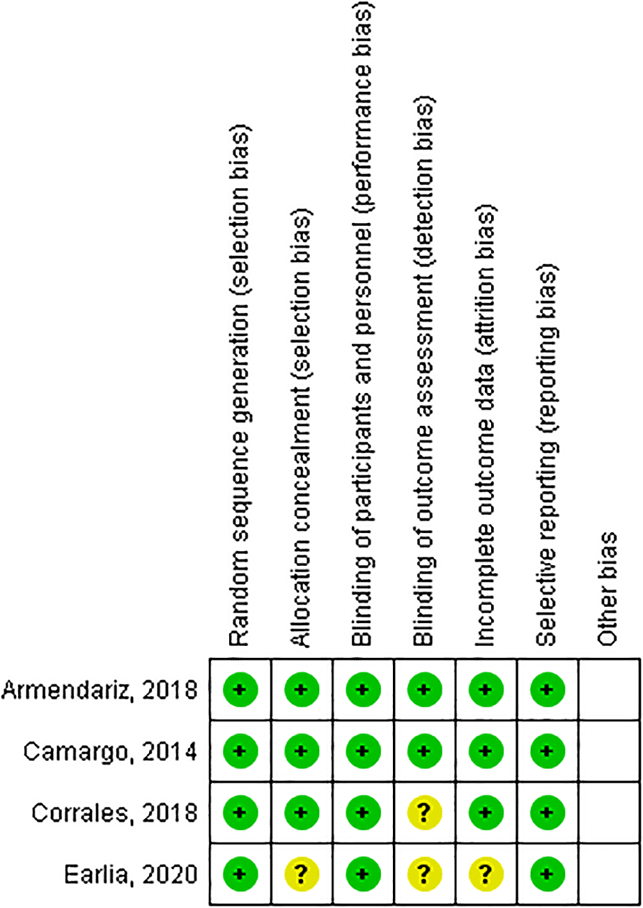
The covidence quality assessment template was customized for this study and the quality of each study was assessed by three authors (ANH, SS and DWS) independently by using the Centre for Evidence-Based Medicine’s RCT (Randomized Control Trial) worksheet, by conducting a critical appraisal to determine validity, importance and applicability. Validity was assessed based on Recruitment, Allocation and Measurement Blinding Outcome. Importance was assessed based on clinical data as well as statistical data. Applicability was assessed by answering several questions related to the author’s setting (Table 2). Critical appraisal for prospective (cohort) study was conducted using critical appraisal skill programme worksheet (Table 3). Another author resolved any disagreement between them (CRSP, DMI, and DD). Quality analysis of the interventional studies had showed three studies scoring randomized double-blind clinical trials with adequate randomization and blinding. These were Refs. 13, 14 and 15. The other study did not mention randomization but confirmed the blinding of both participants and researchers.16
| Camargo, 201414 | Galli, 201519 | Lara-Corrales, 201813 | Sanchez-Armendariz, 201815 | Earlia, 202016 | Mansour, 202020 | ||
|---|---|---|---|---|---|---|---|
| Recruitment | Randomization | Yes | Unclear | Yes | Yes | Unclear | Yes |
| Similarity | Yes | Yes | Yes | Yes | Yes | Yes | |
| Allocation | Treated equally | Yes | Yes | Yes | Yes | Yes | Yes |
| Minimum loss to follow up | Yes | Yes | Yes | Yes | Yes | Yes | |
| Measurement blinding outcome | Yes | Yes | Yes | Yes | Yes | Yes | |
| Importance | Clinical | + | - | - | + | + | - |
| Statistical | p=0.04 | P=0.5 | p=0.07 | p=0.02 | P<0.001 | p=0.039 | |
| Applicability | Yes | Yes | Yes | Yes | Yes | Yes |
| Filippo, 201521 | Raj 202022 | |
|---|---|---|
| Representative of the population | Yes | Yes |
| Methods for exposure objective and consistent | Yes | Yes |
| Subjects/outcome accessor blinded | Unclear | Unclear |
| Sufficiency of follow up | Unclear | Yes |
| Overcoming confounding factor | Yes | No |
| Importance | Yes | Yes |
| Applicability | Yes | Yes |
The risk of bias in RCT studies was assessed with The Cochrane Collaboration's tool for assessing risk of bias17 by ANH, SS and DWS, then we discussed the outcome until we were in agreement. The assessment results were categorized as “yes” for low-risk bias, “unclear”, and “no” for high-risk bias (Figure 1). Cohort studies was assessed with Newcastle-Ottawa Scale (NOS)18 and comprised several items including comparability of the groups (2 points), and ascertainment of exposure (3 points). Each study was interpreted to be low quality (scores <4), moderate quality (scores of 5–6), or high quality (scores ≥7) that was shown in Table 4.
We performed the data with Review Manager (RevMan, Cochrane, London, UK) version 5.4.1. Three authors, DWS, IC, and SA conducted statistical analysis and presented the result in a forest plot and funnel plot. Statistical analysis was done by calculating the standardized mean differences (SMDs), with 95% CIs, of pre- and post-intervention in both groups, and the standard deviation of each study, and was also calculating risk ratio (RR), with 95% CIs, by counting the number of events in each group with a dichotomy table (Table 5). Significance of RRs was determined using the Z test (p<0.05 was considered statistically significant). We assessed the heterogeneity among the studies using I2 (considered heterogeneity existed if I2 > 25%), then Random Effect Model was adopted. For publication bias, we used funnel plot and it can be seen that the four studies are distributed symmetrically, that is, the distribution of research is balanced on the left and right of the centre line boundary. This means that there is no potential for publication bias regarding the conclusions.
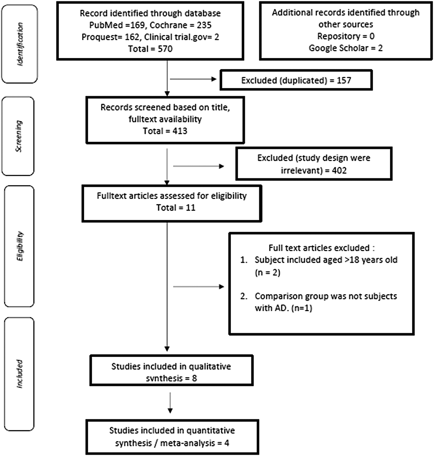
570 articles were initially retrieved, and the results of the evaluation of duplicate articles by title showed 157 articles with similar titles and were subsequently excluded from this study. The next evaluation was carried out by reviewing the title of each piece of literature that had been searched based on keywords. There were 11 literatures by excluding 402 literature that was irrelevant with the study design. We did further evaluation of 11 kinds of literature based on eligibility criteria, critical appraisal, and quality assessment, and excluded two articles with subject included aged > 18 years old and one article with non-AD subjects as a comparison group. Qualitative synthesis then resulted two studies that could not be included in the meta-analysis due the fact that no standard deviation was reported19 and.20 A further two studies were also excluded due to the study design which was single-arm cohort21 and22 so that the final results were four articles which were then analysed in this study.
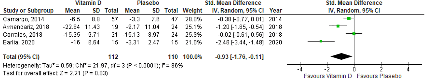
Four articles were included in meta-analysis, as described in Figure 2. The years of publication for all studies were ranging from January 2010 to October 2020. Three studies were conducted with paediatric participants only13,14 and16 and one study was conducted with adult and paediatric participants.15 There were different doses and durations in supplementing vitamin D among studies. One study reported that vitamin D supplementation did not significantly improve the severity of the disease,13 but the other three studies reported otherwise. This study only included AD participants with deficiency or insufficiency status of serum vitamin D. We summarized all studies including population, sample size, intervention, and mean difference outcome of both groups (Vitamin D and placebo groups). All outcomes listed as positive (p<0.05) or negative (p>0.05) are shown in Table 7.
Four randomized controlled trials assessed the efficacy of vitamin D supplementation. The characteristics of the included studies are summarized in Table 6. Three studies measured the SCORAD indexes, whereas only one of the included studies assessed the efficacy of vitamin D supplementation by using EASI. One study used both adults and children as a participant, so we contacted the author to obtain the data associated with the children only. A meta-analysis of four trials showed that the SCORAD index and EASI score decreased significantly after vitamin D supplementation (standardized mean difference = -0.93, 95% CI = -1.76 to -0.11). We observed statistical heterogeneity among the studies (I2 > 25%; Figure 3). We also assessed the potency of the publication bias in those included studies with funnel plot and the result was symmetrical indicated that there was no potency of the publication bias in the four included studies.
| Author | Country | Dose, frequency and duration | Study design | Mean age (years) | Outcome scale | AD severity | Supplementation giving method | Study population | Result |
|---|---|---|---|---|---|---|---|---|---|
| Camargo et al., 201414 | Mongolia | Vitamin D3 1000 IU/day for 1 month | RCT | 9 | EASI | Mild, moderate, severe | orally | Paediatric patients with winter-associated AD, EASI between 10-72, based on randomization with a random number generator. | Vitamin D supplementation showed clinical improvement |
| Lara-Corrales et al., 201813 | Canada | Vitamin D 2000 IU/day for 3 months | RCT | 7.4 | SCORAD | Mild, moderate, severe | orally | Paediatric patients with AD, with vitamin D deficiency or insufficiency status | Vitamin D supplementation did not significantly improve severity |
| Sanchez-Armendariz et al., 201815 | Mexico | Vitamin D3 5000 IU/day for 3 months | RCT | 12.6 | SCORAD | Mild, moderate, severe | orally | Paediatric and adult patients with AD according to the criteria of Hanifin Rajka, which were randomized (simple randomization) with a software. | Vitamin D3 can be considered as adjuvant therapy in AD |
| Earlia et al., 202016 | Indonesia | Vitamin D 600 IU/day for 1 month | RCT | 5 | SCORAD | Mild, moderate, severe | orally | Paediatric patients with AD who seek for treatment at the Dermatology Clinic for a certain period | Vitamin D supplementation for 1 month was more effective in reducing the severity of AD in children than standard therapy. |
| Author | n-(experimental group) | n-(control group) | Mean difference of intervention group (pre and post Vitamin D supplementation); standard deviation | Mean difference of intervention group (pre and post placebo supplementation); standard deviation | Duration | p value | Other outcomes | Adverse effect |
|---|---|---|---|---|---|---|---|---|
| Camargo et al., 201414 | 57 | 47 | -6,5 (8,8) | -3,3 (7,6) | 1 month | 0,04 | IGA Score in the experimental group was smaller than the placebo group | None |
| Lara-Corrales et al., 201813 | 21 | 24 | -15,35 (9,71) | -15,13 (8,97) | 3 months | 0,07 | Patients with severe AD have low 25(OH) D levels | None |
| Sanchez-Armendariz et al., 201815 | 19 | 24 | -22,84 (11,43) | -13,45 (11,04) | 3 months | 0,02 | Serum vitamin D levels in all experimental groups reached >30ng/ml at the end of the study. | Not reported |
| Earlia et al., 202016 | 15 | 15 | -16 (6,64) | -3,31 (2,47) | 1 month | <0,001 | None | Not reported |
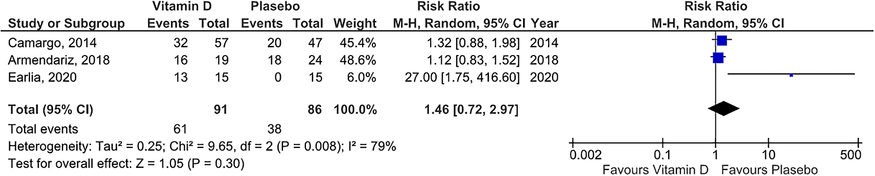
We used the three studies that provided raw data so that the risk ratio of those studies could be measured. The forest plot (see Underlying data33) showed that statistically, there was no difference risk ratio between vitamin D group and placebo group (risk ratio =1.46 , 95% CI = 0.72 to 2.97). We observed statistical heterogeneity among the studies (I2 > 25%; Figure 4) so Random Effect Model was adopted.
Vitamin D can modulate the innate immune system and also increases the phagocytic ability of immune cells and strengthens the barrier function of epithelial cells.6
An in vitro study reported that cathelicidin and defensin (which are antimicrobial-like peptides) increased after vitamin D supplementation.23 Another clinical trial also demonstrated that cathelicidin production could be increased and LL-37 expression could be induced by vitamin D supplementation. Thus, vitamin D could increase antimicrobial activity and external tolerability against pathogens.24
Vitamin D stimulates the production and regulation of skin antimicrobial peptides, such as cathelicidins which exert direct antimicrobial activity and induce host cellular responses to produce cytokine release, inflammation, and angiogenesis, thus, based on the above theory, vitamin D deficiency may predispose to secondary infection in AD patients.25 This is following what was reported by Haridas, Udompataikul et al. who found a reduction in S. aureus colonization in a paediatric population, as well as Rahmawati et al, who have reported that there was a significant difference in the reduction of S. aureus colonization after vitamin D3 supplementation in children with AD Refs. 26 and 27.
This theory is following the results of the meta-analysis of our forest plot. Our findings showed a statistically significant difference between the vitamin D supplementation group and the placebo group. In our study, we found high heterogeneity and we assumed that it was caused by variation of the doses and duration. The meta-analysis published by Kim in 201628 reported the same results but for the paediatric and adult population, as well as the meta-analysis reported by Haridas in 2018. To our knowledge, our study is the first one that reported vitamin D supplementation efficacy only in children population with AD as the meta-analysis.
The outcome of cure rate is one of the risk ratio, wherein this study the risk ratio calculated is the comparison of the probability of recovered participant between vitamin D and placebo. In the forest plot with risk ratio output, three squares were obtained, each represented 3 studies, with a weight of 45.4%; 48.6%, and 6%. All of these studies have heterogeneity above 50% and p-value <0.05 so that the forest plot used the Random Effect Model as seen from the heterogeneity test results and with the eyeball test. Diamond, the result of all studies is on the left side, with a pooled result of 1.46 (CI between 0.72 to 2.97) and touched the vertical line, which means that statistically, there was no difference in cure rate in the vitamin D group and the placebo group. Previously, there were no published meta-analysis with a forest plot with risk ratio outcomes so to our knowledge, our finding is the first meta-analysis with the risk ratio outcome, by point of interest “cure rate” in the experimental group compared to placebo.
In this study, the clinical significance could only be calculated from 3 studies that provided data on the proportion of subjects who recovered and did not recover or had persistent symptoms from the start of the study to the end. As a determination of the criteria for recovery, we had referred to a journal that mentioned MCID (Minimal Clinically Important Difference) in AD, MCID could be described as a clinical improvement that significantly along with reduction of SCORAD of 9 points and EASI by 6 points and IGA score reduced by 1 point.29 From Table 5, Figure 1 it can be calculated that the CER or incidence in the control group (placebo) was 38/86, which is 44%, means that 44% of cases were cured in the group of subjects who were given placebo and EER or the incidence in the experimental group was 61/91, which is equal to 67%, which means that 67% of cases of cure were found in the vitamin D group. ARR (Absolute risk reduction) in both groups was enabled by reducing CER and EER by 23%, and NNT (number needed to treat) was the amount subjects who must be treated at one time to prevent 1 adverse outcome. In these studies, NNT = 4.34 or required 5 subjects to be treated to prevent 1 unwanted event.
If toxicity occurs, there will be an increase in 25(OH) D levels which can trigger hypercalcemia by increasing calcium absorption and bone resorption. Hypercalcemia can lead to hypercalciuria, and persistently elevated calcium levels can lead to polyuria and dehydration.30 Vitamin D toxicity is caused by hypercalcemia, which is described by the appearance of symptoms in several organs that can be involved, such as the central nervous system (lethargy, apathy, depression to coma), heart and blood vessels (hypertension, heart rhythm disturbances), gastrointestinal (vomiting), recurrent abdominal pain, anorexia, constipation, and weight loss), and kidney (hypercalciuria is an early symptom, polyuria, polydipsia, nephrocalcinosis, up to life-threatening symptoms such as dehydration and kidney failure requiring haemodialysis).31 The diagnosis of vitamin D toxicity was established based on a detailed examination and history of taking medication, as well as supporting examinations. Laboratory tests show suppression of parathyroid hormone, which results in increased levels of 1,25(OH)2D.32
Dose and duration among studies are not similar, and not all studies have observed vitamin D levels before and after supplementation so it has not been seen whether there is an increase in vitamin D levels that exceeds the limit, which could potentially cause signs of vitamin D toxicity in several organs.
The limitation of our study was that we did not perform sub-group analysis outcome according to the measuring scale and the severity of AD due to the limitations of the studies included, so that the result of our study should be used carefully.
Our study has showed that statistically, vitamin D supplementation can improve the outcome of atopic dermatitis in children as assessed by SCORAD, EASI or IGA Score and clinically, vitamin D supplementation can increase the cure rate in AD patients. Observation of side effects and monitoring of 25(OH) D levels in AD patients are required as the toxicity can lead into morbidity. The recommendation of the proper dose of vitamin D supplementation cannot be determined yet because there were no studies with the same dose and duration of administration of the vitamin D supplementation.
Figshare: Data for Efficacy of vitamin D supplementation on the severity of atopic dermatitis in children: A systematic review and meta-analysis. https://doi.org/10.6084/m9.figshare.19091474.v2.33
This project contains the following underlying data:
Figshare: Data for Efficacy of vitamin D supplementation on the severity of atopic dermatitis in children: A systematic review and meta-analysis. https://doi.org/10.6084/m9.figshare.19091474.v2.33
This project contains the following extended data:
Figshare: PRISMA checklist for ‘Efficacy of vitamin D supplementation on the severity of atopic dermatitis in children: A systematic review and meta-analysis’. https://doi.org/10.6084/m9.figshare.19091474.v2.33
Data are available under the terms of the Creative Commons Attribution 4.0 International license (CC-BY 4.0).
| Views | Downloads | |
|---|---|---|
| F1000Research | - | - |
|
PubMed Central
Data from PMC are received and updated monthly.
|
- | - |
Provide sufficient details of any financial or non-financial competing interests to enable users to assess whether your comments might lead a reasonable person to question your impartiality. Consider the following examples, but note that this is not an exhaustive list:
Sign up for content alerts and receive a weekly or monthly email with all newly published articles
Already registered? Sign in
The email address should be the one you originally registered with F1000.
You registered with F1000 via Google, so we cannot reset your password.
To sign in, please click here.
If you still need help with your Google account password, please click here.
You registered with F1000 via Facebook, so we cannot reset your password.
To sign in, please click here.
If you still need help with your Facebook account password, please click here.
If your email address is registered with us, we will email you instructions to reset your password.
If you think you should have received this email but it has not arrived, please check your spam filters and/or contact for further assistance.
Comments on this article Comments (0)