Keywords
Apoptotic markers, Khamira abresham, Isoprenaline, Myocardial necrosis, α/β-MHC expression, Nanoformulation, Unani formulation
Apoptotic markers, Khamira abresham, Isoprenaline, Myocardial necrosis, α/β-MHC expression, Nanoformulation, Unani formulation
Historical evidence indicates that the use of medicinal plants dates back to the Paleolithic age and continues to be eminent in the modern age; over 60% of the world population relies on the use of traditional medicine, for the treatment of numerous lifestyle disorders.1,2 Unani system of medicine is amongst the oldest and effective traditional systems of medicine. Popular in South Asian countries, it is also hovering interest in other parts of the world, both amongst commoners as well as researchers.3 Countries like Bangladesh, India, the Islamic Republic of Iran, Pakistan are only a few names where the Unani system is popular. Cardiovascular diseases (CVD) are globally considered as the leading cause of death with 80% of CVD-related deaths being reported from low and middle-income countries like India despite the plethora of modern drugs available at hand.4–6 The Unani System of medicine offers treatment of cardiovascular diseases by compound (murakkabat) medicine rather than a single (mufrrajat) drug. One such compound preparation is “Khamira Abresham Hakim Arshadwala” (KAHAW). The word “Khamira” indicates a fermented confection, first introduced by the Hakeems of the Mughal age. It is a semisolid preparation, a type of majoon, which is prepared by adding a joshandah (decoction) of herbal drugs or powdered drugs to a base (Qiwam) made up of sugar or sugar with honey.7 Khamira Abresham Hakim Arshadwala is a reputed Unani formulation with 22 ingredients from plant, animal, and mineral origin, and is extensively used as a cardiotonic agent.8,9 With its innumerable benefits, the presence of sugar base (as a preservative) makes it non-compliant for the diabetic groups which are concomitantly affected with cardiac disorders.
The current piece of work aims at adding a feather to the cap of Unani formulation by transforming it into novel nanoformulations, making it patient compliant, and screening the developed formulation against an established model for myocardial necrosis with a comparative evaluation with a clinical standard drug.
Isoprenaline is a synthetic catecholamine with β- adrenergic agonist that affects the myocardium with rigorous stress causing an infarct-like injury in the myocardium. Pathophysiological changes that occur in ISO-induced myocardial infarction are similar to those occurring after the attack of myocardial infarction in humans.10 Isoprenaline is testified to cause augmented reactive oxygen species through auto-oxidation and subsequent oxidative stress, which causes myocardial necrosis.11 Increased level of catecholamines induces breakdown of the reserve energy of cardiac muscle cells, thus causing structural changes and complex biochemical changes that result in irreversible cellular injury and finally necrosis.12
Metoprolol is a highly effective FDA-approved drug for cardiovascular disorders; it has been selected as a standard drug for comparative evaluation of the traditional formulations and its novel nanoformulation on experimental models for myocardial necrosis. Consecutively, we have developed and tested the cardioprotective potential of a nanoformulation of KAHAW and compared it with the standard drugs on the experimental models for myocardial necrosis. Various enzymatic, non-enzymatic, and histopathological parameters have been evaluated in the current study, including Western Blot of a cardioselective protein α/β Myosin Heavy Chain Kinase (α/β MHC), Cell Apoptotic markers, Troponin-T (Gold marker test for Myocardial necrosis) along with antioxidant. The study reinstates that the developed herbal nanoformulations will provide a better, cost-effective, and potent treatment approach for myocardial necrosis and related ailments with minimum side effects and high patient compliance.13
The plant materials including Abresham (Silk Cocoon, cut), Ood Hindi (Aquilaria agallocha), Balchar (Nardostachys jatamansi), Post turanj (Citrus medica), Burada sandal safed (Santalum album), Sazaj hindi (Cinnamomum tamala), Qaranfal (Caryophyllus) and Dana heel khurd (Elattaria cardamomum) were purchased from the local market and authenticated from the Central Institute of Medicinal and Aromatic Plants (CIMAP), Lucknow, and other ingredients of the formulation were procured from the local market. Khamira Abresham Hakim Arshadwala (Hamdard Laboratories, B.No. KA265B) was purchased from Unani Medical Store.
Standard Metoprolol and Isoprenaline were procured from Sigma Aldrich and Ramipril and Low molecular weight Chitosan was procured from Yarrow Chem products. Other chemicals/reagents/diagnostic kits utilized in this research work were procured from different manufacturers/suppliers and were of research/analytical grade.
The male Wistar rats (4-week-old) (150-180gm) were acquired lawfully as per CPCSEA guidelines from the animal house center of Central Drug Research Institute (CDRI), Lucknow. Good laboratory practices (GLP) for the animal facility were intended for quality maintenance of housing, feeding, and safety of animals while conducting the approved experimental protocols as per CPCSEA guidelines. The research protocol was affirmed by prior approval from the Institutional Animal Ethical Committee (IAEC) of Integral University, Lucknow (U.P.), India, with approval number (IU/IAEC/19/03). The male Wistar rats (4-week-old) (150-180 g) were housed in a room where the temperature was maintained around 23 ± 5°C and relative dampness of 50 ± 20%, in a 12 h light-dark cycle. Animals were kept on a standard pellet diet and drinking water ad libitum. All efforts were made to ameliorate any suffering of animals, hence, at the end of the study the experimental animals were euthanized by cervical decapitation under anaesthesia for assessment of various study parameters.
All experimental units were added to the individual groups as per experimental design a priori in the beginning of the study and no animals/groups were excluded from the protocol during entire study period.
Abresham (Silk Cocoon, cut) 89.6 g, Ood Hindi (Aquilaria agallocha) 2.24 g, Balchar (Nardostachys jatamansi) 2.8 g, Post turanj (Citrus medica) 2.80 g, Burada sandal safed (Santalum album) 3.36 g, Sazaj hindi (Cinnamomum tamala) 2.80 g, Qaranfal (Caryophyllus) 2.80 g, Dana heel khurd (Elattaria cardamomum) 2.80 g were coarsely powdered and mixed and extracted in 80% Ethanol by hot extraction method using soxhlet apparatus at below 40oC in solvent sample ratio of 1:4 for 72 hours followed by filtration and lyophilization.
Chitosan nanoparticles were prepared by ionic gelation method followed by Servat-Medina L. using lyophilized extract followed by mixing other ingredients in a predetermined ratio.14
Rats were randomly divided, based on the animal body weight, into seven groups (control and treatment groups) with six rats in each (n=6), namely, Normal Control (NCG), Isoprenaline Control Group (ICG), Test Nanoparticle group (TNG), Test Khamira Group (TKG), Standard Group Metoprolol (SMG), Standard Group Ramipril (SRG) and Per Se Group (PNG). This randomization was done so as to minimize any potential confounders like weight bias and in order to minimise standard deviation in the readings for each group, the animals with closely similar body weights were allocated the same group. After following the treatment protocol given in Table 1 for 30 days, on the morning of the 31st day, the animals were euthanized under anesthesia by cervical dislocation. Blood was collected in heparinized and non-heparinized centrifuge tubes, while the heart was excised, weighed, and stored in a 10% formalin solution. 10 % tissue homogenates (auricle and ventricles) were prepared, centrifuged at 12000 rpm at 4oC, and the supernatant was collected and stored at -80oC until further use. Various parameters were recorded to evaluate the cardioprotective effect of the developed nanoformulation, where ICG was compared with NCG and all the other Treatment groups were compared to ICG, The PNG was the satellite group to assess the any other effects of the developed formulation in non disease group per se. All the procedures, experimental design, data collection and analysis were conducted under the supervision of the Principal Investigator of the project.
At the end of the study, different physical parameters including Gross Morphology of the wall thickness of the left ventricle, right ventricle, and intraventricular septum15 were assessed to evaluate the pathological changes in the heart anatomy of different treatment groups.
Cardiac gold marker; Troponin-T and enzymatic parameters including lipid peroxidation (TBARS, Lipid hydroperoxide and conjugated dienes16–18 in plasma as well as heart tissue and non-enzymatic biomarkers like vitamin C,19 vitamin-E,20 glutathione21 and CK-MB were assayed in plasma/serum, enzymatic biomarkers like Catalase (CAT),22 Superoxide dismutase (SOD), Glutathione reductase (GR),23 Glutathione peroxidase (GPx),24 and Glutathione-S-Transferase (GST)25 in the heart were assayed. Total protein, albumin, globulin A/G ratio levels in different treatment groups were determined to compare the effect of nanoparticles on ISO-induced cardiac damage with standards; Metoprolol and Ramipril.26 Serum lipid profile and heart tissue lipid profile in treatment groups were determined as per standard protocol.27–29 Lysosomal hydrolases and mitochondrial enzymes were assayed in heart tissue as per protocol.30–33
The highlight of the current research was the assessment of apoptotic markers; α/β-MHC Expression through Western Blot following the standard procedure with few modifications followed by cell size.13,34 Flow Cytometry to assess cell death using annexin V/PI and Cardiac Collagen Content.35,36 The assessment was determination and histopathological studies of isolated heart samples from each group.
The heart was cut down longitudinally and opened in two halves, followed by measurement of left ventricular wall thickness (LV), right ventricular wall thickness (RV), and Intraventricular septum thickness (IVS) (Figure 1 & Figure 2).
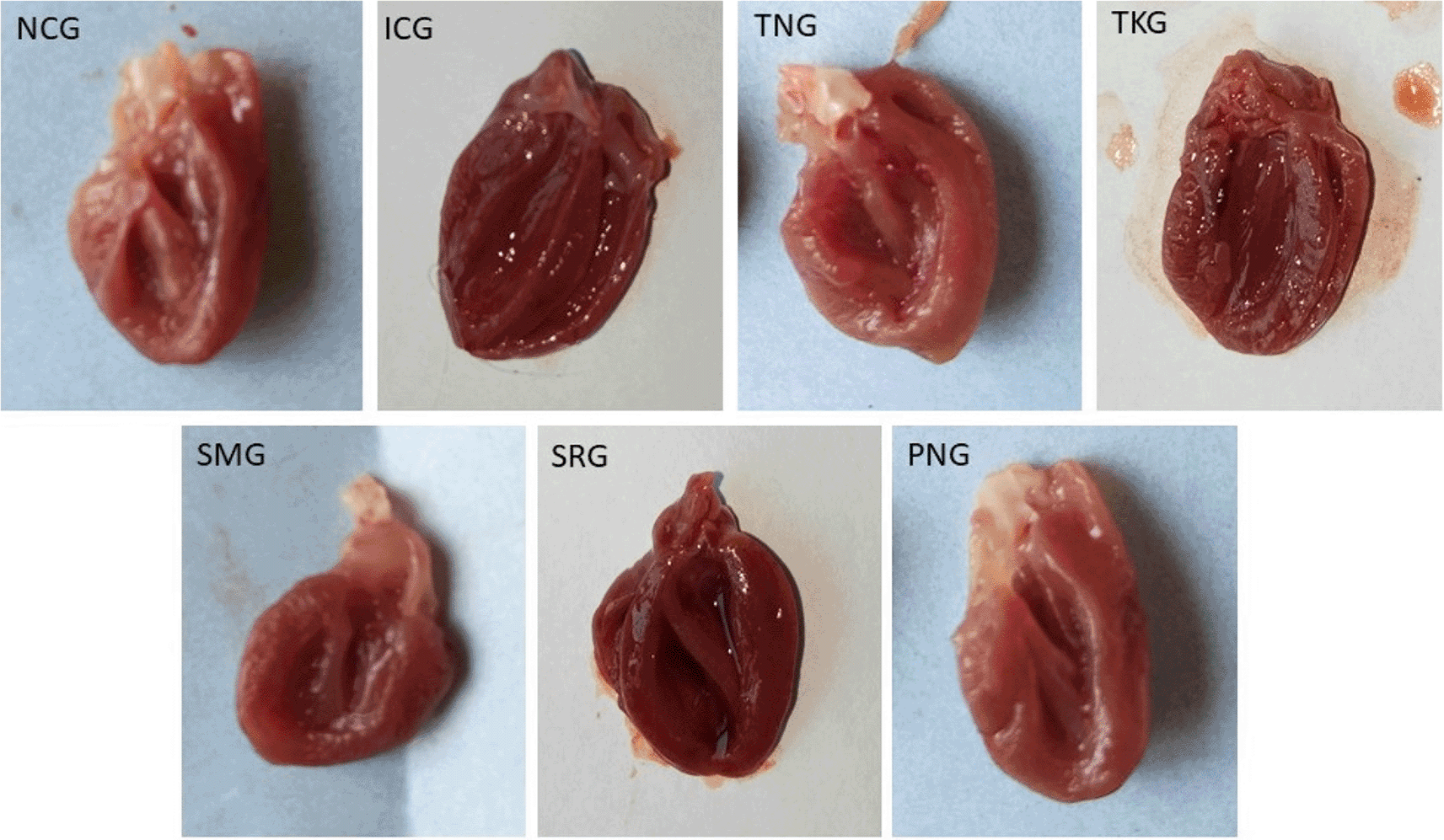
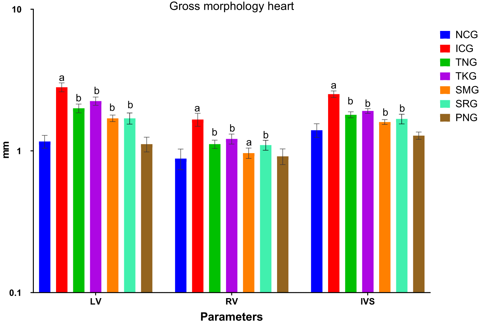
All values were expressed as mean±SD; (n=6). Statistical analysis was done by one-way ANOVA followed by Dunnett’s t-test where TNG, TKG, SMG, and SRG groups were compared to ICG, while ICG and PNG were compared to NCG and the values were found to be statistically highly significant at p<0.001, statistically very significant at b p<0.01 respectively. Where LV is Left Ventricular Wall Thickness, RV is Right ventricular wall thickness, IVS is Intra ventricular septum thickness, TNG is Test Nanoparticle Group, TKG is Test Khamira Group, SMG is Standard Metoprolol Group, SRG is Standard Ramipril group, ICG is Isoprenaline Control Group, PNG is Per se Nanoparticle Group and NCG is Normal Control Group.
Troponin T-test results52 were observed as a double line for positive and a single line for negative as shown in Table 2 and Figure 3.
| Treatment group | Number of animals | |||||
|---|---|---|---|---|---|---|
| 1 | 2 | 3 | 4 | 5 | 6 | |
| NCG | -ve | -ve | -ve | -ve | -ve | -ve |
| ICG | +ve | +ve | +ve | +ve | +ve | +ve |
| TNG | -ve | -ve | -ve | -ve | -ve | -ve |
| TKG | -ve | -ve | -ve | -ve | -ve | -ve |
| SMG | -ve | -ve | -ve | -ve | -ve | -ve |
| SRG | +ve | -ve | +ve | -ve | -ve | -ve |
| PNG | -ve | -ve | -ve | -ve | -ve | -ve |
Lipid peroxidative parameters in plasma provide significant information on lipid peroxidation during myocardial injury. Lipid peroxidation is a phenomenon by which degradation of lipids occurs in the presence of oxygen. Thiobarbituric acid (TBARS), lipid hydroperoxides, and conjugated dienes were estimated and the results were shown in Figure 4.
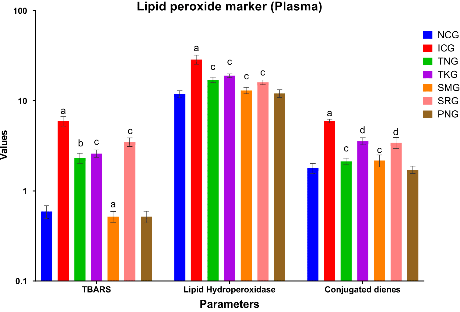
All values were expressed as mean±SD; (n=6). Statistical analysis was done by one-way ANOVA followed by Dunnett’s t-test where TNG, TKG, SMG, and SRG groups were compared to ICG, while ICG and PNG were compared to NCG and the values were found to be statistically very highly significant at p<0.0001, statistically highly significant at b p<0.001, statistically very significant at c p<0.01 and statistically significant at d p<0.05. Where TBARS: Thiobarbituric acid reactive substances. TNG is Test Nanoparticle Group, TKG is Test Khamira Group, SMG is Standard Metoprolol Group, SRG is Standard Ramipril group, ICG is Isoprenaline Control Group, PNG is Per se Nanoparticle Group and NCG is Normal Control Group. TBARS, lipid hydroperoxides, and conjugated diene values were expressed in nmol/dL, mmol/dL, and mmol/dL, respectively. Log10scale was used to plot the graph.
The estimation of non-enzymatic parameters present in the plasma indicates the degree of injury in the myocardium. The non-enzymatic antioxidant markers i.e, Vitamin C, Vitamin E, Glutathione, and CK-MB were estimated and the results were shown in Figure 5.
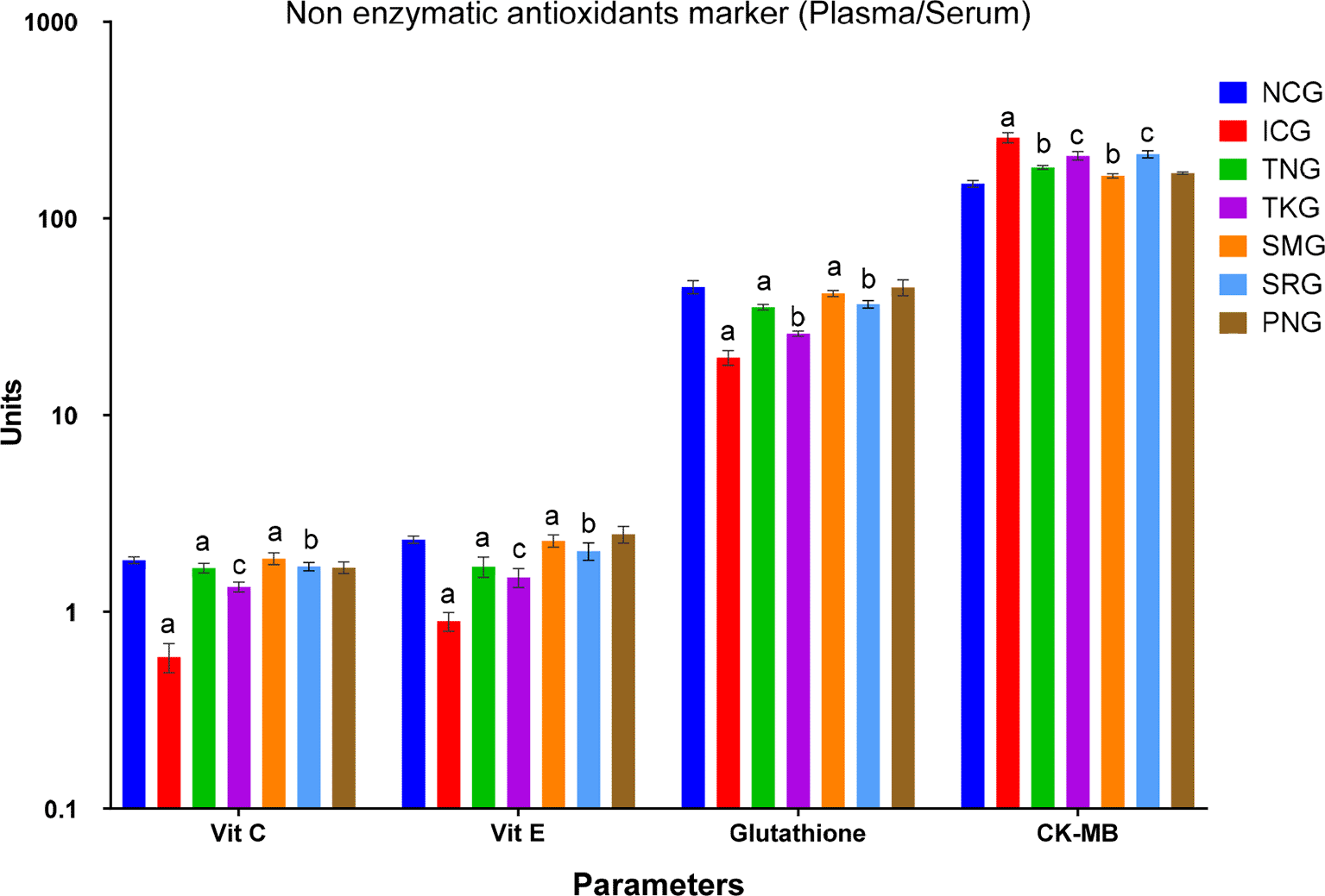
All values were expressed as mean±SD; (n=6). Statistical analysis was done by one-way ANOVA followed by Dunnett’s t-test where TNG, TKG, SMG, and SRG groups were compared to ICG, while ICG and PNG were compared to NCG and the values were found to be statistically highly significant at aP<0.001, statistically very significant at bP<0.01 and statistically significant at cP<0.05. Where TNG is Test Nanoparticle Group, TKG is Test Khamira Group, SMG is Standard Metoprolol Group, SRG is Standard Ramipril group, ICG is Isoprenaline Control Group, PNG is Per se Nanoparticle Group and NCG is Normal Control Group. Log10scale was used to plot the graph. Units of Vit C, Vit E, and glutathione are mg/dl and of CK-MB is IU/L.
The estimation of non-enzymatic parameters present in the tissue indicates the degree of injury in the myocardium. The non-enzymatic antioxidant markers in heart tissue i.e, vitamin C, Vitamin E, and glutathione were estimated and the results were shown in Figure 6.
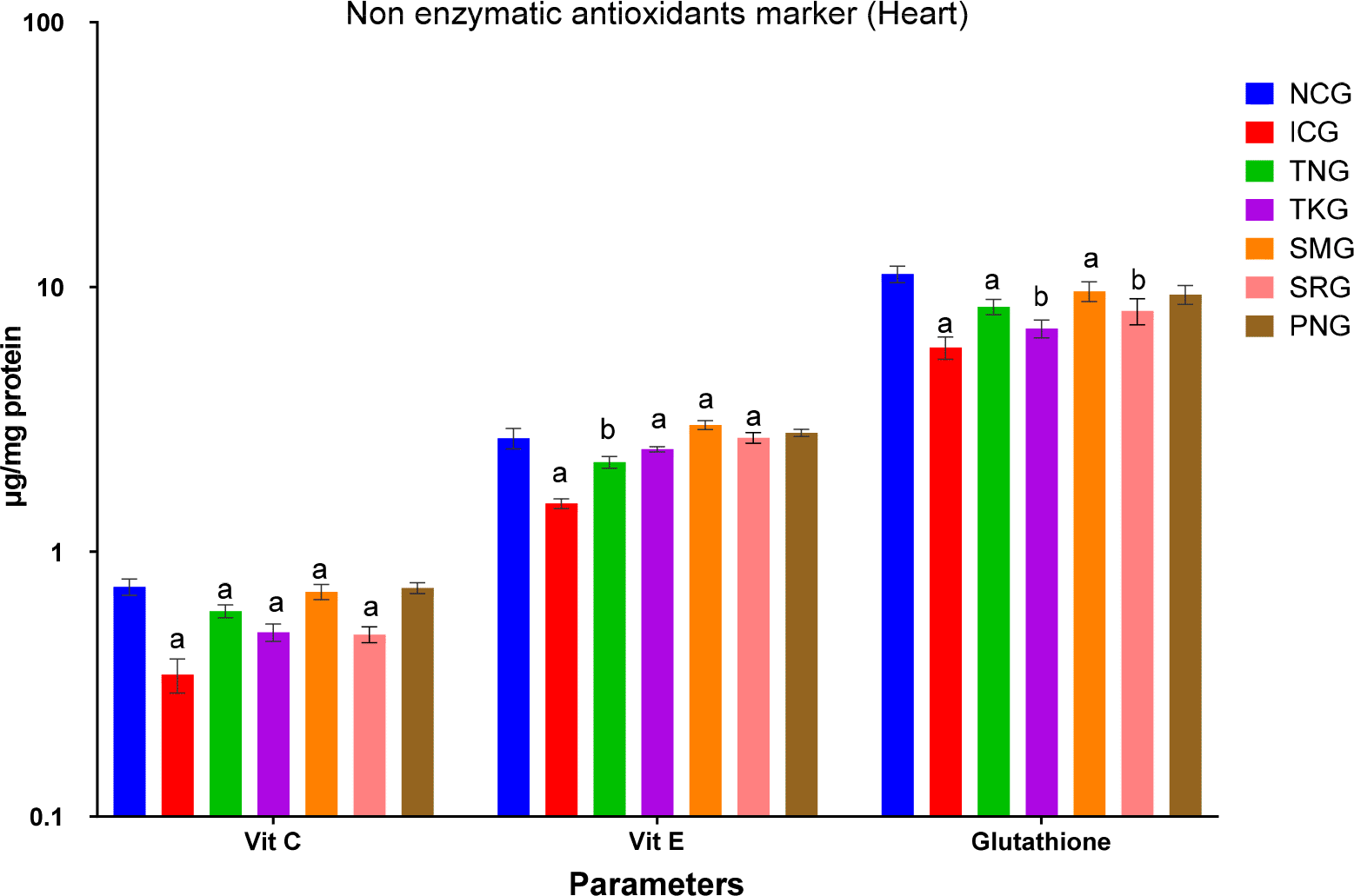
All values were expressed as mean±SD; (n=6). Statistical analysis was done by one-way ANOVA followed by Dunnett’s t-test where TNG, TKG, SMG, and SRG groups were compared to ICG, while ICG and PNG were compared to NCG and the values were found to be statistically highly significant at p<0.001 and statistically very significant at b p<0.01. Where TNG is Test Nanoparticle Group, TKG is Test Khamira Group, SMG is Standard Metoprolol Group, SRG is Standard Ramipril group, ICG is Isoprenaline Control Group, PNG is Per se Nanoparticle Group and NCG is Normal Control Group. Log10scale was used to plot the graph.
The activities of Catalase (CAT), Superoxide dismutase (SOD), Glutathione reductase (GR), Glutathione peroxidase (GPx), and Glutathione-S-Transferase (GST) in the heart tissue were assayed and expressed in Figure 7.
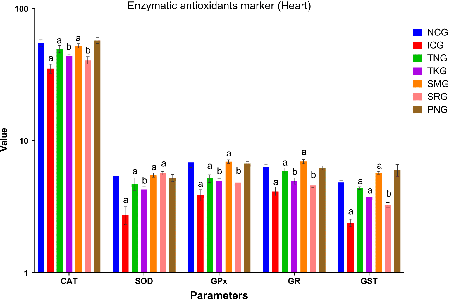
All values were expressed as mean±SD; (n=6). Statistical analysis was done by one-way ANOVA followed by Dunnett’s t-test where TNG, TKG, SMG, and SRG groups were compared to ICG, while ICG and PNG were compared to NCG and the values were found to be statistically highly significant at ap<0.001 and statistically very significant at bp<0.01. Where TNG is Test Nanoparticle Group, TKG is Test Khamira Group, SMG is Standard Metoprolol Group, SRG is Standard Ramipril group, ICG is Isoprenaline Control Group, PNG is Per se Nanoparticle Group and NCG is Normal Control Group, CAT is Catalase, SOD is Superoxide dismutase, GPx is Glutathione peroxidase, GR is Glutathione reductase, GST is Glutathione S-transferases. Log10scale was used to plot the graph. Units:Superoxide dismutase (SOD): The enzyme activity of one unit was considered as the enzyme reaction which gave 50% of the inhibition of nitroblue tetrazolium (NBT) reduction in one minute. Catalase (CAT): μmole of hydrogen peroxide decomposed/min/ml Glutathione peroxidase (GPx): μmole of GSH utilized/min/mg protein Glutathione-S-transferase (GST): μg of CDNB conjugate formed/min/mg protein Glutathione reductase (GR): μg of reduced glutathione formed/min/mg protein.
Protein estimations were carried out, the amount of TP, AL, and GL and A/G ratios was evaluated and shown in Figure 8.
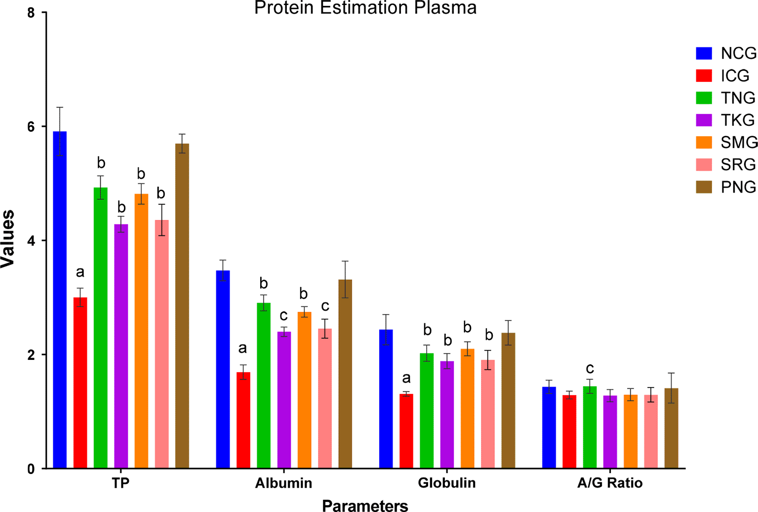
All values were expressed as mean±SD; (n=6). Statistical analysis was done by one-way ANOVA followed by Dunnett’s t-test where TNG, TKG, SMG, and SRG groups were compared to ICG, while ICG and PNG were compared to NCG and the values were found to be statistically highly significant at ap<0.001, statistically very significant at bp<0.01 and statistically significant at cp<0.05. Where TNG is Test Nanoparticle Group, TKG is Test Khamira Group, SMG is Standard Metoprolol Group, SRG is Standard Ramipril group, ICG is Isoprenaline Control Group, PNG is Per se Nanoparticle Group and NCG is Normal Control Group, TP is Total Protein. Units of TP, Albumin, Globulin were gm/dl, while A/G ratio was a numeric value.
Lysosomal hydrolase enzymes perform a significant part in the inflammatory process. Isoprenaline-induced MI results in improved lysosomal hydrolase action that may be accountable for infarcted heart and tissue damage. It is postulated that stabilization of myocardial cells or tissue membranes, predominantly lysosomal membranes, may extend the life of ischemic cardiac muscle and help in preventing MI.
The lysosomal hydrolases enzymes (α–Galactosidase, β–Galactosidase, β–Glucosidase, Cathepsin-B, Cathepsin-D) were estimated, and results were expressed in Figure 9.
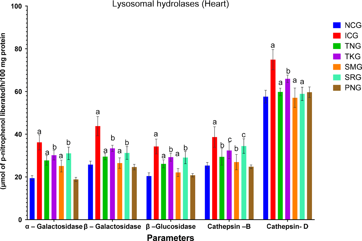
All values were expressed as mean±SD; (n=6). Statistical analysis was done by one-way ANOVA followed by Dunnett’s t-test where TNG, TKG, SMG, and SRG groups were compared to ICG, while ICG and PNG were compared to NCG and the values were found to be statistically highly significant at ap<0.001, statistically very significant at bp<0.01 and statistically significant at cp<0.05. Where TNG is Test Nanoparticle Group, TKG is Test Khamira Group, SMG is Standard Metoprolol Group, SRG is Standard Ramipril group, ICG is Isoprenaline Control Group, PNG is Per se Nanoparticle Group and NCG is Normal Control Group.
Numerous types of metabolic enzymes is present in mitochondria, such as Isocitrate dehydrogenase (IDH), α-Ketoglutarate dehydrogenase (KDH), Malate dehydrogenase (MDH), and Succinate dehydrogenase (SDH) which were quantified in the various treated heart groups, and levels of enzymes were expressed in Figure 10.
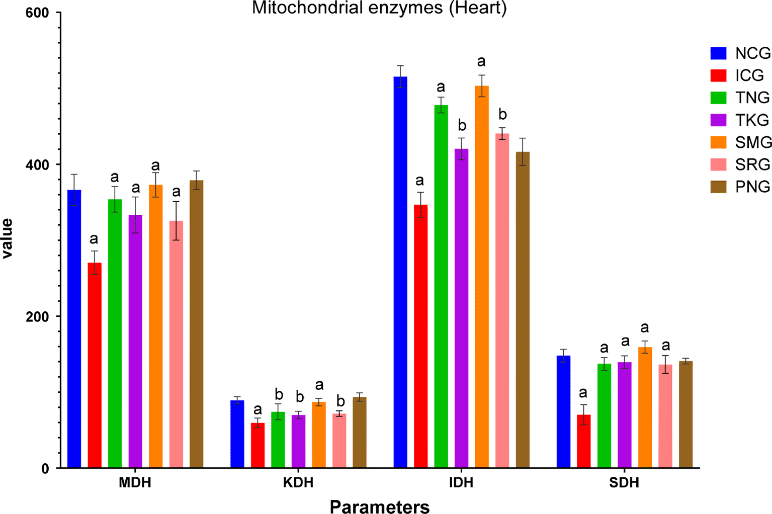
All values were expressed as mean±SD; (n=6). Statistical analysis was done by one-way ANOVA followed by Dunnett’s t-test where TNG, TKG, SMG, and SRG groups were compared to ICG, while ICG and PNG were compared to NCG and the values were found to be statistically highly significant at ap<0.001 and statistically very significant at bp<0.01. Where TNG is Test Nanoparticle Group, TKG is Test Khamira Group, SMG is Standard Metoprolol Group, SRG is Standard Ramipril group, ICG is Isoprenaline Control Group, PNG is Per se Nanoparticle Group, NCG is Normal Control Group, MDH isMalate Dehydrogenase, KDH is alpha-ketoglutarate dehydrogenase, IDH is isocitrate dehydrogenase and SDH is Succinate Dehydrogenase. Units of Isocitrate Dehydrogenase: n moles of α-ketoglutarate formed/hr/mg protein; α-Ketoglutarate Dehydrogenase: nmoles of ferrocyanide formed/hr/mg protein; Succinate Dehydrogenase: nmoles of succinate oxidized/min/mg protein; Malate dehydrogenase: n moles of NADH oxidized/min/mg protein.
α- and β-myosin heavy chain (α, β-MHC) Expression
After administration of isoprenaline (85 mg/kg), we significantly decreased the expression of α-MHC and increased the expression of β-MHC in rats compared to normal control (NCG). Treatment of NP, KAHAW (TNG at 200 mg/kg), and KAHAW (TKG at 800 mg/kg) significantly increased the expressions of α-MHC and decreased the expression of β-MHC compared to the isoprenaline Control Group (ICG) as shown in Figure 11.
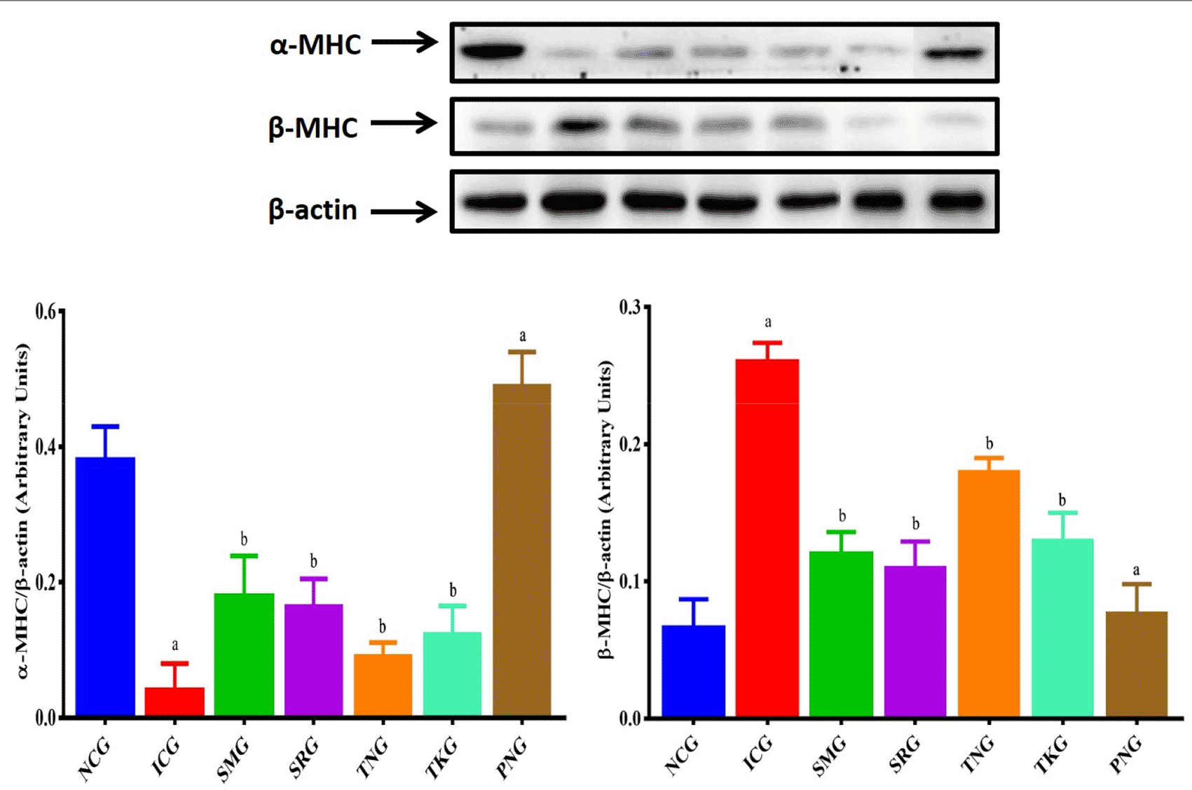
Graph shows an arbitrary value of α, β-MHC proteins vs different treatment groups. All values were expressed as mean±SD. aP<0.01 vs. NCG, bP<0.05 vs. ICG. NCG is Normal Control Group; ICG is Isoprenaline Control Group; SMG=Standard Metoprolol Group; SRG is Standard Ramipril Group; TNG is Test nanoparticle Group; TKG is Test Khamira Group; PNG is Per Se Group.
Effect of NP, KAHAW (TNG at 200 mg/kg) and KAHAW (TKG at 800 mg/kg) group on isoprenaline (85 mg/kg), changes in the percentage of apoptotic cells in flow cytometry and the level of expression of α-MHC and β-MHC (Figure 12). The percentage of apoptotic cells and the level of expression of α-MHC was significantly decreased and the expressions of β-MHC were significantly increased in isoprenaline rat heart tissue in the study design compared to Normal Control Group (NCG). NP, KAHAW (TNG at 200 mg/kg), and KAHAW (TKG at 800 mg/kg) group attenuated a significant decrease and increase in the percentage of apoptotic cells α-MHC and β-MHC in the isoprenaline rat heart in the study respectively (Figure 13).
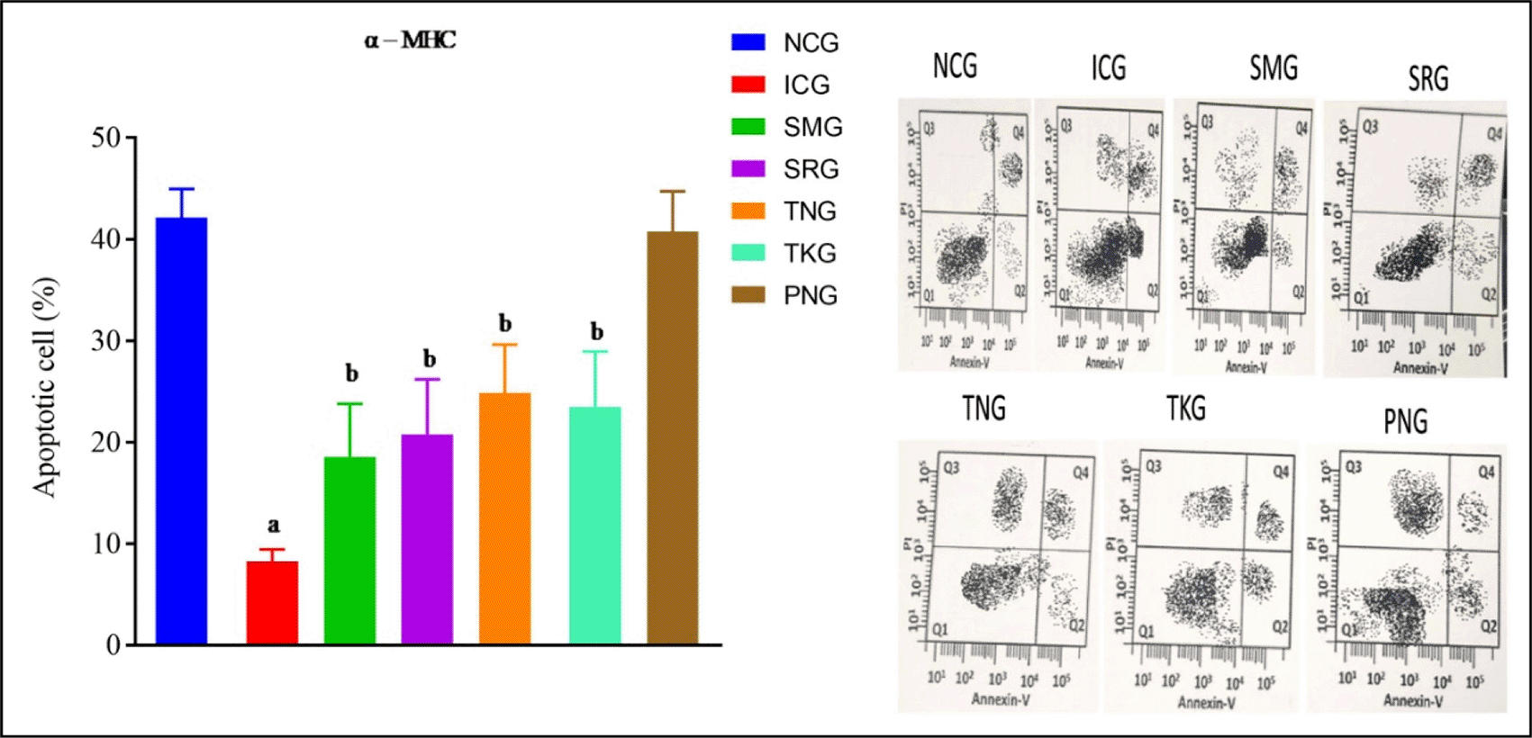
All values are mean±SD. aP<0.01 vs. NCG, bP<0.05 vs. ICG. NCG is Normal Control Group; ICG is Isoprenaline Control Group; SMG=Standard Metoprolol Group; SRG is Standard Ramipril Group; TNG is Test nanoparticle Group; TKG is Test Khamira Group; PNG is Per Se Group.
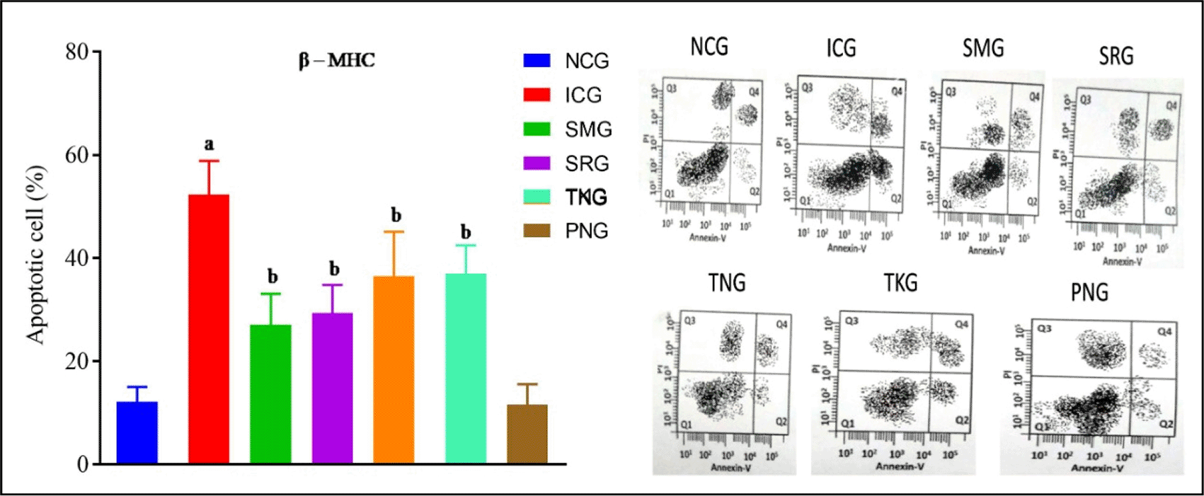
All values are mean±SD. aP<0.01 vs. NCG, bP<0.05 vs. ICG. NCG is Normal Control Group; ICG is Isoprenaline Control Group; SMG=Standard Metoprolol Group; SRG is Standard Ramipril Group; TNG is Test nanoparticle Group; TKG is Test Khamira Group; PNG is Per se Group.
Collagen is an important structural protein that forms the acellular matrix of the heart. Total Myocardial Collagen content was estimated and the results were shown in Figure 14.
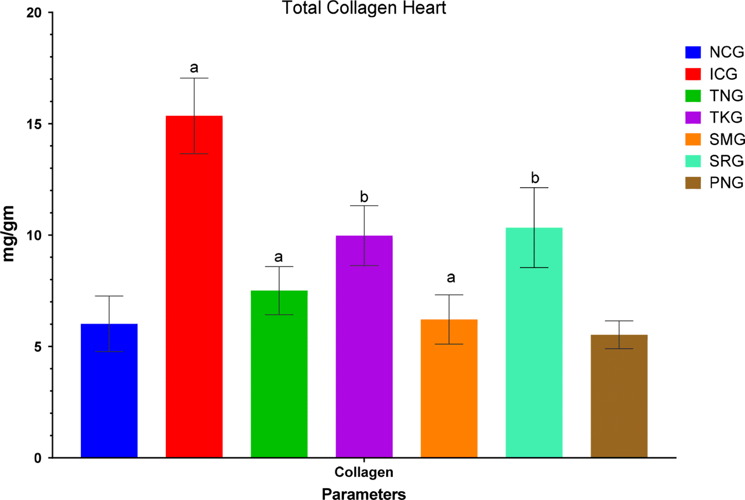
All values were expressed as mean±SD; (n=6). Statistical analysis was done by one-way ANOVA followed by Dunnett’s t-test where TNG, TKG, SMG, and SRG groups were compared to ICG, while ICG and PNG were compared to NCG and the values were found to be statistically highly significant at ap<0.001 and statistically very significant at bp<0.01. Where TNG is Test Nanoparticle Group, TKG is Test Khamira Group, SMG is Standard Metoprolol Group, SRG is Standard Ramipril group, ICG is Isoprenaline Control Group, PNG is Per se Nanoparticle Group, NCG is Normal Control Group.
The histopathology of the myocardium was done followed by staining with hematoxylin and eosin Y dye of different treatment groups later subjected to Image J software and the size of the cell was determined and shown in Figure 15.
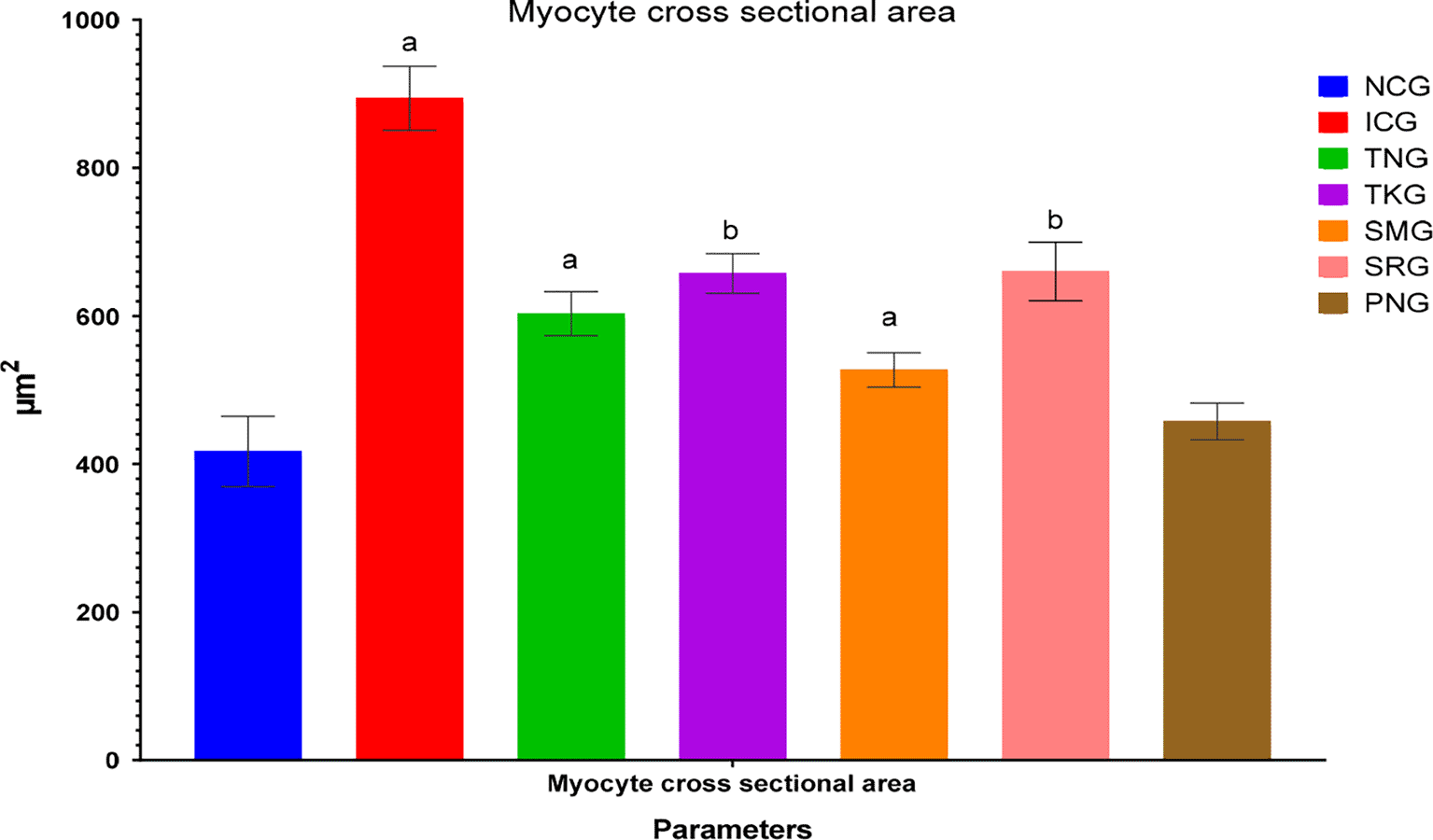
All values were expressed as mean±SD; (n=6). Statistical analysis was done by one-way ANOVA followed by Dunnett’s t-test where TNG, TKG, SMG and SRG groups were compared to ICG, while ICG and PNG were compared to NCG and the values were found to be statistically highly significant at ap<0.0001 and statistically very significant at bp<0.001. Where TNG is Test Nanoparticle Group, TKG is Test Khamira Group, SMG is Standard Metoprolol Group, SRG is Standard Ramipril group, ICG is Isoprenaline Control Group, PNG is Per se Nanoparticle Group, NCG is Normal Control Group.
Heart tissue histopathology53: H&E stain of the heart of various groups. In NCG: treated with normal saline considered as normal control showed the normal orientation of epicardium, endocardium, and myocardium as well as papillary muscles and vasculature. Isoprenaline group (ICG) showed infarcts with occasionally acute aneurysm and mural thrombi. The other groups were compared with the isoprenaline group (ICG) in which the test nanoparticle group (TNG) and Standard metoprolol group (SMG) showed normal intact myocardial tissue with no infiltration, while the test khamira group (TKG) and standard ramipril group (SRG) showed focal lesions with no sign of myonecrosis, myophagocytosis, and lymphocytic infiltration. The per se group (PSG) showed normal architecture of myocardial tissue, an organized pattern of myofiber striations with central nuclei, and with no vacuolization (Figure 16).
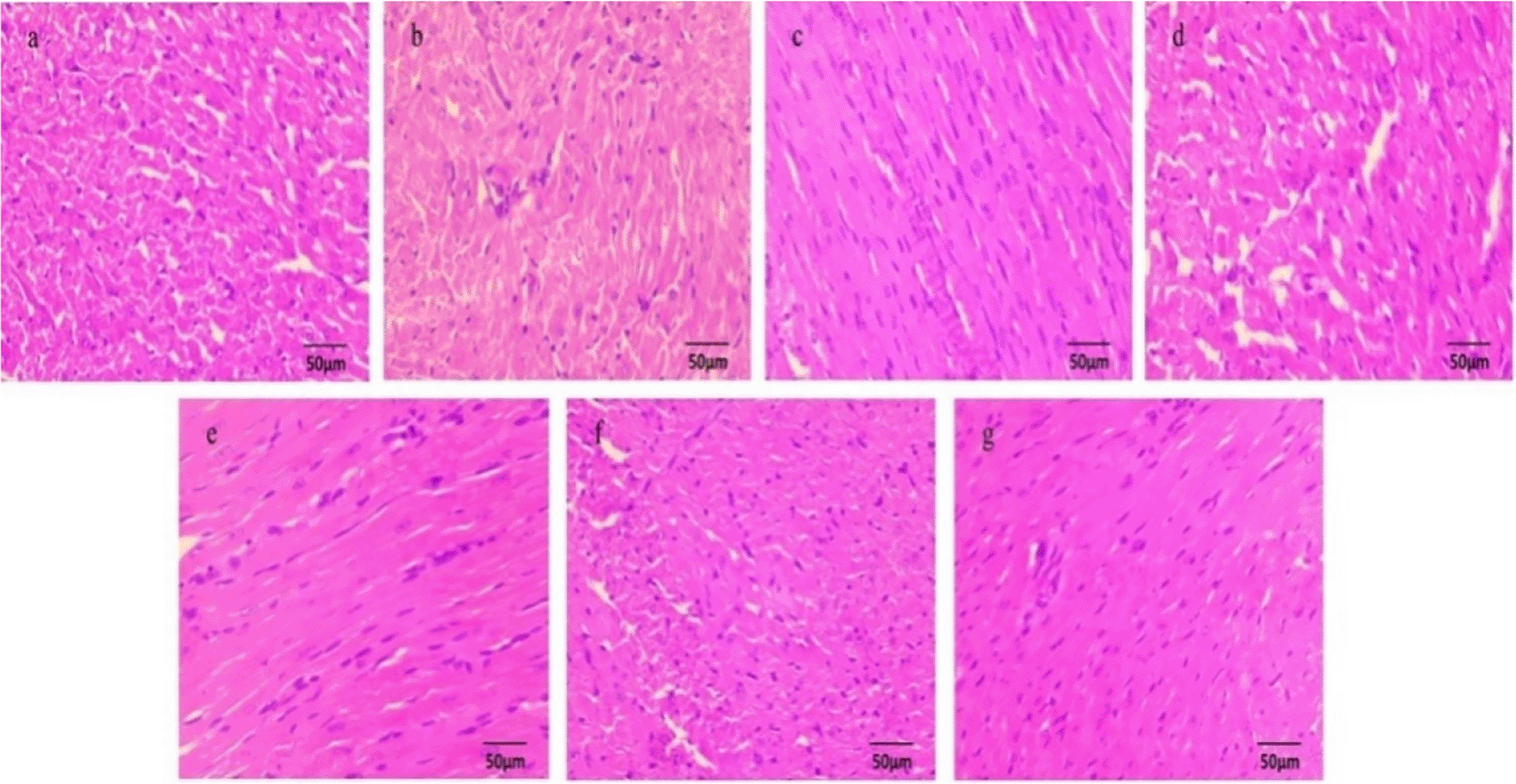
Where a is Normal Control (NCG), b is Isoprenaline control (ICG), c is Test nanoparticle treated (TNG), d is khamira treated (TKG), e is standard metoprolol treated (SMG), f is standard ramipril treated (SRG) and g is Per se nanoparticle treated (PNG).
Myocardial necrosis has been the chief pathological event behind most cardiac disorders including myocardial infarction and associated ailments. Cellular necrosis results from chronic inadequacies of vitamins and other vital nutrients that are cellular energy carriers, antioxidants, and coenzymes. Chronic diminution of these necessary nutrients in vascular smooth muscle and endothelial cells impairs their capacity to function appropriately. Several other parameters viz, Gross morphology, Serum level of cardiovascular troponins (cTn), TBARS, LHP and CD, Creatine Kinase (CK-MB), α/β Myosin Heavy Chain (MHC), Flow cytometry, Mitochondrial enzyme estimations, and Histopathological examinations, etc have been diligently profiled in this study to circumnavigate the holistic effect of developed nanoformulation and comparatively studied against the effects of traditional and modern therapy (Carvedilol and Ramipril).
Gross morphology estimation of the Right ventricle (RV), Left ventricle (LV), and Intraventricular septum (IVS) explains suggest that Isoprenaline in (ICG) group showed highly significant (***p<0.001) enhancement in LV, RV, and IVS mass when compared with normal control group (NCG). Test nanoparticles showed a decline in LV, RV, and IVS mass when compared with the Isoprenaline group (ICG). Prevention of the increase in LV, RV, and IVS mass along with the preservation of cardiac function by novel nanoformulation (TNG) is indicative of its therapeutic effect on the pathogenesis of cardiac hypertrophy. The per se group (PNG) showed no significant changes in LV, RV, and IVS when compared to the Normal control group (NCG).
Serum levels of cardiovascular troponins (cTn), especially troponin T (cTnT), are generally utilized in the identification of intense myocardial necrosis tissue and a variety of other cardiovascular ailments.37 In the current study, (TNG and TKG) and standard group (SMG and SRG) animals showed the absence of troponin T by stabilizing the cellular membrane, thus reducing the degradation of myofibrils which leads to a reduction in the release of troponin T in the blood following an infarct in the myocardium. The per se group (PNG) showed no release of troponin T showing no development of cardiac injury.
The rise in the level of lipid peroxidative markers demonstrates a rise in oxygen-free radicals, either by amplified production or decreased metabolism. In our study, ISO-treated rats also showed a significant rise(***p<0.0001) in the amount of lipid peroxidation products (TBARS, CD, and LHP) in the plasma and heart. Test (TNG and TKG) and Standard group (SMG and SRG) showed a highly significant (**p<0.001) decreased lipid peroxidation level when compared to the Isoprenaline group (ICG). The reverse lipid peroxidative action of sericin in ISO administered rats might be due to the diminished formation of superoxide anion and hydroxyl radical. The per se group (PNG) showed no significant increase in lipid peroxidative markers when compared to the Normal control group (NCG).
Vitamin E is a potent prohibitor of the autocatalyzing procedure of lipid peroxidation in cell membrane fatty acids and can in a straight line react with hydroxyl radicals, superoxides, and nascent oxygen.38,39 The decreased level of vitamin C in ISO-induced rats (ICG) may be due to increased use of antioxidants, protection against increased ROS, and decreased levels of glutathione, as glutathione is required for ascorbic acid recycling. The reduced level of GSH could lead to the reduction in the removal of free radicals, and these radicals could cause a variety of adverse reactions to the myocardium.40 Because of the above qualities of these two vitamins C, E and glutathione have been used more for the counteraction of isoprenaline facilitated free radicals and lipid peroxidation phenomena, and hence the decreased level of vitamin C, vitamin E, and glutathione were observed in ISO treated rats (ICG) when compared to the normal control group (NCG).
Test (TNG and TKG) and Standard group (SMG and SRG) animals showed a significant (*p<0.01) surge in vitamin C, E, and glutathione levels both in plasma and tissue when compared to isoprenaline administrated Group (ICG) rats. The per se group (PSG) showed normal release of Vitamin C, E, and glutathione when compared to the Normal control group (NCG).
Assay of action of isoenzyme Myoglobin (MB) of Creatine kinase (CK-MB) in plasma shows the persistence and extent of elevation. It is therefore helpful in assessing the degree of infarction.41 A marked increase in this enzyme level was detected in isoprenaline-induced rats (ICG) when compared to normal control rats (NCG). The magnitude of CK-MB action correlates with the size of the infarct. Thus, Test (TNG and TKG) and Standard group (SMG and SRG) animals significantly decreased the enzyme level (CK-MB) when compared to the ISO group.
The first line of cellular defense against oxidative stress are the free radical scavenging enzymes, such as Superoxide dismutase (SOD), Catalase (CAT), Glutathione peroxidase (GPx), Glutathione-S-Transferase (GST), and Glutathione reductase (GR).
In Isoprenaline Group (ICG) rats, the activity of heart tissue antioxidant enzymes which were found to be highly significantly (**p<0.001) decreased when compared to Normal control (NC) rats.42 Test Nanoparticle (TNG) and test khamira (TKG) rats exhibited a marked increase (**p<0.01) in these parameters when compared to isoprenaline Group (ICG) rats. Deori et al. have reported that sericin (a chief constituent of KAHAW and nanoparticles) accomplishes its antioxidant role at numerous levels of oxidative steps by maintaining antioxidant and cellular oxidant balance.43 The per se group (PSG) showed a significant (*p<0.05) increase in enzymatic antioxidant markers when compared to the Normal control group (NCG).
The disintegration of the myocardial membrane noticed in the current study may be because of the increased outflow of lysosomal hydrolases into the cytosol from the enclosed sac. This is in respect to an earlier description44 that the enzyme, cytosolic acid hydrolases released from lysosomes and those released from the sarcoplasmic reticulum cause the destruction and dysfunction of sarcolemma, mitochondria, and other cellular organelles. The reduced level in the heart or lysosomal fraction and increased levels in serum of these catalytic enzymes are observed in the current study; it may be either due to the intrusion of inflammatory cells or may be due to isoprenaline-induced lysosomal fragility due to Ca2+ overload. As it is stated that the release of lysosomal enzymes is vital in the pathogenesis of ischemic cardiac injury and the associated inflammatory cycle,45 the decrease of release of such enzymes would likely prove to be beneficial, indirectly approving the salubrious effect of sericin. Thus, Pre-treatment with nanoparticle (TNG) very significantly decreased (**p<0.001) the level of hydrolytic enzymes in the heart. The per se group (PNG) showed normal levels of hydrolase enzymes in the myocardium, showing that nanoparticles may inhibit the release of these enzymes into the blood, thus maintaining the level of enzymes in the myocardium when compared to the normal control group (NCG).
Ischemia is related to a loss of myocardial energy production, diminished oxygen uptake, and mitochondrial respiration.46 The mechanisms behind these variations are studied by evaluating the actions of dehydrogenases of the TCA cycle and various respiratory enzymes like IDH, SDH, MDH, and α-KDH. ISO has been stated to cause tissue hypoxia where this action is caused by increasing oxygen demand.47 In the present study, the actions of TCA cycle enzymes were highly decreased (***p<0.001) in the ISO-administered group (ICG). Pre-treatment with nanoparticle (TNG) group and standard group (STG) rats significantly increased (**p<0.01) the level of enzyme when compared to the isoprenaline group (ICG). The per se group (PSG) showed an increase (*p<0.01) in the level of mitochondrial enzymes in the myocardium, when compared to the normal control group (NCG).
Myosin is a molecular motor protein that produces muscular strength and contraction.34 Myosin forms natural complexes with actin, the principal component of the thin filaments. This contact is important for force production which pushes the thick and thin filaments past each other. Structural subunits of myosin are a large fibrous molecule (~ 500kD), composed of two large subunits called myosin heavy chains (MHC) and four small subunits called myosin light chains (MLC). Myosin heavy chain comprises of two isoforms i.e α and β MHC. α isoform (α-MHC) is predominantly a myocardial protein which is having a molecular weight of 224 KD present in humans. It is programmed by the MYH6 gene. This, α isoform is different from the slow/ventricular isoform of the myosin heavy chain, MYH7, also termed as MHC-β. α-MHC isoform is expressed largely in human cardiac atria, showing the very least present in the human cardiac ventricles. It is one of the main proteins forming the thick filaments of cardiac muscle and functions in the contraction of cardiac muscle.48
Exposure to isoprenaline in high doses leads to degradation of myosin heavy chain which is the core myofibrillar protein. This is in correspondence with some research that has also drawn attention towards reduced myosin heavy chain (MHC) under diverse muscle wasting circumstances.34 Test nanoparticles (TNG), however, preserved the myosin heavy chain composition of cardiac muscle, which is apparent in the expression of protein MHC in western blot outcomes. The prevention of both these proteins was significantly (*p<0.01) done by metoprolol in the standard group (SMG & SRG) when compared to the Isoprenaline control group (ICG). The level of protein was found to be intact within the myocardium as indicated by western blot bands in per se group (PNG) when compared with the normal control group (NCG). This augmented MHC content is related to an enhanced number of muscle fibers after nanoparticle administration under the condition of hypobaric hypoxia as revealed in the histological slices of rat cardiac muscle. In the present study, we found that isoprenaline attenuated the expression of α, β-MHC in isoprenaline treated rats, and the expression of α, β-MHC can be increased in the myocardium by pre-treatment with nanoparticle and rescue condition of cardiac hypertrophy induced by isoprenaline.
The major protein ingredient of the extracellular cardiovascular matrix is collagen. Collagen fibers of the heart are a network between myocytes, hence maintaining the structure of the ventricles and spreading the contractile force from all cells of the myocardium to the ventricular lumen. It plays a key part in maintaining cardiac tissue architecture by supporting the function and geometry of heart chambers. Collagen in the heart is described as types I and III, among both collagen I have a significant effect on ventricular rigidity due to its rigid and tensile features.49 Type I and type III collagen were the essential constituents of total collagen. Collagen I has vital tensile strength, while collagen III plays a key role in upholding the elasticity of the entire myocardial matrix complex.50 Collagen in the heart causes rigidity in the ventricle due to its ductile nature.51 In the current study, rats treated with nanoparticle (TNG) and standard group (SMG and SRG) inverted isoprenaline-induced hypertrophic growth of myocardium markedly (***p<0.001) when compared with the Isoprenaline group (ICG). Consistent with the estimation of myocardial hypertrophy, the quantitative evaluation of collagen noticeably showed that isoprenaline administration in the isoprenaline group (ICG) induced amplified collagen accumulation and promoted fibrosis in rat hearts. However, the treatment with nanoparticle (TNG) reduced collagen accumulation and fibrosis configuration significantly (**p<0.01) in the heart. Reduction in hydroxyproline level caused by administration of nanoparticles additional confirmed that the developed nanoparticles could reduce isoprenaline-induced collagen accumulation. The per se group (PNG) showed reduced collagen accumulation when compared to the normal control group (NCG).
The mechanisms concerned in cardiac remodelling in the left ventricle (LV) after myocardial infarction (MI) are still not clear. Cardiac hypertrophy is explained as an extension and enlargement in the size and magnitude of the whole heart or of a specific cardiac LV chamber related to body size. Left ventricular hypertrophy (LVH) is a compensatory process for the heart to work more efficiently in response to oxidized or mechanical stress and overactive sympathetic action, or as a result of genetic abnormalities. LVH is a main pathologic marker of the hypertrophic disorder of the myocardium. There was an extension in the size of myocardium cells in the isoprenaline treated group (ICG), which was depicted by an increase in cardiomyocyte size, with augmented protein synthesis and alterations in the structure of the sarcomere. These changes were subsided in treatment groups (TNG and TKG) and standard groups (SMG and SRG). The per se group (PNG) also showed a similar result, i.e., no significant changes in the size of myocardium cells when compared to the normal control group (NCG). The result of this study indicates that this defense of myocardial necrosis could have been because of the antioxidant properties of the active components.
Based on the data expressed in the contemporary study, it could be stated that the oral administration of nanoparticles at the dose of 200 mg/kg has a potent cardioprotective action against isoprenaline induced experimental myocardial necrosis and hypertrophy, thus reducing oxidative stress and inflammatory reactions that resulted in enhanced myocardial activity and attenuated heart damage after myocardial ischemia, and the possible daily consumption of nanoparticle formulation might be considered as a cardiotonic drug for human beings in future. Having the properties of KAHAW and being sugar-free, this nanoparticle formulation may further expand its patient compliance.
All data in the experiment were expressed as mean±SD. Data analyses were performed using the Graphpad Prism 8 software package. Statistical analysis was assessed by the analysis of variance (ANOVA) followed by the Dunnett’s t-test for multiple comparisons. The differences were considered statistically significant at the value of *p<0.05, **p<0.01 and ***p<0.001. The data collected analysed under the current study met the assumptions of the statistical approach, thereby approving of the hypothesis designed for the study.
Figshare: Raw Data F1000.xlsx. Dataset. https://doi.org/10.6084/m9.figshare.18551225.v1.52
Figshare: Histopathological Images. Figure. https://doi.org/10.6084/m9.figshare.19207176.v1.53
Figshare: ARRIVE Author Checklist. https://doi.org/10.6084/m9.figshare.19235436.v1.54
Data are available under the terms of the Creative Commons Zero “No rights reserved” data waiver (CC0 1.0 Public domain dedication).
Good laboratory practices (GLP) for the animal facility were intended for quality maintenance of housing, feeding, and safety of animals while conducting the approved experimental protocols as per CPCSEA guidelines. The research protocol was affirmed by prior approval from the Institutional Animal Ethical Committee (IAEC) of Integral University, Lucknow (U.P.), India, with approval number (IU/IAEC/19/03). All efforts were made to ameliorate any suffering of animals, hence, at the end of the study the experimental animals were euthanized by cervical decapitation under anaesthesia for assessment of various study parameters.
This research has been funded by the Ministry of AYUSH, Govt of India as a part of a project sanctioned under the EMR Scheme vide Approval Letter No. Z.28015/08/2016-HPC-(EMR)-AYUSH-C. The authors are highly thankful to Honorable Founder and Chancellor, Prof. Syed Waseem Akhtar, Integral University and Vice-Chancellor, Prof. Javed Musarrat, Integral University, for providing an excellent research environment and facilities. The manuscript communication number provided by the university is IU/R&D/2021-MCN0001184.
| Views | Downloads | |
|---|---|---|
| F1000Research | - | - |
|
PubMed Central
Data from PMC are received and updated monthly.
|
- | - |
Is the work clearly and accurately presented and does it cite the current literature?
No
Is the study design appropriate and is the work technically sound?
Partly
Are sufficient details of methods and analysis provided to allow replication by others?
Yes
If applicable, is the statistical analysis and its interpretation appropriate?
Yes
Are all the source data underlying the results available to ensure full reproducibility?
Yes
Are the conclusions drawn adequately supported by the results?
Yes
Competing Interests: No competing interests were disclosed.
Reviewer Expertise: Plant Biotechnology, phytochemistry and phytomedicines
Is the work clearly and accurately presented and does it cite the current literature?
Yes
Is the study design appropriate and is the work technically sound?
Yes
Are sufficient details of methods and analysis provided to allow replication by others?
Yes
If applicable, is the statistical analysis and its interpretation appropriate?
Yes
Are all the source data underlying the results available to ensure full reproducibility?
Yes
Are the conclusions drawn adequately supported by the results?
Yes
Competing Interests: No competing interests were disclosed.
Reviewer Expertise: Drug Development: Preclinical and clinical research and Toxicology
Is the work clearly and accurately presented and does it cite the current literature?
Yes
Is the study design appropriate and is the work technically sound?
No
Are sufficient details of methods and analysis provided to allow replication by others?
No
If applicable, is the statistical analysis and its interpretation appropriate?
Yes
Are all the source data underlying the results available to ensure full reproducibility?
Yes
Are the conclusions drawn adequately supported by the results?
Yes
Competing Interests: No competing interests were disclosed.
Reviewer Expertise: Cardiovascular Pharmacology
Alongside their report, reviewers assign a status to the article:
| Invited Reviewers | |||
|---|---|---|---|
| 1 | 2 | 3 | |
|
Version 1 09 Mar 22 |
read | read | read |
Provide sufficient details of any financial or non-financial competing interests to enable users to assess whether your comments might lead a reasonable person to question your impartiality. Consider the following examples, but note that this is not an exhaustive list:
Sign up for content alerts and receive a weekly or monthly email with all newly published articles
Already registered? Sign in
The email address should be the one you originally registered with F1000.
You registered with F1000 via Google, so we cannot reset your password.
To sign in, please click here.
If you still need help with your Google account password, please click here.
You registered with F1000 via Facebook, so we cannot reset your password.
To sign in, please click here.
If you still need help with your Facebook account password, please click here.
If your email address is registered with us, we will email you instructions to reset your password.
If you think you should have received this email but it has not arrived, please check your spam filters and/or contact for further assistance.
Comments on this article Comments (0)