Keywords
Breast cancer, cardiac metastasis, mitral stenosis, acute heart failure
Breast cancer, cardiac metastasis, mitral stenosis, acute heart failure
CT: computed tomography
LA: left atrium
LSPV: left superior pulmonary vein
MRI: magnetic resonance imaging
Pts: Phyllodes tumors
TTE: trans thoracic echocardiography
Phyllodes tumors (PTs) represent a rare category of breast neoplasm, with a prevalence accounting for <1% of all breast tumors.1 PTs predominantly occur in women aged 35-50 years,2 and they range from benign to malignant forms according to the histological features.3 Malignant PTs account for 16% to 30% of all PTs and they have an inherent recurrence and/or metastasis potential.4,5 Cardiac metastases are more frequent than primary cardiac tumors.6 Herein, we report a case of concomitant cardiac and pulmonary metastases of malignant PTs, causing severe mitral valve obstruction.
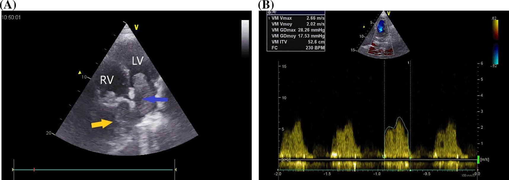
A 37-year-old Maghrebian female patient was presented to the cardiology department due to complaints of dyspnea, progressing over one month. She had a dry cough and had been resistant to symptomatic treatment. The patient was diagnosed with borderline breast PTs ten years earlier, which was treated by surgical excision. Upon examination, her dyspnea was classified as class IV on the New York Heart Association Functional Classification with orthopnea. Her transcutaneous oxygen saturation was 92%, and pulmonary auscultation revealed bibasilar crackles. Additionally, the patient’s chest x-ray showed a homogeneous opacity located in the basal part of the right lung. Transthoracic echocardiography (TTE) revealed 5 × 4 cm homogenous mass occupying nearly all the left atrium (LA), resulting in severe mitral valve obstruction (mean gradient = 17 mmHg) (Figure 1).

LV: left ventricle, MV: mitral valve, RV: right ventricle.
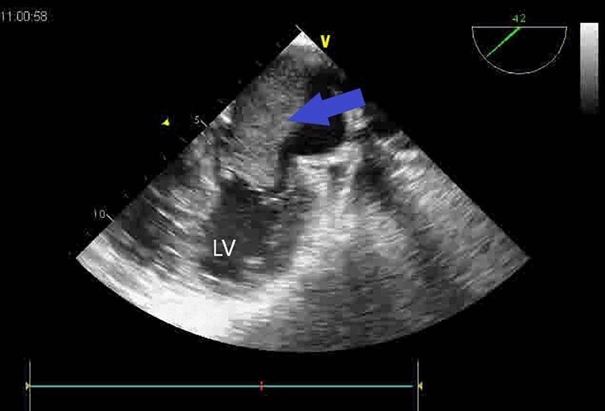
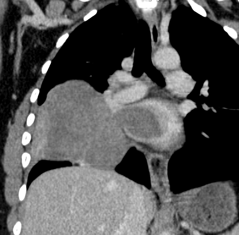
A second huge mass compressed the right atrium posterior wall. Following respiratory stabilization, transesophageal echocardiography confirmed TTE results and revealed an extended mass into LA via the right inferior pulmonary vein (RIPV) (Figure 2). Cardiac computed tomography (CT) revealed a large (100 × 70 × 100) mediastino-pulmonary mass extending to LA via RIPV (Figure 3).


The Cardiac magnetic resonance imaging (MRI) results showed low signal on T1-weighted imaging and high signal on T2-weighted imaging of the mediastino-pulmonary mass (Figure 4). The patient accepted to undergo an urgent mass resection surgery to avoid total mitral valve obstruction and sudden death. The histological study of the resected mass confirmed the metastatic spread of malignant PTs to LA (Figure 5). The patient was discharged from the hospital after having an echocardiographic check-up, which demonstrated no residual tumor. However, three months after the surgery, she died from a huge relapse of mediastinal mass with tracheal invasion.
PTs or cystosarcoma is a rare breast neoplasm.1 These types of tumors are commonly manifested in the breast tissue and are usually benign; however, they might rarely be malignant.2,3 A malignant tumor has a potential to metastasize to distant organs, such as lung, bone, and liver.8 Our case revealed concomitant pulmonary and cardiac metastases, which is unusual, and it is associated with poor prognosis. It has been reported that cardiac invasion could be caused by hematogenous spread, direct extension, or via the lymphatic route.9 In the case of this patient, direct extension from pulmonary metastasis to RIPV is the probable route of metastasis. Reported cases of cardiac metastasis are mostly located in the right heart with the possibility of right ventricle outflow tract obstruction.10 To the best of our knowledge, this is the first case of LA location, complicated by severe mitral obstruction and acute heart failure. The clinical expression of cardiac metastasis is mainly dependent on the tumor burden and location.6 As in the case of our patient, cardiac metastasis can manifest with dyspnea and chest pain, or it can be asymptomatic. Previously, malignant cardiac metastasis had poor prognosis and very rare cases were identified at autopsy.11 However, advances in imaging tools such as echocardiography allows for detection and confirmation of intra-cardiac mass and eventual valve or cavity obstruction. However, echocardiography is limited in the differentiation between PTs, myxoma, fibroadenoma, and thrombus.11 In our case, echocardiography revealed severe mitral obstruction by an intra-LA mass. Cardiac CT and MRI provide multiple views in different axes with a precision of limits as well as intra, and extra cardiac extension, thus allowing a better distinction between the thrombus and other masses.12 The results of the echocardiography, cardiac CT, and MRI for our patient confirmed the intra and extra cardiac location of the tumor and its LA access from RIPV to the mitral valve. Therapeutic approaches, including chemotherapy, radiotherapy, and hormonal therapy are still controversial.7 The surgical excision of cardiac metastasis from a malignant PTs was described in few reports.13 This type of intervention could be an urgent life-saving therapeutic strategy in case of right ventricle outflow obstruction or mitral obstruction, and it can also improve the patient’s quality of life in the short term, as it was in our case.14,15 However, intra-operative mass manipulation could cause tumor dissemination, thus leading to a risk of further metastasis development.11,16 This may explain the hudge relapse of mediastinal mass with tracheal invasion in our patient. In this case report the major limitations were the delay in diagnosing cardiac and pulmonary metastases and the lack of immunohistochemical analysis of the tumor.
Cardiac metastases from PTs are rare. Tumor surgical excision might be indicated to avoid sudden death and to improve the patient’s quality of life despite the extremely unfavorable prognosis. Nevertheless, urgent surgical removal could be unavoidable in case of valve obstruction. Early diagnosis and immunohistological analysis of PTs, especially the malignant type, is imperative given that there is little effective treatment for metastatic disease.
All data underlying the results are available as part of the article and no additional source data are required.
NA, AA and AB were actively involved in data collection and processing. IC and RK were involved in manuscript preparation. CK, SJ and FM were involved in manuscript reviewing. All authors have read and approved the manuscript.
| Views | Downloads | |
|---|---|---|
| F1000Research | - | - |
|
PubMed Central
Data from PMC are received and updated monthly.
|
- | - |
Is the background of the case’s history and progression described in sufficient detail?
Yes
Are enough details provided of any physical examination and diagnostic tests, treatment given and outcomes?
Yes
Is sufficient discussion included of the importance of the findings and their relevance to future understanding of disease processes, diagnosis or treatment?
Partly
Is the case presented with sufficient detail to be useful for other practitioners?
Yes
Competing Interests: No competing interests were disclosed.
Reviewer Expertise: breast and plastic surgery
Is the background of the case’s history and progression described in sufficient detail?
Yes
Are enough details provided of any physical examination and diagnostic tests, treatment given and outcomes?
Yes
Is sufficient discussion included of the importance of the findings and their relevance to future understanding of disease processes, diagnosis or treatment?
Yes
Is the case presented with sufficient detail to be useful for other practitioners?
Yes
Competing Interests: No competing interests were disclosed.
Reviewer Expertise: Cardiology
Alongside their report, reviewers assign a status to the article:
| Invited Reviewers | ||
|---|---|---|
| 1 | 2 | |
|
Version 2 (revision) 25 Jul 22 |
read | |
|
Version 1 14 Mar 22 |
read | read |
Provide sufficient details of any financial or non-financial competing interests to enable users to assess whether your comments might lead a reasonable person to question your impartiality. Consider the following examples, but note that this is not an exhaustive list:
Sign up for content alerts and receive a weekly or monthly email with all newly published articles
Already registered? Sign in
The email address should be the one you originally registered with F1000.
You registered with F1000 via Google, so we cannot reset your password.
To sign in, please click here.
If you still need help with your Google account password, please click here.
You registered with F1000 via Facebook, so we cannot reset your password.
To sign in, please click here.
If you still need help with your Facebook account password, please click here.
If your email address is registered with us, we will email you instructions to reset your password.
If you think you should have received this email but it has not arrived, please check your spam filters and/or contact for further assistance.
Comments on this article Comments (0)