Keywords
NF2, Caesarean Section, Fetal
NF2, Caesarean Section, Fetal
Neurofibromatosis is an autosomal dominant genetic diseasewhich consists of three classifications, Neurofibromatosis type 1 (NF1), Neurofibromatosis type 2 (NF2), and Schwannomatosis.1 NF2 is caused by a mutation on chromosome 22 (22q12.2), the incidence rate 1:33,000-40,000 in general population, while the prevalence is 1:57,000.2 A retrospective study in Japan showed that patients with NF2 had survival rates of 5 years, 10 years, and 20 years from diagnosis of 85%, 67%, and 38%, respectively.3 Pregnant women with neurofibromatosis have an increased risk of morbidity due to the progressive growth of meningiomas and schwannomas, hypertensive/cardiovascular complications, but not death during pregnancy. Additionally, pregnant women with NF2 have a high risk of developing hypertension, preeclampsia, cardiovascular problems, and Intrauterine growth restriction (IUGR).4 They should be notified by their physician that this is an inherited disease and there is a 50% chance that their baby will Inherit this disease. Appropriate steps and supervision during pregnancy up to the birth process are needed to reduce pregnancy morbidity.5
A 26-year-old female at 29-30 weeks of gestation, had limb weakness, decreased hearing and vision since six months pregnant. She had one normal child age 4 years old, no family members suffer the same symptoms or had history of NF. Initially, the patient felt lumps appearing all over her body at the age of nine. These lumps continued to proliferate and increase in size by the age of 17, and she had weakness in her legs at 23 years of age. But, she never checked and got any medication or intervention. In 2016, she delivered her first child without any complications. At the end of 2017, the patient began to feel weakness in her legs. Towards the end of 2019, her neurological examination showed decreased vision (ODS > 2/60), and hypoesthesia of approximately L1 and below. MRI examination of her spine indicated multiple mass Intradural extramedullary (IDEM) cervical level 6 and thoracolumbar (VC2 2.2 × 1.5 cm; VC4-5 0.8 × 0.8 × 0.9 cm; VTh 10-11 3.5 × 1 × 0.7 cm; VL1 1.1 × 1 × 0.8 cm) (Figure 1).

Multiple intradural extramedullary solid masses seen as high as VC level 4-5 (A,B,C), VTh levels 10-11 (D,E, F) and VL 1 (G, H, I) levels which show strong contrast enhancement are consistent with the IDEM mass picture. A solid, intramedullary mass appears as high as the VC 6 level (J, K, L), which strongly enhances contrast following the IDIM image.
During her pregnancy, treatments were carried out under the supervision of the Department of Obstetrics and Gynecology, the Department of Neurology, Neurosurgery, and Radiology at RSUD dr. Soetomo hospital, Surabaya. The routine fetal evaluation showed normal fetal growth with no signs of IUGR. A cardiovascular evaluation revealed no abnormalities and symptoms of gestational hypertension or preeclampsia. The decline in neurological function and vision worsened when entering the 20th gestational age until the third trimester. It was stagnant until term, without any deterioration of neurological deficit, hydrocephalus, preeclampsia and IUGR. Based on our evaluation and multidisciplinary discussion, we decided to terminate the pregnancy by cesarean section under General Anesthesia at 37 weeks. A male infant was born with a birth weight of 2600 grams, without any abnormalities. Head MRI with contrast evaluation after delivery was performed, which showed bilateral vestibular schwannoma, meningioma, and schwannoma (Figures 2–5).

Multiple extra-axial masses showing strong contrast enhancement suggest meningiomatosis.
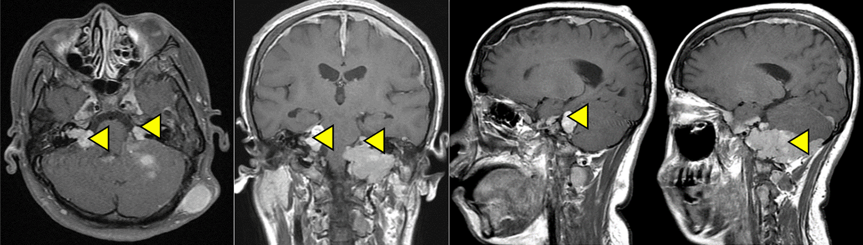
Extra-axial solid mass was seen in the right and left cerebellopontine angles. The picture showed a strong contrast enhancement. According to the image, bilateral vestibular schwannoma extends to the right and left side of the intra-canaliculi and pushes the left cerebellar hemisphere to the superior side. There was also a subcutaneous nodule in the left occipital region.

The lesion appears posterior to the right and left orbits, suggesting a right-left retinal detachment.
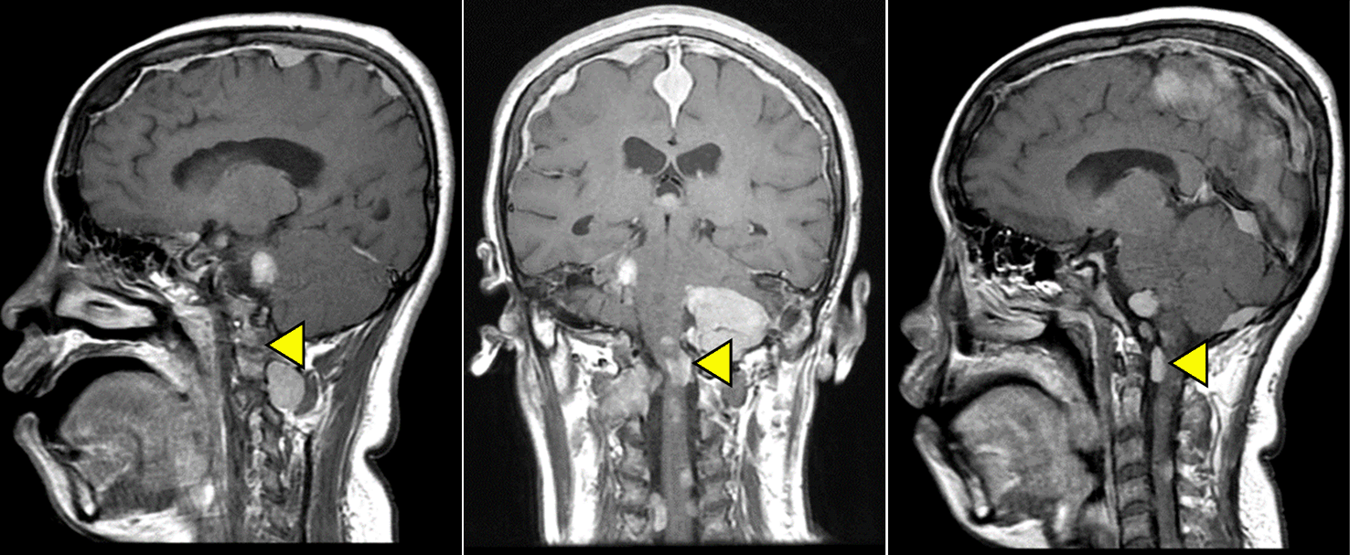
Multiple extra-axial solid masses as high as VC 1 to VC 5 showing strong contrast enhancement suggested a schwannoma dd meningioma image.
The clinical diagnosis of neurofibromatosis for NF1/NF2 differentiation often overlaps because both have almost the same clinical condition. The key differences between NF1 and NF2 is that Lisch nodules (protruding and pigmented hamartomas of the iris) are common in NF1 and are not seen in significant numbers in NF2 cases. Additionally, schwannoma is rare in NF1 and common in NF2 cases. Schwannomas in NF1 are at risk for malignant transformation into neurofibrosarcoma (Malignant Peripheral Nerve Sheath Tumor (MPNST)). Spinal Root Tumors seen in NF2 are schwannomas and neurofibromas seen in NF1.6 NF2 is not associated with cognitive impairment that is often seen in NF1.6
NF2 (22q11.2) encodes the tumor suppression protein merlin, which is responsible for the repair of Schwann cells, which, when disturbed, causes a schwannoma. Loss of merlin function could lead to the activation of the proto-oncogene signal as in NF2.1,7 Genetic mosaicism was found in 30-60% of de novo NF2 mutation cases, which is more common in tumor cells.1,6 Genetic mosaicism generally leads to a milder form of the disease, either NF1 or NF2 cases.1 Based on the pedigree data, similar clinical illness was not found in other family members. Therefore, mosaicism and the de novo mutation process explained the NF2 disease in this case.
The MRI Spine examination was done in October 2019 and found multiple IDEM (Intradural Extramedulla), cervical, thoracolumbar, and cerebellum. Many reported cases mention a delay in diagnosis due to the asymptomatic properties of the disease, especially complaints related to central and peripheral nervous disorders before the age of 20 years.8 The same was true in our patient with the addition that the patient's family's lack of knowledge on NF has also contributed to the delay in diagnosing the disease.
Diagnosis of NF2 mainly uses radiological modalities such as CT scan or MRI. The choice of radio-diagnostic modality can be made based on the availability of facilities and specific medical indications, such as pregnancy.9 CT scans in the first trimester to 20 weeks of gestation have a very high risk of abortion and teratogenic effects.10 Many studies have reported that the use of MRI during pregnancy is safe, with no teratogenic effect on the fetus.11 MRI 1.5 Tesla is safe to use in all trimesters, however MRI 3 Tesla has not been proven safe in pregnancy.12 Therefore pregnant women are advised to use MRI 1.5 Tesla.12
Contrast media used for imaging is known to cross the placental barrier and enter fetal circulation and amniotic fluid in pregnant patients.13 Several investigations stated that the utilization and repetition of high doses of gadolinium-based contrast agent (GBCA) in experimental animals had a teratogenic effect, whereas others have not found this effect in animal studies.13 The American College of Radiology does not recommend the use of contrast agent during pregnancy.14 However, Food and Drug Administration (FDA) classifies GBCA as category C agent in pregnancy; which should be given in conditions where the benefits far outweigh the risks to the fetus and should only be done once during pregnancy.13,14
Tumor growth often becomes more rapid in pregnancy. Schwannomas are generally slow-growing, but in some cases, it has been reported that development and neurological manifestations increase during pregnancy.15 The mechanism of tumor growth acceleration during pregnancy is still unclear, however two mechanisms are widely proposed. First, the increase in blood volume during pregnancy causes vascular congestion, contributing to tumor growth. Second, the direct hormonal effects of progesterone receptors (PR) and estrogen receptors (ER) mediate tumor growth. Several studies report that pregnancy-related hormones can influence tumor growth and the development of neurological symptoms.15 A study conducted on 16 vestibular schwannomas (VS) patients who had microsurgery showed an increase in estrogen receptors Erα and ErbB.16 These patients had a positive effect with the administration of antiestrogen therapy and ErbB inhibitors. In patients with NF2, there is an increase in tumor growth due to the rise in the hormone estrogen during pregnancy.16 In the case of our patient, several mass locations were suspected to be schwannoma due to the course of the patient's NF2 disease, Vestibular Schwannoma, and Spinal Schwannoma.
A study conducted on nine women of childbearing age with VS showed no ER/PR on immunohistochemistry, while expression of VEGF was found.17 The growth of schwannomas based on periodic MRI evaluations can be categorized: 60% offer prolonged growth to no development, 30% have a growth rate of about 0.2 cm per year, and 10% overgrow at 1 cm or more per year.17 On the other hand Kawaguchi et al reported the immunohistochemical staining of spinal schwannoma resulted in ER-positive and PR-negative expressions.15 However, further investigation regarding the type of ER (Erα/Erβ) and their expressions during pregnancy requires further studies. In this case, there was an increase in complaints of neurological deficits, especially when entering 6 months of pregnancy, which could be due to a rise in the growth/size of spinal schwannoma.
Another study mentioned changes in follicle-stimulating hormone, luteinizing hormone, and human chorionic gonadotropin hormone during pregnancy inhibits tumor cell proliferation, whereas human placental lactogen hormone and prolactin hormone stimulate tumor growth.18 Changes in plasma hormone concentrations during pregnancy affect meningioma growth, mainly in the second and third trimesters. Consequently, these conditions may lead to critical situations. The study continues to report that ER is not expressed in most meningiomas. However, the effect of the PR protein expression and higher mitotic rates on the expression of this protein has been investigated in many studies.18
Progesterone, estrogen, and its metabolites increase during pregnancy and reach a peak levels in the third trimester. Progesterone increases about 6-8 times compared to a regular menstrual cycle, while estrogen increases 15-75 times compared to the luteal phase and 60-300 times compared to the follicular phase of a normal menstrual cycle.19 Most meningiomas are PR-positive and ER-negative, and the hormonal effects depend on the balance between estrogen and progesterone. Progesterone is mitogenic in breast cancer and meningioma cells. Based on this hypothesis, rapid growth cases of meningiomas during pregnancy is sometimes followed by spontaneous post-partum involution.19
Pregnant patients with neurofibromatosis may have complications such as hypertension, preeclampsia, cardiovascular problems, and IUGR babies. NF1 causes more maternal and fetal complications than NF2.2,20,21 Individuals with NF1 can suffer from various cardiovascular disorders, congenital heart disease, vasculopathy, and hypertension. Echocardiographic data showed that 27% of patients with NF1 had cardiovascular disease, 50% of cases had pulmonary artery stenosis.22 A low prevalence rate is expected because the diagnosis is carried out when symptoms begin to appear. However, complications due to neurofibromas in pregnancy are more likely due to vasculopathy, the pathogenesis of which is generally unknown.22 Terry AR et al. report that a more common complication in NF2 patients is preeclampsia, which is caused by a decrease in the ability of vascular dilators.4 In their study, periodic evaluation of the fetus by assessing the biophysical profile, fetal weight growth, and Doppler velocimetry assessment, showed no signs of impaired fetal development. Additionally, maternal evaluation of cardiovascular disorders and preeclampsia risk which is routinely checked, did not show any of such diseases and complications.
Based on their results, Terry AR et al. stated that antenatal examination and treatment of pregnant patients with neurofibromatosis along with a thorough evaluation of the medical history and a complete physical assessment should be performed in a Tertiary Hospital. Moreover, multidisciplinary treatment of any clinical findings in the eyes, skin, neurological, cardiac, and digestive systems must also be carried out. Most importantly, genetic counselling should be provided. Obstetricians should obtain information regarding history of seizures, hearing loss, neurological disorders, or visual disturbances. For these patients, a complete neurologic examination should focus on balance, sensory, motor, coordination, and nystagmus. Patients with extensive kyphoscoliosis should undergo pulmonary function tests. Appropriate fetal evaluation should be performed to monitor fetal well-being.2
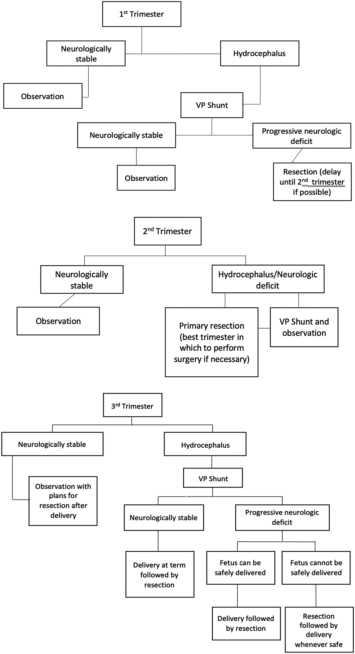
In the case of our patient, the clinical evaluation and course of the disease was considered stagnant and without any rapid progression, besides a clinical decline in her neurologic and vision from 20 weeks of gestation. Based on the results of periodic evaluations, the progression of neurological deficits was stable. According to the algorithm presented in Figure 6, approaching the third trimester with stable neurologic deficits and no sign of hydrocephalus, tumor resection can be postponed after termination of pregnancy.23 Periodic evaluation of mother and fetus did not indicate any risks of hypertension, preeclampsia, and no IUGR. Hence it was decided to terminate at gestational age of 37 weeks, when the fetus enters term age with a weight > 2500 g.
The mode of delivery in neurofibromatosis is determined primarily by obstetric indications. The rate of Cesarean section (C-section) is relatively increased in patients with neurofibromas due to an increase in the rate of pregnancy complications. Pelvic neurofibroma can cause labor dystocia, as such abdominal termination is an option in these cases.24
The choice of modality for termination of pregnancy in patients with neurofibromatosis depends on the findings obtained during pregnancy. Evaluation of spinal and intracranial masses/lesions is needed to determine contraindications to labor related to the risk of increased intracranial pressure (ICP) during the labor and type of anesthesia if surgery is carried out.2,9 The patient had an intracranial and spinal mass at the level of the neck (cervical); the process of straining could be at risk of increasing ICP; therefore, it was decided to terminate the pregnancy by C-section under general anesthesia. The decision of delivery.2,9,20,23
Early diagnosis of NF is possible due to the clinical manifestation of this disease between the ages of 18-20 years. If a patient with NF is pregnant or is planning to have children, it is vital for him/her to have preimplantation genetic tests along with prenatal diagnosis to evaluate the risks and early detection of this disease in their child. Despite the benefits of early diagnosis, prenatal and preimplantation genetic test are not done routinely, many families consider evaluation after birth for their children.6,25 It would be highly beneficial to combine these diagnostic tests with genetic counselling to fully inform these patients that NF is a genetic neurological disorder which their children may inherit. Additionally, the patient would be advised that if a mutation is found in one member of the family, it is recommended that all family members be tested for this disease. After the counselling, if the family decides to continue with the pregnancy, preventive efforts with postnatal diagnostics and early treatment will be the main goals to reduce morbidity, if the child is affected with NF.7,25
Genetic counselling for couples (patients with neurofibromatosis) planning to have children should be carried out thoroughly. In some countries, it is recommended to undergo assisted technology pregnancy using an ovum and sperm donor, if NF is diagnosed in one or both partners.5 In vitro fertilization (IVF) will help in deciding the genetic inheritance of this disease. However, the possibility of de novo mutations during the fertilization process can still occur in the general population without the genetic mutation of NF.6
Although the primary diagnosis of NF is clinical, surgery and examining mutations through lymphocytes and tumors are the best methods of confirming diagnosis. For example patients with mosaicism carry small genetic mutations in lymphocyte cells that can be used as a marker for this disease.7 In addition, an immunohistochemistry examination can be performed to evaluate the ER and PR to assess the possible response to pregnancy.15 In the case of our patient, surgery and molecular diagnostics were not performed because the she was unwilling to undergo post-partum surgery.
NF2 is a severe disease as several central nervous system (CNS) tumors are asymptomatic and often recur after treatment. Survival rates for NF2 patients based on long-term follow-up data have not been reported. However, some studies estimate that the mean possible survival time for NF2 patients is approximately 15 years after diagnosis. More than 40% of patients are expected to die by the age of 50 years.3 The study by Otsuka et al. concluded that the long-term survival rate of patients with NF2 proved poor. Patients with symptoms before the age of 20 have poorer survival rates. Therefore, based on such studies informing and counseling should be done to assist NF patients in understanding the prognosis of this disease.3
The case reported here is classified as severe NF2 because the patient had experienced symptoms before the age of 20 years. Because of her symptoms the patient is now unable to take care of herself and in need of surgery to improve her quality of life. However, the patient has decided not to undergo surgery which will further worsen her quality of life and survival rate. Our case did not perform genetic testing on either patient, her child and other family members to confirm the diagnosis due to limitations and refusal of the patient.
Neurofibromatosis can be diagnosed based on the evaluation of clinical symptoms, pedigree analysis, and radiological assessment. Pregnant women with NF2 can experience clinical deterioration due to rapid tumor growth during pregnancy. Patients with neurofibromatosis are advised to undergo genetic, prenatal, and antenatal counseling to better plan pregnancy and prevent lower the risk of inherited disease. NF tumor genetic and molecular testing should be performed to confirm the diagnosis. Multidisciplinary management in tertiary hospitals should be carried out in pregnant women with neurofibromatosis to fetal and maternal morbidity and mortality.
All data underlying the results are available as part of the article and no additional source data are required
Written informed consent for publication of their clinical details as obtained from the patient and her family.
Nanda Bagus P
Roles: Conceptualization, Data Curation, Formal Analysis, Resources, Supervision, Investigation, Validation, Visualization, Writing – Original Draft Preparation, Writing – Review & Editing
Hermanto TJ
Roles: Conceptualization, Formal Analysis, Methodology, Resources, Project Administration, Supervision, Validation, Visualization, Writing – Original Draft Preparation, Writing – Review & Editing
Cininta N.I.
Roles: Conceptualization, Formal Analysis, Methodology, Supervision, Validation, Visualization, Writing – Original Draft Preparation, Writing – Review & Editing
Djohan Ardiansyah
Roles: Conceptualization, Formal Analysis, Methodology, Supervision, Validation, Visualization, Writing – Original Draft Preparation, Writing – Review & Editing
Rahadian Indarto S
Roles: Conceptualization, Formal Analysis, Methodology, Supervision, Validation, Visualization, Writing – Original Draft Preparation, Writing – Review & Editing
Widiana Ferriastuti
Roles: Conceptualization, Formal Analysis, Methodology, Supervision, Validation, Visualization, Writing – Original Draft Preparation, Writing – Review & Editing
| Views | Downloads | |
|---|---|---|
| F1000Research | - | - |
|
PubMed Central
Data from PMC are received and updated monthly.
|
- | - |
Provide sufficient details of any financial or non-financial competing interests to enable users to assess whether your comments might lead a reasonable person to question your impartiality. Consider the following examples, but note that this is not an exhaustive list:
Sign up for content alerts and receive a weekly or monthly email with all newly published articles
Already registered? Sign in
The email address should be the one you originally registered with F1000.
You registered with F1000 via Google, so we cannot reset your password.
To sign in, please click here.
If you still need help with your Google account password, please click here.
You registered with F1000 via Facebook, so we cannot reset your password.
To sign in, please click here.
If you still need help with your Facebook account password, please click here.
If your email address is registered with us, we will email you instructions to reset your password.
If you think you should have received this email but it has not arrived, please check your spam filters and/or contact for further assistance.
Comments on this article Comments (0)