Keywords
HRCT thorax, low dose, dose reduction, matrix size, tube voltage
This article is included in the Manipal Academy of Higher Education gateway.
HRCT thorax, low dose, dose reduction, matrix size, tube voltage
The prevalence of respiratory diseases in India is substantially high as per the recent global burden of disease survey data (Dandona et al., 2017). Chronic respiratory diseases, especially asthma and obstructive pulmonary disease, are of particular importance for having wide variations in morbidity and mortality in different Indian states (Salvi et al., 2018). Additionally, the novel COVID-19 outbreak declared by the World health organization (WHO) has increased the burden of respiratory disease in India as well as globally (Cucinotta & Vanelli, 2020; To et al., 2020). The outbreak of COVID-19 led to an increase in the demand for image modalities for the diagnosis of the disease. A chest radiograph plays a crucial role in diagnosing suspected respiratory diseases. High resolution computed tomography (HRCT) of the thorax is one such examination in high demand in routine use and for initial and rapid diagnosis of COVID-19 pneumonia with the low rate of missed diagnosis (Kashyape & Jain, 2021; Li & Xia, 2020). Although reverse-transcription polymerase chain reaction (RT-PCR) is the preferred tool for diagnosing novel COVID-19 infection, HRCT thorax has better sensitivity (Kalra, Homayounieh, Arru, Holmberg & Vassileva, 2020). The benefit of HRCT is not only limited to disease confirmation, but it plays a significant role in determining disease progression, treatment response, management, and decision on hospitalization and discharge of the COVID-19 patient through subsequent scans performed during these periods (Kalra et al., 2020; Rubin & Ryerson & Haramati, 2020; Azadbakht et al., 2021; Davarpanah et al., 2020).
The use of imaging modalities during the COVID-19 pandemic indeed has maximum benefit, but the fact that radiation exposure increases the risk of cancer should not be ignored. The linear-no threshold (LNT) model describes cancer risk even with a low dose of radiation (Shah, Sachs & Wilson, 2012; Mullenders, Atkinson, Paretzke, Sabatier & Bouffler, 2009). The risk of radiation-induced cancer during the pandemic is of concern, primarily due to the increase in COVID-19 infection and the need for multiple scans required during the recovery period of the disease (Wang, Liu, Yang & Chen, 2020). As recommended by International Commission on Radiological Protection (ICRP), the radiation dose delivered to the patient should be as low as reasonable achievable (ALARA) even during the pandemic, and the dose optimization for COVID-19 patients is necessary as there is no low dose HRCT protocol reported (Azadbakht et al., 2021; Yeung, 2019). There are several techniques to optimize the radiation dose for HRCT, such as reducing tube voltage, tube current, scan length, high pitch techniques, automatic exposure control (AEC), and iterative reconstruction techniques (Azadbakht et al., 2021). The feasibility of matrix size with different tube voltage in the HRCT protocol of thorax is unknown. Therefore, this study aimed to compare the effect of matrix sizes and tube voltage on image quality and radiation dose on adult HRCT thorax. The working hypothesis of this study was that the change in tube voltage could influence the image quality.
The study approval was obtained from the institutional ethics committee (IEC: 168/2019), Kasturba hospital, Manipal. The study was carried out in department of Radio-diagnosis and Imaging, Kasturba Hospital, Manipal. An experimental study was initially tested on Phantom to understand the influence of tube voltage and matrix size on the quality of the image and dose. The outcome of the results was then tested on the patient’s population.
This experiment was conducted in a 32 cm body CT polymethyl metacrylate (PMMA) phantom (Unfors, serial no. 0143) using an iCT (Philips Medical Systems) 128-row multiple slice CT scanner. The phantom was positioned on the CT table. The height of the phantom was adjusted using the position of laser light. The experiment was performed at two different tube voltage settings (120 kVp and 100 kVp) and three matrix sizes (512, 768 & 1024) available in the CT scanner (Table 1). All the other parameters, Reconstruction mode iDose4 level 2, collimation 64*0.625, gantry tilt of 0 degrees, the pitch of 1, rotation time: 0.5, slice thickness: 1 mm, reconstruction increment of 0.7500, a field of view (FOV) of 350 mm, and the adaptive filter were kept constant. The automatic tube current modulation was enabled during the procedure. An axial image with 1 mm thickness was obtained. Each protocol was scanned with a 130 mm scan length. Phantom CT image obtained using different parameters was analysed for dose and quantitative image quality. Signal-to-noise (SNR) and contrast-to-noise (CNR) ratios were analysed by drawing five regions-of-interest (ROIs) of 100 mm2. Out of five ROIs, four were positioned in peripheral location (3, 6, 9, 12 o’clock) and one in the centre location of the phantom along with one ROI in the background to encompass homogeneity of measured regions (Figure 1).
| Protocol | Tube voltage settings | Matrix size |
|---|---|---|
| A | 120 | 512 × 512 |
| B | 120 | 768 × 768 |
| C | 120 | 1024 × 1024 |
| D | 100 | 512 × 512 |
| E | 100 | 768 × 768 |
| F | 100 | 1024 × 1024 |

A total of 180 patients (Mean age 54.50 ±15.81, Male 55.06 ±17.15, Female 53.85±14.17) who were 18 years and above and prescribed for the HRCT Thorax were considered for this study (See Underlying data) (Sukumar, 2022). The written informed consent was obtained from the participant. All the scan was performed in iCT (Philips Medical Systems) 128-row multiple slice CT scanner with six different scanning protocol similar to phantom (Table 1). The automatic tube current modulation (Dose Right Index: 17) was enabled during the procedure.
After acquiring the image using a different protocol, the subjective image assessment of the patient’s population was performed according to the European Guidelines on Quality Criteria for CT (Menzel et al., 2000). Two radiologists with a mean experience of 12.00 ± 1.41 years, reviewed images independently. The radiologists were blinded to the scanning protocols and the procedure. Images were displayed in the HRCT window (-600 WL, 1600 WL) on an Advantage Workstation (Philips Healthcare). Radiologists’ evaluation for the diagnostically acceptable image quality for HRCT thorax was considered (See Underlying data) (Sukumar, 2022). The diagnostic acceptability of the image quality was rated on five-point rating scale were, 1 = poor for diagnosis, 2 = suboptimal for diagnosis, 3 = good for diagnosis, 4 = very good for diagnosis, 5 = excellently. The streak artefacts seen anywhere on the CT image was rated on a five-point rating scale, were 1= artefact causing diagnosis impossible, 2 = artefacts affecting diagnostic information, 3 = major artefacts affecting visualization of major structures but diagnosis still possible, 4 = minor artefacts not interfering with diagnostic decision-making, and 5 = no artefacts.
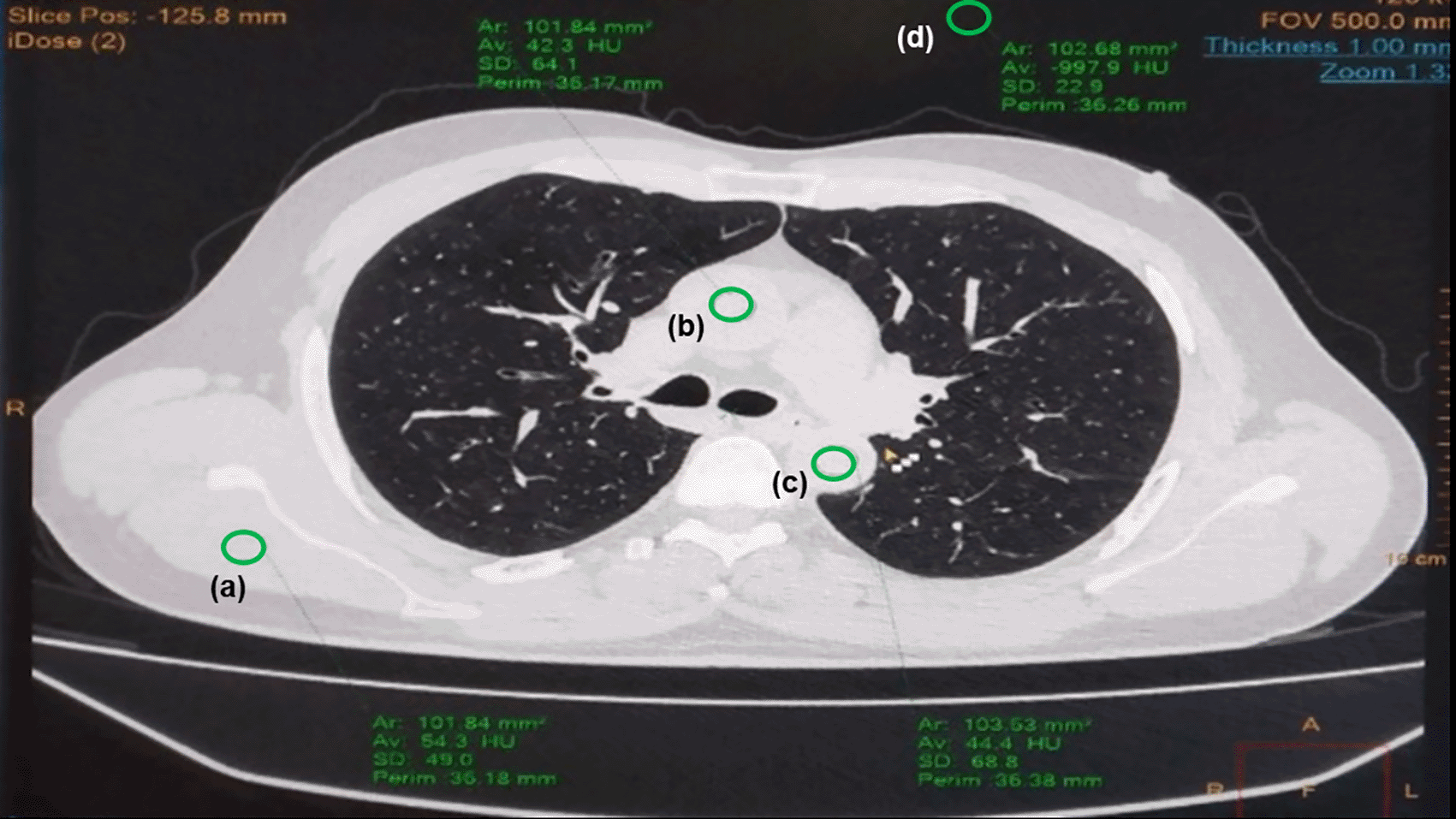
The quantitative image quality assessment was assessed by drawing 100 mm2 ROIs on high density structures of thorax, such as subscapularis muscle, ascending aorta, and descending aorta (See Underlying data) (Sukumar, 2022) (Figure 2).
The SNR, CNR, and Figure of Merit (FOM) ratios for the phantom and the participant population were analysed by the following equation (Chang, Hsu, Lin & Hsu, 2017) (See Underlying data) (Sukumar, 2022).
Where CTROI and CTBackground are the average Hounsfield unit of the corresponding ROI, and SDROI and SDBackground are the standard deviations of the corresponding ROI.
The dose data (CTDIvol and DLP) for both phantom study and patient population study were collected from the dose info of the system. For the study population the obtained DLP was further used to calculate the effective dose by multiplying with a conversion factor of 0.014 (Deak, Smal & Kalender, 2010) (See Underlying data) (Sukumar, 2022).
Statistical Package for the Social Sciences (SPSS) Statistics for Windows, Version 16 (Kumar, Pai & Vineetha, 2020) was used to perform the statistical analysis. For the study population the obtained data were not normally distributed. Therefore, the median and quartiles are reported, and for comparison of quantitative image analysis and dose matrix, Mann Whitney U (Kumar, Pai & Vineetha, 2020) test was performed. Mean score for each protocol was calculated for subjective image assessment. P<0.05 was considered statistically significant.
The influence of various combinations of tube voltage (120 and 100 kVp) and matrix size (512, 768 and 1024) on image quality (SNR and CNR) and dose matrix (CTDIvol and DLP) are summarized in Table 2. The difference noted (in percentage) when comparing different protocols are outlined in Table 3. In this study, the phantom was scanned with two tube voltage techniques combined with three different matrix sizes (a total of six scanning protocols). When comparing the image quality of 120 kVp with various matrix sizes, protocol A showed the highest values compared to protocols B and C. The dose changes noted were very minimal.
Similarly, when comparing the image quality of 100 kVp with various matrix sizes, protocol D showed higher values than protocol E and protocol F, and no changes were noted in terms of dose matrix (Table 2). The result of the phantom study showed that, as the matrix size increases, the SNR and CNR decreases, except for the case of protocol C, where the SNR and CNR values were slightly higher than protocol B. Therefore, when comparing the image quality of the 120 kVp technique and 100 kVp technique with various matrix sizes, the matrix size of 512 (protocol A and protocol D) produced better image quality than another matrix size. When comparing among two tube voltages with the same matrix size of 512 (protocol A and protocol D), protocol A showed better result than protocol D when considering image quality where the difference (in percentage) among these two protocols for SNR and CNR was 8% and 23.80% respectively, which is minimal as compared to the difference of other protocol (Table 3). However, the difference noted in CTDIvol and DLP among these two protocols was 15.64% and 15.62%, respectively (Table 3), which shows that 100 kVp techniques have reduced radiation dose.
Similar to the phantom study, the influence of tube voltage and matrix size on image quality and radiation dose was performed in the patient population. The collected patient data were not normally distributed. Hence the median and quartiles for image quality (SNR and CNR), radiation dose matrix (CTDIvol and DLP), and Figure of merit (FOM) are summarized in Table 4. The statistically significant value (p<0.05) for comparison of median among various protocols are outlined in Table 5.
| Protocols comparison | p-value | ||||
|---|---|---|---|---|---|
| CTDI | DLP | SNR | CNR | FOM | |
| A vs B | 0.567 | 0.196 | <0.001* | <0.001* | <0.001* |
| A vs C | 0.267 | 0.395 | <0.001* | <0.001* | <0.001* |
| A vs D | <0.001* | <0.001* | <0.001* | <0.001* | 0.003* |
| A vs E | <0.001* | <0.001* | <0.001* | <0.001* | <0.001* |
| A vs F | <0.001* | <0.001* | <0.001* | <0.001* | <0.001* |
| B vs C | 0.756 | 0.030* | 0.773 | <0.012* | 0.034* |
| B vs D | <0.001* | <0.001* | 0.641 | 0.255 | 0.067 |
| B vs E | <0.001* | <0.001* | <0.001* | <0.001* | 0.033* |
| B vs F | <0.001* | <0.001* | <0.001* | <0.001* | 0.038* |
| C vs D | <0.001* | <0.001* | 0.953 | 0.255 | <0.001* |
| C vs E | <0.001* | <0.001* | <0.001* | 0.007* | 0.906 |
| C vs F | <0.001* | <0.001* | 0.001* | 0.004* | 0.796 |
| D vs E | 0.110 | 0.156 | <0.001* | <0.001* | <0.001* |
| D vs F | 0.387 | 0.171 | <0.001* | <0.001* | <0.001* |
| E vs F | 0.549 | 0.756 | 0.976 | 0.745 | 0.859 |
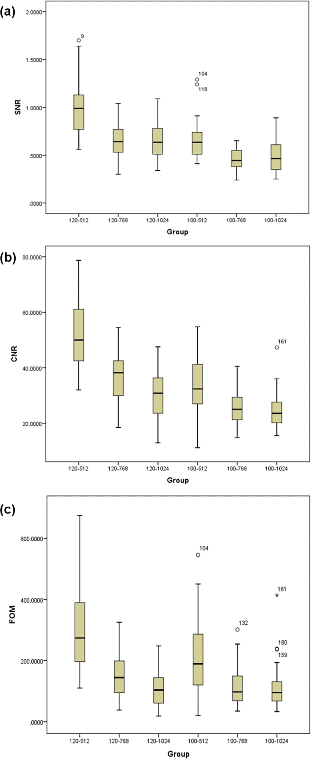
When comparing the image quality of 120 kVp technique (standard dose) with three different matrix size, protocol A showed highest value when compared with protocol B and protocol C. The difference noted was statistically significant between protocol A and protocol B, and protocol A and protocol C but for protocol B and protocol C, difference in SNR was not statistically significance whereas CNR was statistically significance. There was no difference noted for radiation dose when comparing within the protocols, except for the protocol B and protocol C where DLP showed statistical significance difference and FOM showed significant difference for all the comparisons (Table 5). For 100 kVp technique (low dose), the result was in line with 120 kVp techniques, that is protocol D showed highest value in terms of image quality when compared with protocol E and protocol F, and the difference noted were statistically significant for all the comparison except for protocol E and protocol F. Similarly, there were no statistical difference noted in radiation dose matrix and FOM for all the comparisons except for comparison between protocol E and protocol F (Table 5). The overall result showed that, with the increase in matrix size, the SNR, CNR, and FOM decrease for both standard 120 kVp and a low dose of 100 kVp techniques (Figure 3).
The image quality for 512 × 512 matrix size with tube voltage settings of 120 kVp showed superior quality as compared to other combinations of tube voltage and matrix size, but when radiation dose was considered, the dose for 120 kVp with 512 matrix size (protocol A) was significantly higher than 100 kVp with 512 matrix size (protocol D) and other combinations (Table 5). Nevertheless, the mean score for the subjective image assessment did not indicate a preference for any of the protocols in terms of diagnostic acceptability or streak artefacts (Table 6).
The radiation dose in the HRCT thorax is high compared to the conventional lung imaging and exceeds 100 times the radiation dose of the Chest X-ray (Ambrosino, Genieser, Roche, Kaul & Lawrence, 1994). Hence, optimising radiation dose and diagnostic accuracy of obtaining HRCT images is essential. The result of the present study state that with the use of a low dose, that is 100 kVp with the combination of 512×512 matrix size, the radiation dose to the patient can be reduced without compromising image quality.
The present study's findings on the effect of matrix size were limited to quantitative image quality, and no significant difference was noted on radiation dose. The result of the study revealed that the increase in matrix size decreased the SNR and CNR values, leading to noisier images for higher matrix size images. Similarly, Hata et al. (2018) reported the increase in noise and streak artefacts with larger matrix size, which reduces the application of larger matrix size in CT. Although the matrix size influences the spatial resolution of the image, increasing the matrix size seldom makes the spatial resolution better since the system's spatial resolution usually depends on the focal spot size of the X-ray tube and the size of the detector elements (Hata et al., 2018). Also, the data size of the acquired images is larger with high matrix size images. Therefore, using a smaller matrix size, the images of HRCT thorax can be achieved without compromising spatial resolution.
In this study, the influence of tube voltage on image quality and radiation dose was analysed and revealed that using a low tube voltage of 100 kVp significantly reduced the radiation dose compared to 120 kVp. When comparing the effect of tube voltage along with matrix size, the study revealed that low tube voltage with low matrix size could achieve images without compromising diagnostic confidence. The institute protocol for HRCT thorax uses 120 kVp with 728 × 728 matrix size as a standard protocol. Compared with a low dose protocol of 100 kVp with a lower matrix size of 512 × 512, a significant reduction in radiation dose was achieved with no significant changes noted in quantitative image quality (Protocol B and D, Table 5). However, reducing the tube voltage below 100 kVp can be beneficial if it is tailored with patient weight, as reported by Desmet J. et al. (2021), where they used 80kVp for the patient weighing below 50 kg.
The effect of tube current on image quality and radiation dose was not performed in the present study as the protocol make use of automatic tube current modulation, which modulates the tube current according to the patient size. Although the radiation dose decreases linearly with the reduction of tube current but has limited use in HRCT as reducing tube current reduces the resolution of the images (Azadbakht et al., 2021; Karabulut, Törü, Gelebek, Gülsün & Ariyürek, 2002). However, this can be overcome with the recent advancement in the reconstruction techniques, where filtered back projection (FBP) is being widely replaced by iterative reconstruction (IR) (Honda et al., 2011). Literature reports that the use of low tube current with the combination of IR, such as Model-based Iterative Reconstruction (MBIR) and Adaptive Statistical Iterative Reconstruction (ASIR), can improve the compromised image quality due to the result of reduced tube current (Yanagawa et al., 2014; Katsura et al., 2012; Tsukada et al., 2016). However, selecting the lowest possible tube current settings has benefited only over the selective examination, and it decreases the detectability of low-contrast details (Karabulut et al., 2002; Neroladaki, Botsikas, Boudabbous, Becker & Montet, 2013). Therefore, it is preferred to use automatic tube current modulation for HRCT thorax.
The low dose HRCT thorax protocol with 100 kVp and 512 × 512 matrix can be recommended for COVID-19 patients. However, according to the webinar by International Atomic Energy Agency (IAEA) on CT practices and dose optimization, it was suggested to use HRCT thorax to diagnose COVID-19 only when the availability of RT-PCR is limited (Kalra et al., 2020). Nevertheless, the use of HRCT thorax has several benefits when assessing disease progression and management of COVID-19 patients (Davarpanah et al., 2020; Rubin et al., 2020). Also, there is an increasing demand for repeat scans for the patient with comorbidities, for the recovered individuals in evaluating pulmonary complications, and for research purposes (Mahdavi et al., 2020).
The current study has a few Limitations. The present study was focused on SNR, CNR, and FOM. The correlation between the dose and quality of the image based on Body Mass Index (BMI) and chest circumference was not reported. The overall subjective diagnostic acceptability of the lung was reported. The visibility of the nodule, Broncho vascular bundle thickening, interlobular reticulation, and bronchiectasis was not reported individually.
This study showed that 120 kVp with 512 × 512 matrix size produces superior image quality than 100 kVp techniques. However, the tube voltage of 100 kVp with the matrix size of 512 × 512 reduced almost 40% of radiation dose compared to 120 kVp techniques. Therefore, 100 kVp with 512 × 512 matrix size can be preferred for HRCT adult lung to achieve the optimal diagnostic image quality. This low dose protocol may be well adapted for the patients requiring routine HRCT and follow-up examination.
Harvard Dataverse: Low dose protocol for high resolution CT thorax: influence of matrix size and tube voltage on image quality and radiation dose.
https://doi.org/10.7910/DVN/P3BL0F (Sukumar, 2022)
This project contains the following underlying data:
que 1(B). (Ratings for diagnostic acceptability of the image quality)
que 1(E1-R2). (Combined ratings of two radiologists for diagnostic acceptability of the image quality)
que 2(B). (Ratings for streak artefacts seen on image)
que 2(R1-R2). (Combined ratings of two radiologists for streak artefacts seen on image)
ROI. (Quantitative values)
SNR, CNR. (Demographic details, radiation dose value and SNR and CNR values)
Data are available under the terms of the Creative Commons Zero “No rights reserved” data waiver (CC0 1.0 Public domain dedication).
All the authors have substantially contributed to the conception or design of the work. Data collection: Navish Kumar, Data analysis and Interpretation: Rajagopal Kadavigere and Abhimanyu Pradhan, drafting of article: Abhimanyu Pradhan and Suresh Sukumar, Critical Revision of the article: Suresh Sukumar and Rajagopal Kadavigere, Final Approval of the version to be published: Rajagopal Kadavigere.
| Views | Downloads | |
|---|---|---|
| F1000Research | - | - |
|
PubMed Central
Data from PMC are received and updated monthly.
|
- | - |
Provide sufficient details of any financial or non-financial competing interests to enable users to assess whether your comments might lead a reasonable person to question your impartiality. Consider the following examples, but note that this is not an exhaustive list:
Sign up for content alerts and receive a weekly or monthly email with all newly published articles
Already registered? Sign in
The email address should be the one you originally registered with F1000.
You registered with F1000 via Google, so we cannot reset your password.
To sign in, please click here.
If you still need help with your Google account password, please click here.
You registered with F1000 via Facebook, so we cannot reset your password.
To sign in, please click here.
If you still need help with your Facebook account password, please click here.
If your email address is registered with us, we will email you instructions to reset your password.
If you think you should have received this email but it has not arrived, please check your spam filters and/or contact for further assistance.
Comments on this article Comments (0)