Keywords
asthma, children, endothelial dysfunction, microparticles
This article is included in the Research Synergy Foundation gateway.
This article is included in the Cell & Molecular Biology gateway.
asthma, children, endothelial dysfunction, microparticles
Asthma is still a global health problem with an increasing prevalence, especially in children.1 The prevalence of asthma has continued to increase in both children and adults over the last two decades, especially in developing countries with low incomes.2,3 Asthma is defined by a history of respiratory symptoms and is accompanied by airflow limitation, with heterogeneous clinical manifestations, and varied intensity of airway inflammation and remodeling.4
Asthma is usually characterized by chronic airway inflammation associated with type 2 cytokines, which promote airway eosinophilia, mucus overproduction, bronchial hyperresponsiveness, and immunoglobulin E synthesis, with a potential systemic impact,1,5,6 and an important mechanism of susceptibility to asthma exacerbation.7 Although the previous studies identified increased Th2 inflammation in 70% of asthmatic patients, 30% of the cohort did not present evidence of airway Th2 inflammation.8
Cytokines produced by CD4+ Th2 play a role in bronchial inflammation and remodeling, whereas eosinophils and myofibroblasts have an impact on airway damage and remodeling.9 Previous studies have focused on several cytokines, such as IL-4, IL-5, IL-13 and IL-17, whereas IL-6 was previously known to be the result of ongoing inflammation in respiratory airways. IL-6, along with other inflammatory markers in the lung, can be a modulator of an immune response.10 the production of IFN-α and IL-6 was also found to be significantly higher in dendritic cells (DCs) with age.11
Inflammation in asthma is characterized by the infiltration of inflammatory cells and differs in the inflammatory process.12 Several diseases characterized by chronic inflammation have been linked to enhanced atherosclerosis,13 one of them being allergic asthma, which is associated with distinct atherosclerotic artery changes.5 Asthma is related to an increased cardiovascular disease (CVD) risk in adults, but the effect on CVD risk in children is inadequately established.14 In a large multiethnic cohort, persistent asthmatics had a higher CVD event rate than non-asthmatics.15
A decrease in the elastic properties of the walls of blood vessels, and a violation of the endothelial structure is seen in children with asthma, which is a pathological process. The extensive impact of the manifestations of the bronchial obstruction on the functional state of the vascular endothelium is proved by the presence of an association with pulmonary function.16 Allergic asthma and atopic children had higher right carotid bifurcation (RCB) intima-media thickness (IMT) compared to those without these conditions.14
Development endothelial dysfunction is accompanied by activation of endothelial cells, which activates mediators of inflammation and adhesion molecules.16,17 Inflammation leads to endothelial cells activation, endothelial cells play a role and are controllers of the inflammatory process and intracellular adhesion molecules.16 Increased endothelin-1 (ET-1) expression and decreased endothelial nitric oxide synthase (eNOS) can impact the proliferation and pulmonary vascular vasospasm induced by endothelial dysfunction due to pulmonary arterial hypertension (PAH). Study results have shown an association between endothelial function and vasculature remodeling in PAH.17
The evidence for the cause of endothelial dysfunction in asthma is still unclear. In patients with asthma or chronic bronchitis, increased vascular endothelial growth factor (VEGF) and vascular remodeling in the airways may have a more critical role.18 The endothelial dysfunction in asthmatic children has been defined in exacerbation and remission. The severity of the disease leads to a degree of damage to endothelial dysfunction.16 Previous research has found that there is a correlation between a history of childhood asthma on arterial stiffness and its progress in young adults with overweight, obese or hypertension.19
Previous research has proven that endothelial dysfunction also appears in asthma and involves the regulation of endothelial progenitor cells. The inflammation mechanism may cause alterations in the endothelium; some treatments could target these mechanisms and enhance underlying endothelial function.18 The presence of tissue damage, cellular activation, and apoptosis releases microparticles (MP). MPs in circulation come from several cell types, namely endothelial cells, monocytes, leukocytes, platelets, T cells, and neutrophils. Bioactive molecules, including receptors, ligands, functional RNA, and enzymes, may be activated by MPs based on their origin.20
MPs also have potential roles in patients with asthma, diffuse parenchymal lung disease, thromboembolism, lung cancer, and pulmonary arterial hypertension.21 Endothelial cell activation with TNF-α increases the production of CD62E, CD54, and CD106. Levels of CD31, CD105, and CD144 were found to increase in endothelial cells undergoing apoptosis. The CD31 (PECAM-1) release tends to be stimulated by apoptosis of damaged endothelial cells.22 A previous study found that the high E-selectin endothelial microparticles (EMPs) levels predict rapid FEV1 decline.23
EMPs release is triggered by various stimulations followed by various pathways that can collectively promote atherogenesis; increased EMP levels in circulation are a biomarker of alteration in vascular function. Endothelial dysfunction is a critical initiating event in atherosclerotic plaque formation,24 smooth muscle cells (SMCs) are known to contribute to increased airway thickening and narrowing during airway remodeling. Several studies have suggested that the exact mechanisms of SMC activation and phenotypic changes apply to both the airway and vasculature. SMC migration and proliferation are features of atherosclerotic lesion intima thickening and airway narrowing.25
Several techniques can evaluate endothelial function; the results of endothelial function investigation have prognostic implications and are a predictor of atherosclerosis progression and cardiovascular events.26 The purpose of this study was to detect the levels and role of CD31 and CD62E, which are EMP markers of endothelial dysfunction and a critical occurrence event in atherosclerosis formation in clinically stable children with intermittent asthma. The evidence that EMPs are a biomarker of endothelial dysfunction in asthma is scarce. Further research is needed to identify biomarkers, as well as the mechanism of atherosclerosis in children with asthma.
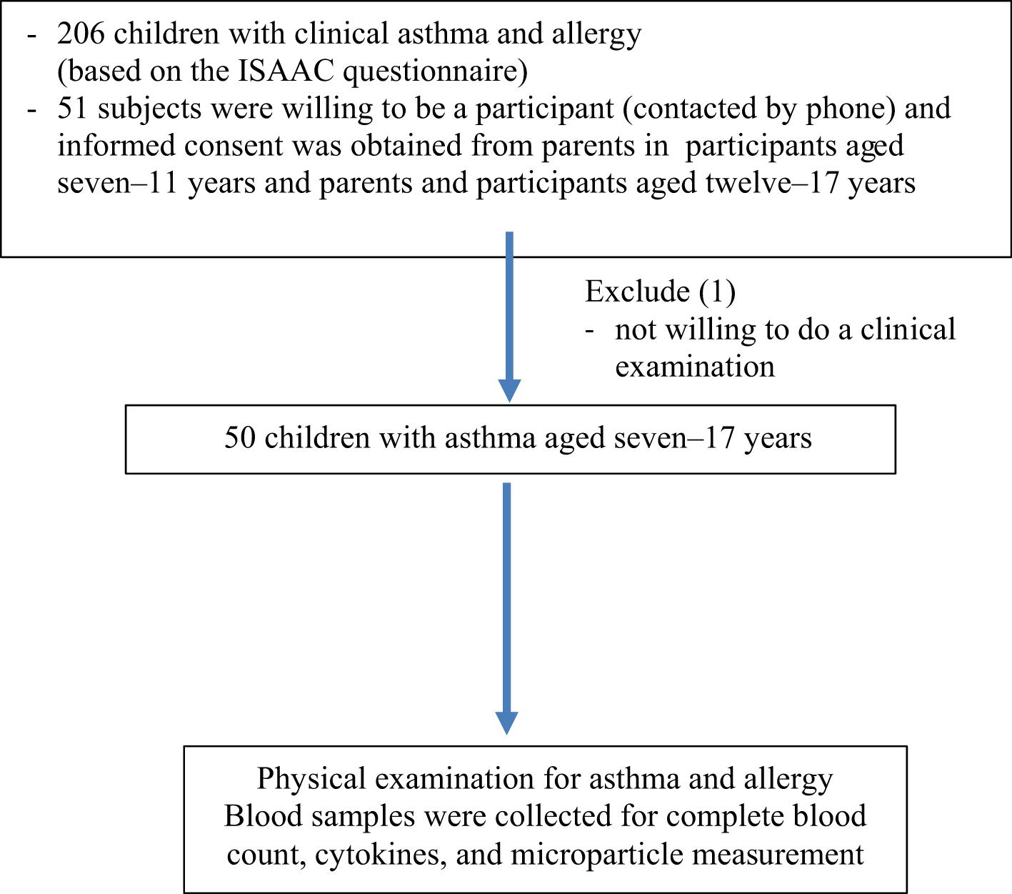
This study was conducted in a government school in Bandung City, West Java Province, Indonesia. A cross-sectional study in children with asthma was conducted from September 2020 to March 2021. The inclusion criteria were children with confirmed asthma aged seven–17 years willing to participate. The exclusion criteria in this study were children who were experiencing asthma exacerbations, inflammation (cough, runny nose, fever, or diarrhoea), and were taking oral or inhaled steroids during the study. The minimum sample size of 49 children was calculated to represent the mean population in the region. The sample was collected based on a purposive sampling procedure resulting in 50 children with asthma. Research subjects with clinical asthma and allergy based on the ISAAC questionnaire27 were contacted about their willingness to participate and written informed consent was signed by the parents for children aged seven–11 years, and informed consent was obtained from adolescents aged twelve–17 years as well as their guardians. The diagnosis of asthma used peak expiratory flow (PEFR) examination to determine the value of reversibility test with an increase in PEFR >15% after 15 minutes of administration of salbutamol 200-400 mcg,28 with the use of an Ultechnovo peak flow meter by a pulmonologist. The spirometry examination was not used in this study to avoid aerosol transmission of viruses during the coronavirus disease 2019 (COVID-19) pandemic. The schematic flowchart for the selected subject is shown in Figure 1.
Clinical data
The PEFR values were determined after three examinations with a maximum difference of 20 points. The highest value was taken, and the PEFR percentage was determined based on Godfrey's nomogram for boys and girls aged five–18 years. The diagnosis and severity of asthma were conducted based on the National Guidelines for Paediatrics Asthma (Pedoman Nasional Asma Anak/PNAA) from The Indonesian Paediatrics Association (IDAI),3 and Global Initiative for Asthma (GINA).1 Nutritional status was measured based on body mass index (BMI) based on the the World Health Organization (WHO) child growth standard (WCGS) using Z-score.
To obtain platelet-free plasma (PFP), MPs were isolated from 3 ml blood samples added with 3.2% sodium citrate, followed by centrifugation at 1,500 rpm for 15 minutes at room temperature, then at 14,000 rpm for two minutes at room temperature. The PFP was centrifuged again at 4,000 RPM for 20 minutes at 4°C to obtain pellets of MPs.29 The MP levels in plasma can be determined using standard beads (YG). The examination was carried out in the Clinical Pathology study laboratory of Cipto-Mangunkusumo Hospital, Jakarta, Indonesia.
100 μL aliquot samples were stained. Two reagents from different antibody combinations were examined, namely PE mouse anti-human CD31-phycoerythrin (PE, clone MBC 78.2 or PECAM-1,2:1,2, 5μl/test) and PE-CyTM 5 mouse anti-human CD62E (clone68-5H11, 20μl/test) were obtained from Becton Dickinson Biosciences (BD Biosciences, San Diego, CA, USA). 10 μL aliquots were stained and added to the bead containing TruCountTM BD (catalogue number 340334). The total volume was obtained from the addition of buffer and double filter and analyzed on the BD Facs Calibur (BD Biosciences). MP size gate range was set between 1 μm and 3 μm calibration (Spherotech, Chicago) by flow cytometry and considered EMPs when they were less than 1.0 μm in diameter. Positive and negative isotypes were used as controls.
Absolute count of EMPs (μL) was determined from the following formula: (number of events in the quadrant containing cell population) / (number of events in absolute count bead region) x total number absolute count beads (47,150 or 47,500) /test volume 100μL. The ratio of CD62E+/CD31+ EMP population rather than absolute count, was described as a criterion for distinguishing activation versus apoptosis. A ratio ≥10 identified activation while a ratio ≤1.0 identified apoptosis.30
The enzyme-linked immunosorbent assay (ELISA) method was used to examine pro-inflammatory cytokines (IL-6 and TNF-α). The IL-6 concentration was based on the standard curve obtained from the assay procedure according to the Quantikine Elisa human R & D system, USA, while the TNF-α standard curve was obtained from the assay procedure according to Elabscience, USA.
Complete blood count examination was carried out directly from 3 mL of blood sample plus EDTA anticoagulant using automated haematology analyzers; the differential blood count was also measured to determine the percentage of eosinophils in allergic asthma.
The data was analyzed using the IBM Social Science Statistics Package v.20.0 (IBM Corp, Armonk, USA). The numeric variables were examined for a normal data distribution using mean median difference, standard deviation, skewness, kurtosis, and Kolmogorov-Smirnov test. Descriptive data were presented as the mean and standard deviation for numerical data, whereas categorical data were presented as number and percentage. The correlation test between two variables was analyzed using Spearman Rho analysis. The Structural Equation Modelling (SEM) analysis for regression coefficient (p-value) using JASP statistical software version 0.16.1 (Department of Psychological Methods University of Amsterdam, Amsterdam, The Netherlands, https://jasp-stats.org/).
Of a total of 206 children aged seven-17 years who were clinically diagnosed with asthma and allergy in the study region, the parents of 51 children (24.8%) agreed to the test, although one child was not ready for the physical and laboratory examination. Of the remaining 50 children with asthma, most were male, the mean age was 12 ± 2 years, with normal nutritional status and intermittent asthma (Table 1 presents characteristics and clinical features of the research subjects).
Our study also found that most asthmatic children with a history of allergy (76%), no history of rhinitis (60%), had a mean PEFR % of 75 ± 12 liter/minute.
Based on laboratory examination, results found that mean leucocyte, platelet and other differential counts were within normal limits, while mean eosinophil counts were increased (7 ± 4, %). Mean pro-inflammatory cytokine TNF-α counts were 4.2 ± 2.8, and IL-6 was 1.6 ± 1 (pg/mL). Results of this study also showed that in the circulation of children with asthma who were found to have EMPs, the mean levels of CD31+/CD62E+ were higher than CD31+/CD62E- and CD62E+/CD31- EMP, and the average ratio of CD31+/CD62E+ ≤1.0 indicated apoptosis (as shown in Table 2).
Our results also found a significant correlation between the level of TNF-α with CD31+/CD62E+, CD31+/CD62E- EMP (p = 0.001, r = 0.5; p = 0.02, r = 0.3), showing that an increase in TNF-α would be accompanied by an increase in EMPs. Our study also found a significant correlation between the percentage of neutrophils and the level of IL-6 (p = 0.002, r = 0.4), suggesting that an increase in neutrophils would be accompanied by an increase in IL-6.
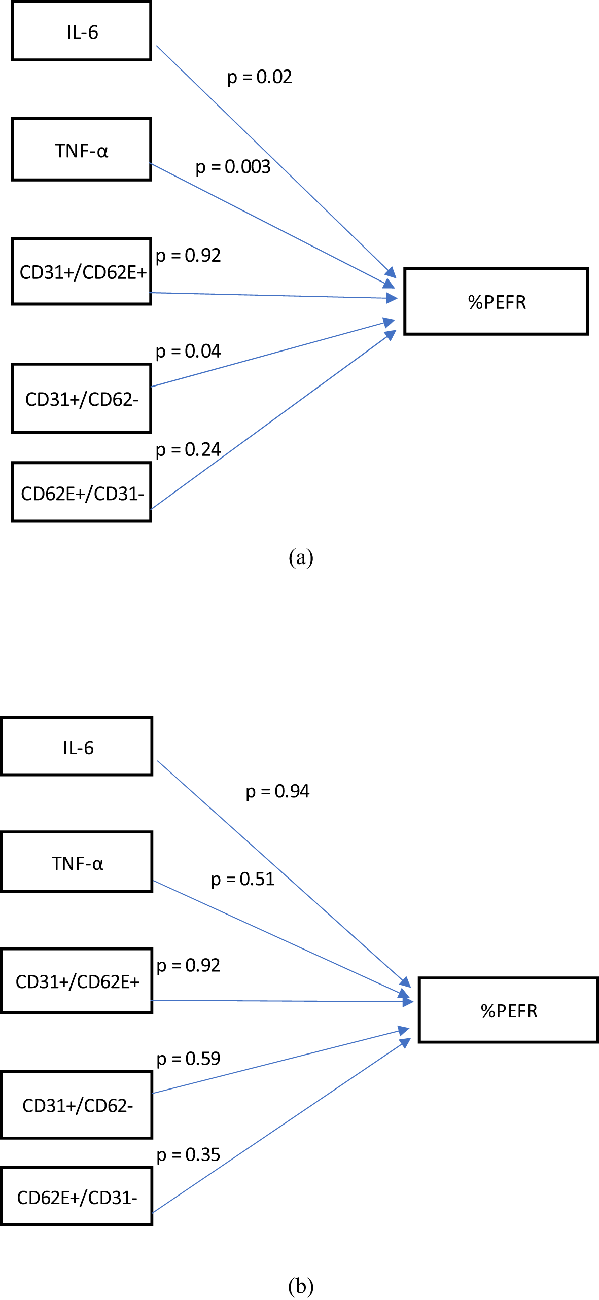
To evaluate which inflammatory factors and EMPs play a role in the PEFR in allergic asthma (38 children) and without allergic asthma (12 children), a SEM analysis was done. Regression coefficient results of the SEM analysis found that TNF-α (p = 0.003), IL-6 (p = 0.02), and CD31+/CD62E- MP (p = 0.04) had an effect on the PEFR in allergic asthma children, compared to TNF-α (p = 0.51), IL-6 (p = 0.94), and CD31+/CD62E- (p = 0.59) in children without allergic asthma. In asthmatic children without allergies, none of the pro-inflammatory cytokines and EMPs affected the PEFR (Figure 2a and 2b).
Previous research in animal models and humans found a higher chance of atherosclerosis event and mean carotid artery intima-media thickness (IMT) in an adult with asthma. To our knowledge, few studies investigating EMPs as a biomarker of endothelial dysfunction in children with asthma, and the results are still controversial. The results of our study show that in the circulation of children with asthma were found have CD31+/CD62E+, CD31-/CD62E+, and CD62E+/CD31- EMPs, with the mean levels of CD31+/CD62E+ were higher than CD31+/CD62E- and CD62E+/CD31- EMPs.
In children with asthma, the levels of CD31+/42b+ and CD31+/42b+/AnV+ platelet MPs were significantly higher even after being analyzed with other confounding factors. The level of CD31+/42b-/AnV+ EMPs (apoptotic EMP) increased significantly but became insignificant after multivariate analysis with other risk factors31; from the results of our study, the average ratio of CD31+/CD62E+ was found to be ≤1.0, indicating apoptosis. Similarly, previous research in diabetic patients found that the ratio of CD62E/CD31 EMP populations reflected an apoptotic process.30 It is known that two cellular processes can trigger the formation of MPs, namely chemical, physical activation, and apoptosis.32 EMPs are small vesicles from activated or apoptotic endothelial cells and are involved in cellular cross-talk mechanism.30
Shear stress, cytokines, thrombin, reactive oxygen species (ROS), oxidized low-density lipoprotein, C-reactive protein, plasminogen activator inhibitor, lipopolysaccharide can induce endothelial cells to release microparticles into the circulation. The release of MPs has detrimental effects such as endothelial activation, arterial stiffness, thrombosis, and inflammation.33 In our study, increased levels of TNF-α were accompanied by increased levels of CD31+/CD62E+ and CD31+/CD62E- EMP. Similarly to previous research, it was found that endothelial cell activation with TNF-α increases the production of CD62E, CD54, and CD106. Production of CD31, CD105, and CD144 were increased in endothelial cells undergoing apoptosis. The CD31 (PECAM-1) release tends to be stimulated by apoptosis of damaged endothelial cells.22
Asthma is also characterized by an increase of circulating pro-inflammatory and Th2 cytokines, indicating that blood vessels are exposed to the inflammation in the lung.34 The best-known phenotype of allergic asthma is caused by an immunological response driven by Th2. Th2 has controversy in relation to its association with atherosclerosis-related CVD in asthma, and classically Th1 has dominated pathologies.34 Our results found proinflammatory cytokine (IL-6 and TNF-α), and CD31+/CD62E- EMPs played a role in PEFR in children with allergic asthma. Similarly to previous research comparing FEV1 with endothelial function in children with asthma, stiffness of vasculature was found to be inversely proportional to FEV1 in children with asthma.35
Interestingly, a higher level of IL-6 may lead to CVD comorbidity in asthma. Other cytokines such as TNF-α have been implicated in both asthma and CVD, at first appears to be a potential target for further study in shared dysregulation in these two pathologies.34 The inflammation mechanism that can result in arterial injury and increased CVD risk are not been understood in asthma and atopic disease.14 Inflammation plays a role in both atherosclerosis and asthma; potential targets are cytokines whose dysregulation have a notable effect in both conditions.34
The current concept of the pathogenesis of asthma is a characteristic chronic inflammatory process involving the walls of the airways, increasing airway reactivity and causing airflow limitation. The hyperreactivity predisposes to narrowing of the airways in response to various stimuli.3,36,37 EMPs are described as 0.1 to 1.0 𝜇m vesicle-like structures released from endothelial cell activation or apoptosis. Endothelial MPs have physiological and pathological effects and may activate oxidative stress and vascular inflammation, released by inducers like angiotensin II, lipopolysaccharide, and hydrogen peroxide, leading to the progression of atherosclerosis.24 Therefore, other inflammatory cells and mechanisms also participate in the pathogenesis of both asthma and atherosclerosis diseases.25
Some limitations of our study are that as an observational study, the described association do not confirm causation. We did not measure lung function parameters from spirometry examination to prevent the spread of aerosols during the COVID-19 pandemic, so the severity of asthma could not be determined accurately. It is essential to investigate further other factors affecting the number of EMPs, such as asthma control, lung function, diet, and environmental conditions; these factors may be associated with endothelial dysfunction, which is a hallmark of the process of atherosclerosis in future studies.
EMPs affect the PEFR in children with allergic asthma. Few studies have investigated EMPs as a biomarker of endothelial dysfunction in children. Further study is needed to investigate the role of these biomarkers in the mechanism of atherosclerosis progression at different asthma severities and with a large number of subjects.
Figshare: Datasheet_Characteristic_Lisa Adhia Garina.csv, https://doi.org/10.6084/m9.figshare.1938256738
Figshare: Datasheet lung function and asthma severity_Lisa Adhia Garina, https://doi.org/10.6084/m9.figshares1938262139
Figshare: Datasheet laboratory examination of research subject-Lisa Adhia Garina, https://doi.org/10.6084/m9.figshare.19394627.v240
Figshare: Datasheet Elisa absorbance data-Lisa Adhia Garina, https://doi.org/10.6084/m9.figshare.19640478.v241
Figshare: Datasheet flow cytometry-Lisa Adhia Garina, https://doi.org/10.6084/m9.figshare.19640493.v142
Figshare: Figure standard curve IL-6_ Lisa Adhia Garina, https://doi.org/10.6084/m9.figshare.19640499.v143
Figshare: Figure standard curve TNF alpha-Lisa Adhia Garina, https://doi.org/10.6084/m9.figshare.19640514.v244
Figshare: Godfrey’s nomogram, http://doi/10.6084/m9.figshares1966882245
Data are available under the terms of the Creative Commons Attribution 4.0 International license (CC-BY 4.0).
| Views | Downloads | |
|---|---|---|
| F1000Research | - | - |
|
PubMed Central
Data from PMC are received and updated monthly.
|
- | - |
Provide sufficient details of any financial or non-financial competing interests to enable users to assess whether your comments might lead a reasonable person to question your impartiality. Consider the following examples, but note that this is not an exhaustive list:
Sign up for content alerts and receive a weekly or monthly email with all newly published articles
Already registered? Sign in
The email address should be the one you originally registered with F1000.
You registered with F1000 via Google, so we cannot reset your password.
To sign in, please click here.
If you still need help with your Google account password, please click here.
You registered with F1000 via Facebook, so we cannot reset your password.
To sign in, please click here.
If you still need help with your Facebook account password, please click here.
If your email address is registered with us, we will email you instructions to reset your password.
If you think you should have received this email but it has not arrived, please check your spam filters and/or contact for further assistance.
Comments on this article Comments (0)