Keywords
Bone defect, SVF, Scaffolds; Osteocalcin, BMP-2, In-vivo study
This article is included in the Cell & Molecular Biology gateway.
Bone defect, SVF, Scaffolds; Osteocalcin, BMP-2, In-vivo study
Bone is naturally regenerable, with a high ability to repair itself, especially in young individuals; this means that most fractures or bone deformities recover spontaneously without requiring a significant intervention.1,2 Despite bones having some capacity for healing and regeneration, massive segmental bone defects cannot be repaired independently.3 Therefore, for bone defect conditions, it is necessary to give a bone graft to promote the healing process.4 In humans, a bone defect occurs when there is a loss of bone components larger than 1 cm in length which contributes to more than 50% of the circumference of the affected bone.5 In contrast, in animal research objects such as rats, a bone defect is considered when there is a loss of bone components up to 3 mm.5
Various bone grafts have been employed to correct these defects, including autografts, allografts, and synthetic grafts.6 To date, autografts are the gold standard for bone grafts due to their histocompatibility and low risk of hypersensitivity reactions.7,8 Moreover, autografts also provide the essential parts for osteoconduction (i.e., osteoprogenitor cells, three-dimensional and porous matrix), osteogenesis (i.e., osteoprogenitor cells), and osteoinduction (i.e., bone morphogenetic proteins [BMPs] and other growth factors). Although autografts are a popular graft strategy, they have certain disadvantages, such as donor site morbidity, muscle weakening, risk of infection, bleeding, nerve damage, and function loss.9–11 Another choice for bone substitution is an autologous bone graft.12 However, the availability of suitable bone is restricted; moreover, harvesting a bone graft is challenging, with a high risk of disease transmission and relatively high cost.13,14
Therefore, in an effort to develop alternative treatments for correcting bone defects and their consequences, bone tissue engineering (BTE) has gained popularity and is nowadays being researched as a potential alternative in bone defect management.15,16 BTE approach is perceived as a preferable solution for bone defect condition due to the healing process being facilitated with the patient’s own tissue and provide a good healing process15,17,18 The BTE theory involves the integration of several collaborating elements: stem cells held together by a three-dimensional biomaterial framework that gives shape and initial mechanical strength, and molecular signals that stimulate progenitor cell differentiation into the osteoblastic phenotype.19 It can be concluded that there are three combined fundamental components in BTE: biomaterials (scaffolds), mesenchymal stem cells (MSCs), and growth factors.20,21
In common tissue engineering procedures, a framework is needed to guide the formation of the tissue formation called a “scaffold”.21,22 Scaffolds are made from synthetic or natural biomaterials that facilitate the proliferation, migration, and differentiation of bone cells for bone repair. Scaffolds themselves are made of a variety of biomaterials and synthetic bone substitutes, including collagen, hydroxyapatite (HA), b-tricalcium phosphate (b-TCP), calcium-phosphate cement, as well as glass-ceramics and bovine HA.23 Other components in BTE include stem cells and growth factors, where both of these components can be found in adipose tissue. Adipose tissue is a multifunctional structure consisting of various cell types, including stromal vascular fraction (SVF) and mature adipocytes. Adult stem cells are abundant and easily extracted from adipose tissue compared to the umbilical cord or bone marrow. The combination of these components is believed to help the healing process of bone defects.24–26 Therefore, this study aimed to observe the effect of bone tissue engineering (SVF and scaffold) in the bone defect healing process based on levels of osteocalcin and BMP-2 in vivo.
This research was performed from March to December 2020. All protocols were approved by the Ethical Committee of Medical Research Faculty of Medicine Universitas Brawijaya with approval number 160/EC/KEPK – PPDS/09/2020, and all subsequent experimental studies followed the ARRIVE guidelines. All animals were housed in certified vivariums under standard procedures with gentle handling, daily cage cleaning, and regular monitoring to avoid animal suffering.
This study was an in vivo experimental research with a randomized posttest-only control group design. The parameters measured in this study resulted from the authors’ intervention. The research started by identifying rats that had finally passed the inclusion and exclusion criteria. The inclusion criteria for this study consisted of male Wistar rats aged three months, or twelve weeks, with a body weight of 200-280 grams, who were fit, active, clear of limb abnormalities, and had no history of therapy or chemical administration.
After identifying the 20 experimental animals that met the inclusion criteria, we used the Federer formula to determine the sample size. Then, we used simple random sampling for each group. After identifying the 20 experimental animals that met the inclusion criteria, these rats were separated into five groups consisting of four rats. The groups were categorized as follows: (1) was a negative group that consisted of normal rats without critical bone defect and without SVF or scaffold application; (2) was a positive group which were murine models with bone defect and without SVF or scaffold application. These two groups were control groups. Then, for an experimental group, we divided into three groups: group (3) was named K-P1: murine models with bone defect and giving porous-carbonated HA application; group (4) was named K-P2: murine models with bone defect and treated with nanocrystalline HA applications; and group (5) was K-P3: murine models with bone defects and treated with bovine HA application. These five groups were followed for 30 days to evaluate osteocalcin and BMP-2 biomarker levels.
Five 12-week-old male Wistar strain rats were sacrificed by cervical dislocation procedure. We then put the rats in a supine position. A broad and longitudinal skin incision was done to expose the abdomen of rats. Then, the testicles and the fat surrounding the epididymal and perirenal fat pad were extracted. By severing the innervation of the retroperitoneal fat pad, adipose tissue from perirenal fat was harvested from the epididymal and perirenal fat pad for collection.
After adipose tissue was obtained, the harvested adipose was washed using a Phosphate-buffered saline (PBS, Sigma-Aldrich, Germany) solution containing a 10% antibiotic-antimycotic solution. The adipose tissue was mashed with a knife. It was then incubated for 30 minutes at 37°C in a 0.075 percent type IA collagenase combination (Sigma-Aldrich) and PBS. After processing the tissue, it was filtered using a 100 mm mesh strainer (Sigma-Aldrich) and centrifuged at 1200 rpm for 10 minutes at 20 °C. The supernatant was removed, leaving a heterogeneous cell suspension with an estimated 2 × 10⁶ cells per gram of adipose tissue
Scaffolding is classified into synthetic porous carbonated-HA, nanocrystalline-HA, and bovine xenograft-HA. These three scaffolds are commonly available at Saiful Anwar hospital, making them accessible. In addition, this scaffold is frequently utilized in other research. These three scaffolds were then administered to fractures with bone defects using a measuring spoon to ensure that each mouse model received an equivalent dose.
After seven days of acclimatization, rats in the positive and intervention groups received a bone deformity. Rats were sedated with 100 mg/kg ketamine injection and 10 mg/kg xylazine hydrochloride intraperitoneally preoperatively. The authors confirmed that rats were sedated by extending the extremities and pinching the web between the toes using the pedal reflex technique. If the rat retreated or twitched a muscle and made a noise, the anesthetic was insufficient. After that, they were given an antibiotic injection of 20 mg/kg Cefazolin in the right leg. Then, the operating area was shaved and disinfected with chlorhexidine. The murine was positioned prone on the surgical table and incised over 3-4 cm, gradually deepening the incision until the bone was visible. Osteotomy was performed with a 3mm Kerrison, resulting in a 3mm broad bone defect. After that, the intervention was carried out according to the designated groups. Finally, plaster of Paris was put from the proximal femur to the ankle, with the knee in 90 degrees of flexion. Analgesia was supplied every eight hours (IM 5 mg/kg Ketorolac), and antibiotics, i.e., 20 mg/kg cefazoline were administered intramuscularly 24 hours after surgery. Monitoring was conducted on a daily basis for 30 days.
Murine models were sacrificed after 30 days. We collected and then extracted the area of bone defect with callus formation. Osteocalcin and BMP-2 levels were determined using the ELISA technique.27
The first steps of the hypothetical comparative test are followed by the data normality test and variant homogeneity test. If the data collected was homogenous and normally distributed, we used ANOVA. However, if these two criteria are not fulfilled, we used a non-parametric Kruskal-Wallis test with a confidence interval of 95%. After the hypothesis test was performed, we conducted a post hoc test to evaluate the significant difference in each group. The resulting test was significant when p<0.05
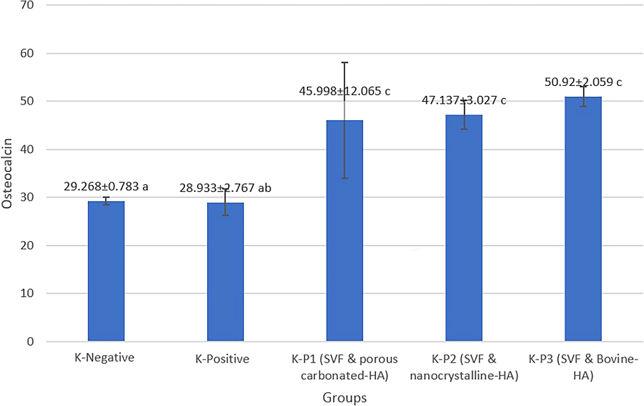
In this research, we used osteocalcin and BMP-2 levels to assess the outcome of using bone tissue engineering (SVF and scaffold) in experimental animals. Osteocalcin and BMP-2 were measured using ELISA, and the results are depicted in Table 1.
| Treatment groups | Mean±SD osteocalcin | Mean±SD BMP-2 | p-value |
|---|---|---|---|
| K-negative | 29.268±0.783 | 224.504±65.686 | p: 0.006a |
| K-positive | 28.933±2.767 | 230.592±38.624 | |
| K-P1 (SVF and porous carbonated-HA) | 45.998±12.065 | 313.337±56.372 | p: 0.001b |
| K-P2 (SVF and nanocrystalline-HA) | 47.137±3.027 | 402.528±122.479 | |
| K-P3 (SVF and bovine-HA) | 50.92±2.059 | 491.572±88.597 |
In this study, we found that the mean level of osteocalcin using bone tissue engineering (SVF and scaffold) was higher when compared to either the positive or negative control groups. The group with the highest osteocalcin level after 30 days was the KP-3 group, which contained SVF and bovine HA, with a mean osteocalcin level of 50.92±2.059 ng/ml while the lowest was KP-1 which contained SVF and porous carbonated-HA (45.998±12.065) ng/ml. Then, we performed a statistical Kruskal-Wallis test; it was found that the significance test result was less than 0.05. Therefore, it can be concluded that applying SVF and scaffolds has a significant effect on osteocalcin levels (Figure 1).
Then, for further comprehension of the difference between in each intervention, the experiment processed to the post hoc test next. From the results of the post hoc test, a significant relationship between positive control and SVF and bovine-HA was found (p=0.006), as well as between negative control and treatment with SVF and bovine HA (p=0.003); therefore, it can be concluded that the use of bone tissue engineering using SVF and bovine HA can significantly increase the level of osteocalcin in experimental animals when compared to the negative and positive groups (Table 2).
| Treatment groups comparison | p-value osteocalcin levels | p-value BMP-2 levels |
|---|---|---|
| K-Negative-K-Positive | 0.811* | 1* |
| K-Negative-K-P2 (SVF & nanocrystalline-HA) | 0.031* | 0.534* |
| K-Negative-K-P1 (SVF & porous carbonated-HA) | 0.017* | 0.044+ |
| K-Negative-K-P3 (SVF & Bovine-HA) | 0.003+ | 0.002+ |
| K-Positive-K-P2 (SVF & nanocrystalline-HA) | 0.046+ | 0.050+ |
| K-Positive-K-P1 (SVF & porous carbonated-HA) | 0.031+ | 0.045+ |
| K-Positive-K-P3 (SVF & Bovine-HA) | 0.006+ | 0.003+ |
| K-P2 (SVF & nanocrystalline-HA)-K-P1 (SVF & porous carbonated-HA) | 0.811* | 0.53* |
| K-P2 (SVF & nanocrystalline-HA)-K-P3 (SVF & Bovine-HA) | 0.403* | 0.044+ |
| K-P1 (SVF & porous carbonated-HA)-K-P3 (SVF & Bovine-HA) | 0.55* | 0.532* |
Furthermore, we evaluated BMP-2 levels in rats with bone defects using bone tissue engineering. It was found that the outcomes of applying bone tissue engineering using SVF and bovine HA (KP-3) led to a significantly higher BMP level (491,572±88,597) pg/ml, more than the results of the other groups. Moreover, the results of the Kruskal-Wallis test proved that there was a significant difference between groups for this marker (p=0.001). In comparison, the lowest concentration of BMP-2 was found in KP-1, which was treated with SVF and porous carbonated HA (313,337±56,372) pg/mL. This finding was similar to the results obtained in the previous osteocalcin level, which was highest for KP-3 and lowest for KP-1 compared to other experimental groups (Figure 2).

We also performed a post hoc test to determine the differences between each group. The post hoc test revealed a significant relationship between positive control and SVF and bovine HA (p=0.003) and a relationship between negative control and SVF and bovine HA (p=0.002) (Table 2).
One of the most extraordinary characteristics of bone is its remarkable capability to recover with almost minimal scarring.7 However, when bone defects reach a critical size, disruptions at the fracture site may affect the repair process. This condition results in nonunion healing.19 Because of so many restrictions associated with bone reconstruction using autografts or bank bones, researchers have explored other methods for bone repair. Recent tissue engineering techniques have regenerated bone by putting MSCs and growth factors contained in SVF from adipose tissue onto porous ceramic scaffolds.
Adipose tissue has long been viewed as useless tissue, and for many years, fat tissue has been regarded as “waste material” in surgical treatments.28 Hollenberg initially described SVF in 1960, and in 2001 SVF was discovered to contain a large number of MSCs.28,29 SVF have many benefits promoting bone healing. These advantages include, first, that fat tissue is relatively simple to extract which may minimize patient discomfort. Second, tissue extraction processes from adipose tissue to SVF with significant number of MSCs can be completed quickly; third, the multipotent cells contained in SVF can adhere quickly to the scaffold material, multiply rapidly, and differentiate into osteogenic elements.30,31 Not only does it have the capability to assist the healing process of bone defects, SVF also can prevent bone bridge formation on growth plate injury, which can be useful for growth plate injury cases. This has been evidenced in our previous studies32
Scaffolds have been developed over the last few decades; they provide a three-dimensional structure for cell support.33 Thus, they have the potential to dictate cell-specific features via the release of numerous growth factors, as well as surface binding and physicochemical proportions. Furthermore, Scaffolds or bone grafts are used to enhance or induce bone formation for fixing bone fractures or connecting two bones along a diseased joint, to replace and regenerate lost bone as a result of trauma, infection, or disease, or to improve the bone healing response and regeneration of bone tissue surrounding surgically implanted devices, such as artificial joints replacements or plates and screws used to maintain bone alignment.16,34
Several studies have reported that SFV and scaffolds lead to promising outcomes for stimulating bone repair in bone defects.13,14,18,19 In this study, we compared the outcome of combining SVF with various types of scaffolds in vivo to assess their effectiveness. We used combinations of SVF and porous carbonated HA, SVF and nanocrystalline HA, last, and SVF and bovine HA. Interestingly, we found the combination of SVF and bovine-HA showed a significantly higher activity of osteocalcin and BMP-2 than other combinations. Moreover, it has been reported previously that bovine HA has a lower toxicity effect than other types; this was confirmed in a study conducted by Kamal et al., who stated that Scaffold IV (bovine HA granule) had the least toxic effect on rat bone marrow.35
In addition, in this study, we used BMP-2 levels to evaluate bone healing, as BMPs levels are widely considered as the most effective group of growth factors for helping in the healing of major bone lesions. However, only BMP-2 has been demonstrated to be required for the osteogenic process, and both BMP-2 and BMP-7 have been approved for clinical usage in the treatment of significant bone abnormalities.36
Bone remodeling is a complex process involving a variety of cellular and molecular events. Bone cells collaborate with other cells to promote the healing process.37 MSCs can be administered in combination with a scaffold to increase bone formation in vivo; autologous MSCs have been effectively used in combination with a HA-based scaffolds to repair critical-sized and bone defects in vivo.38 In another study, Roato, 2018 compared SVF and adipose tissue-derived stem cells (ASCs) combined with bovine scaffold. From the results of the study it was found that the administration of SVF and bovine scaffold had better osteoinductive abilities than ASCs.39 Therefore, the use of SVF in combination with bovine scaffold has a good potential for promoting bone healing in bone defects cases. This theory is relevant to this study, as the combination of SVF and bovine HA scaffold had higher levels of osteocalcin and BMP-2 when compared to the control groups.
The application of SVF (stem cells) and scaffolds enhances the activity and efficiency of the bone healing process, because SVF containing progenitor cells synergizes with the scaffold, which provides a site for colonization of progenitor cells and growth factors such as transforming growth factor (TGF), insulin-like growth factor 1 (IGF1), and platelet-derived growth factor (PDGF, insulin). In addition to fibroblast growth factor 1 (FGF1), FGF2, and PDGF, scaffolds have a role in filling bone defects. The bone healing process becomes more effective when the procedure described previously is used.
We suggest further studies to use a different fixation, such as external or internal fixation, to better aim the study towards various modalities used in orthopedics fields. Different biomarkers could also be assessed in future studies, such as alkaline phosphatase (ALP), osteopontin bone marker, type II collagen among others, as well as from a histological standpoint.
From this study, it can be concluded that the application of Bone Tissue Engineering (SVF and scaffolds) could enhance the healing process in murine models with bone defect, marked by increasing levels of osteocalcin and BMP-2 as bone formation markers.
Zenodo: Bone Tissue Engineering Application on Fracture Healing with Bone Defect as Assessed Through Osteocalcin and Bone Morphogenetic Protein-2 (BMP-2) Biomarker Examination: Experimental Study on Murine Model, https://doi.org/10.5281/zenodo.6361033.40
This project contains the following underlying data:
Data are available under the terms of the Creative Creative Commons Attribution 4.0 International license (CC-BY 4.0).
Zenodo: Bone Tissue Engginering Application on Fracture Healing with Bone Defect as Assessed Through Osteocalcin and Bone Morphogenetic Protein-2 (BMP-2) Biomarker Examination: Experimental Study on Murine Model, https://doi.org/10.5281/zenodo.6361033.40
This project contains the following reporting guidelines:
Data are available under the terms of the Creative Commons Attribution 4.0 International license (CC-BY 4.0).
| Views | Downloads | |
|---|---|---|
| F1000Research | - | - |
|
PubMed Central
Data from PMC are received and updated monthly.
|
- | - |
Provide sufficient details of any financial or non-financial competing interests to enable users to assess whether your comments might lead a reasonable person to question your impartiality. Consider the following examples, but note that this is not an exhaustive list:
Sign up for content alerts and receive a weekly or monthly email with all newly published articles
Already registered? Sign in
The email address should be the one you originally registered with F1000.
You registered with F1000 via Google, so we cannot reset your password.
To sign in, please click here.
If you still need help with your Google account password, please click here.
You registered with F1000 via Facebook, so we cannot reset your password.
To sign in, please click here.
If you still need help with your Facebook account password, please click here.
If your email address is registered with us, we will email you instructions to reset your password.
If you think you should have received this email but it has not arrived, please check your spam filters and/or contact for further assistance.
Comments on this article Comments (0)