Keywords
Adrenocortical carcinoma, Hypokalemia, Hypertension, Aldosterone/Renin ratio
This article is included in the Oncology gateway.
Adrenocortical carcinoma, Hypokalemia, Hypertension, Aldosterone/Renin ratio
Adrenocortical Carcinoma is a cancerous tumor. In 25% of seemingly spontaneous ACC, somatic mutations in the tumor-suppressor gene TP53 are detected. The Li-Fraumeni syndrome is linked to a variety of solid organ malignancies, including ACC, and is caused by TP53 mutations in the sperm. Variations in the Wnt/-catenin system and the insulin-like growth factor 2 (IGF2) clusters are also evident in ACC; IGF2 amplification is reported in 90% of the cases.1
Around 60% of ACCs seem to be adequately secretive in character to manifest as a clinical condition of hormone overabundance.2 Cushing's syndrome (45 percent) or a combination Cushing's and virilization syndrome (oversaturation of both glucocorticoids and androgens) are the most common symptoms in adults with hormone-secreting ACCs (25 percent).3 Cushing's syndrome, virilization, hypertension, hypokalemia, abdominal discomfort, and vomiting are examples of clinical symptoms linked with hormone imbalances. Diagnosis is done by radio-imaging (CECT abdomen and pelvis) or MRI abdomen and pelvis for the size of tumor and local invasion as well as metastasis. The size of the adrenal tumor at its widest point indicates if it is cancerous. Adenomas of the adrenal glands typically have a dimension smaller than 4 cm. When an ACC is detected, it usually has a diameter of larger than 4cm. FNAC cannot differentiate benign from malignant.
We should always rule out pheochromocytoma by using serum and urinary catecholamines before surgery and biopsy. For adrenocortical cancer (ACC) stages I-III, total surgical removal is the only possible therapeutic strategy.4
A 40-year-old serving soldier who has history of hypertension presented initially with decrease in appetite and loss of weight of around two to three kg over 30 days. He had been vomiting for two days about 3-4 episodes, non-projectile, and non-bloody. He also complained of generalized weakness which started after vomiting, however, he could walk and perform daily activities. He had a past history of hypertensive urgency with multiple hospital admissions. Fundoscopy revealed grade III hypertensive retinopathy changes.
His blood pressure was 170/100 mmHg and pulse rate was 56 beats per minute and it was regular, respiratory rate was 18/min, saturation was 96% at room air. His systemic examinations were unremarkable. His initial hematological, renal, and liver function parameters were in the normal range but serum sodium was 146mmol/l and potassium was 2.0 mmol/l. His USG imaging and X-ray chest were unremarkable.
He was admitted for evaluation of hypokalemia. His antihypertensive medications were optimized and started on Losartan, amlodipine, Beta-blocker, alpha-blocker, and diuretics (spironolactone). An extensive evaluation was done to find the cause of hypokalemia. His 24 hours urine analysis revealed potassium 18 meq/dl, osmolality 300 mosm/l, and sodium 54 mmol/dl. His serum calcium was 8.4 mg/dl and Phosphate 4.1mg/dl. The transtubular potassium gradient (TTKG) was 15 (which was 3.5 times normal levels). His aldosterone renin ratio was increased and urinary catecholamines levels were normal as shown in Table 1 and Table 2.
| Result | References | |
|---|---|---|
| Serum Aldosterone | 535 ng/dl | Supine <16 ng/dl, upright 4-31 ng/dl |
| Serum Renin, direct | 0.50 mcgU/ml | Supine 0.2-1.2, upright 0.5-4.0 |
| Aldosterone/Renin Ratio (ARR) | 1070 ng/dl/mcgU/ml | <25 |
The contrast-enhanced CT abdomen showed well defined, smooth marinated, heterogeneously enhancing mass lesion in the left adrenal gland measuring 5.1 cm × 5.9 cm × 5.7 cm with few tiny calcifications in the lesion. The coronal section of the contrast-enhanced CT abdomen is shown in Figure 1 and the transverse section is shown in Figure 2. An overnight dexamethasone suppression test was done, and serum cortisol at 8 AM was 10.61 nmol/l. Color Doppler of bilateral renal arteries showed normal flow. The thyroid function test was normal. ABG showed metabolic alkalosis. Potassium was supplemented from the IV route as well as the oral route and six times hourly potassium was monitored but even after continuous supplement, there was no rise in serum potassium level. Intravenous fluid was given as ½ NS to avoid hypernatremia and serum sodium was monitored along with potassium.

Arrow showing heterogeneously enhancing mass lesion of 5.1 cm × 5.9 cm × 5.7 cm with few tiny calcification in the lesion.

Arrow showing well defined, smooth marinated, heterogeneously enhancing mass lesion.
After confirming our diagnosis of left adrenal incidentaloma with hyperaldosteronism, He underwent laparoscopic unilateral adrenalectomy (left) and intraoperative and postoperative periods were uneventful. The Operated left adrenal mass is shown in Figure 3. Histopathological examination showed atypical cells showing marked pleomorphism exhibiting small cells to giant bizarre-shaped cells indicating adrenocortical carcinoma. The capsular invasion was seen. The immunohistochemistry report is shown in Table 3.
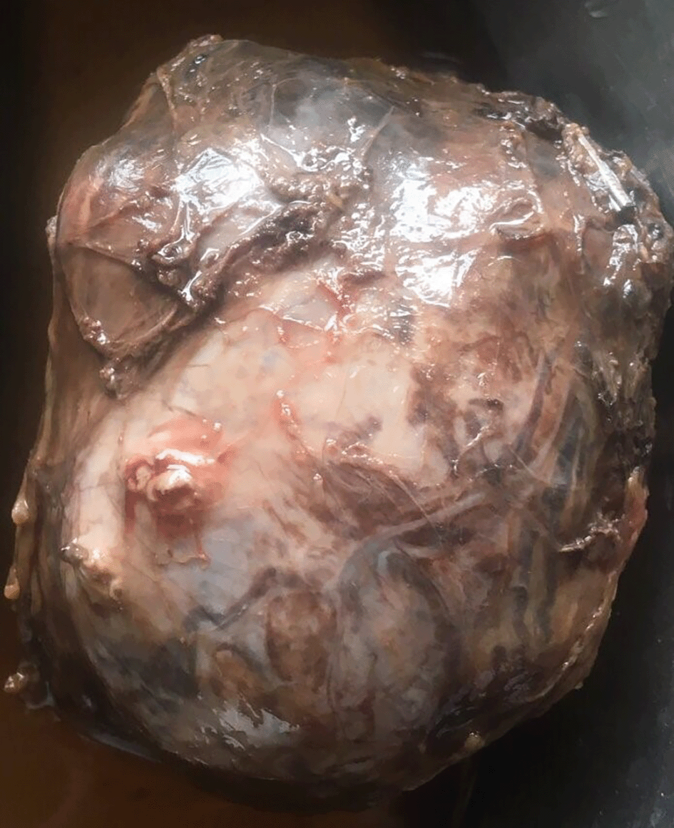
Showing operated left adrenal mass with capsulated, smooth surface, prominent surface vascular marking with maximum diameter of 7 cm and weight of 195 g.
He was discharged with antihypertensive medications (calcium channel blocker and alpha-blocker), analgesics, oral antibiotics planned to tapper off antihypertensive every 2-3 weekly as there is a high chance of normalization of blood pressure.
Adrenocortical carcinoma (ACC) is an uncommon adrenal neoplasm that is regarded as a highly malignant tumor. There are two variants of tumor functional and nonfunctional, functional variants are more common than nonfunctional. Females are generally more affected than males (3:1).5 There are Bimodal peaks, appearing first in the forties and fifties and a second one appearing in their first decade. The prognosis for elderly men is poorer, although younger people with ACC have a better prognosis than adults. The majority of tumors are sporadic in nature.6
ACC can be functional with a sole endocrine condition like Cushing's, or it can be a mixed syndrome with virilization. Virilization is common in youngsters and is associated with a 70% likelihood of cancer. The majority of patients with ACC, particularly those with the non-functional form, have advanced illness with several abdominal or extra-abdominal metastatic masses.7 As a result, tumor diagnosis at an early clinical stage is critical for curative excision.
Adrenal adenoma, Pheochromocytoma, and renal cell cancer are our differential diagnoses. The weight of the tumor distinguishes adenoma from carcinoma; if it weighs more than 95 g, it is typically malignant. Radio-imaging also distinguishes adenoma and carcinoma by the size, carcinoma is usually greater than 4 cm. The tumor in contrast-enhanced CT appeared heterogeneously enhanced mass lesion in the left adrenal gland measuring 5.1 cm × 5.9 cm × 5.7 cm with few tiny calcifications in the lesion. We have Weiss criteria for assessing the prognosis in ACC. If the mitotic figures are less than 20 per HPF, the tumor is low grade; if the mitotic figures are more than 20 per HPF, the tumor is high grade. The tumor is malignant if three of the nine criteria are met.8 We have calculated Weiss criteria which were 4 points out of a total of 9 points (Capsular infiltration, Necrosis, Clear cells <25%, and Mitosis 10/50 HPF).
ENSAT (European Network for the Study of Adrenal Tumors) and the TNM staging system are used for the tumor staging as shown in Table 4. We classified this tumor as stage II (T1N0M0).
The most important differential diagnosis is Pheochromocytoma. A classic zellballen pattern characterized by elevated catecholamine levels in serum and urine, as well as positive for immunohistochemical markers such as chromogranin, will suggest a Pheochromocytoma. Whereas, inhibin, calretinin, and Melan-A, are positive in ACC. Cushing syndrome was ruled by a dexamethasone suppression test. Normal serum cortisol and high aldosterone level ruled out Liddle syndrome, a Syndrome of apparent mineralocorticoid excess (SAME). High serum level of renin is seen in renal artery stenosis (RAS), renin secreting tumor (RST), and malignant hypertension.
We reported a very rare case of adrenocortical carcinoma in an adult who presented with hypokalemia and hypertension and he is living a disease-free postoperative period. Because their prognoses are distinct, it's critical to distinguish it from an adrenocortical adenoma with biochemical, imaging, and histological markers. The most common therapy is surgical resection.
| Views | Downloads | |
|---|---|---|
| F1000Research | - | - |
|
PubMed Central
Data from PMC are received and updated monthly.
|
- | - |
Provide sufficient details of any financial or non-financial competing interests to enable users to assess whether your comments might lead a reasonable person to question your impartiality. Consider the following examples, but note that this is not an exhaustive list:
Sign up for content alerts and receive a weekly or monthly email with all newly published articles
Already registered? Sign in
The email address should be the one you originally registered with F1000.
You registered with F1000 via Google, so we cannot reset your password.
To sign in, please click here.
If you still need help with your Google account password, please click here.
You registered with F1000 via Facebook, so we cannot reset your password.
To sign in, please click here.
If you still need help with your Facebook account password, please click here.
If your email address is registered with us, we will email you instructions to reset your password.
If you think you should have received this email but it has not arrived, please check your spam filters and/or contact for further assistance.
Comments on this article Comments (0)