Keywords
Stiff Skin Syndrome, Case report, Rural tertiary hospital
Stiff Skin Syndrome, Case report, Rural tertiary hospital
Stiff skin syndrome is a rare genetic connective tissue disorder that manifests at birth or in early childhood.1 Patients suffer from rock-hard or rock-hard skin all over their body, with enormous fascia in places like the thighs and buttocks. It causes reduced joint mobility in the affected area and in some cases hypertrichosis.2 In our clinic we present the case of a girl with stiff skin syndrome associated with bone involvement at the femoral head, using the CAse REport (CARE) guidelines as a guiding principle.3
Written informed consent for publication of their clinical details and clinical images was obtained from the parent of the patient. Additionally, ethics clearance for this case report was obtained from the Walter Sisulu University Ethic committee (WSU Ethics approval No: 055/2022).
The patient was a four-year-old South African girl who developed hypertrichosis and skin thickening in the trunk, buttocks (Figure 1), upper and lower limbs in the first year of life (Figure 2). This was associated with arthralgia, or limited mobility, in the left leg. Raynaud’s phenomenon, joint swelling, or pigment changes in the affected area have not been reported. Her developmental milestones were appropriate for her age and she had no neurological symptoms. There was no history of skin diseases, related skin diseases or scleroderma in the patient’s family. She is the result of a singleton conception, a lasting pregnancy, and an uneventful birth and delivery. The patient has no medical or surgical history.
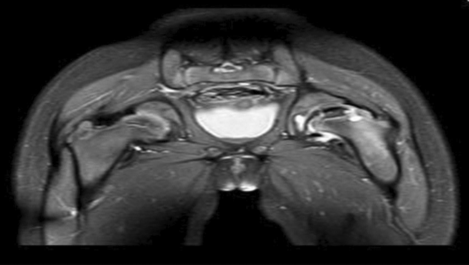
Routine blood tests, imaging, and histology were ordered to confirm the definitive diagnosis. Routine blood tests were requested, including a complete blood count, urea, electrolytes, and liver function tests, all of which were within normal limits. Anti-nuclear antibodies, anti-scleroderma-70, anti-centromere antibodies and anti-ribonucleoprotein antibodies were all tested and all were negative, including the HIV ELISA test. Her chest X-ray and abdominal ultrasound both showed normal results. Magnetic resonance imaging of the patient showed flattening of the upper part of the left femoral head, shortening of the left femoral neck, a fluid-filled metaphyseal cyst with adjacent associated effusion, and mild atrophy of the muscles surrounding the proximal femur (Figure 3).

MRI, magnetic resonance imaging.
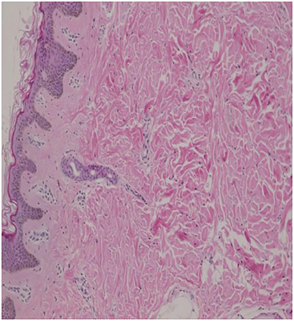
Histopathological examination of a skin biopsy of the lesion revealed epidermal atrophy and orthokeratosis, pandermal fibrosis, and mild loss of perieccrine fat. Within the deep reticular dermis there were increased collagen bundles and slight mucin deposition. There was no evidence of dermal or subcutaneous inflammation (Figures 4 and 5).

Esterly and McKusic1 were the first to describe stiff skin syndrome, also known as congenital fascial dystrophy and mucopolysaccharidosis. It is a rare, genetically inherited connective tissue disease similar to scleroderma. Manifestations of the syndrome typically begin at birth, in infancy or early childhood.2 It is caused by mutations in the ArgGlyAsp (RGD) sequence-encoding domain of the fibrillin-1 (FBN1) encoding gene. As a result of altered transforming growth factor (TGF)4–6 activation and signaling, this promotes profibrotic activation. As a result, the elastic fibers responsible for skin rebound are replaced with fibrotic deposits. Despite cutaneous involvement, the visceral and adjacent muscles are usually unaffected and there are no immunological or vascular changes.7
The exact cause of stiff skin syndrome is unknown. Postulations include abnormal mucopolysaccharide metabolism occurring exclusively in the skin and later thought to be localized in the fascia,8–11 primary fascial dystrophy with consequent collagen overabsorption7 and an inflammatory process.11,12 The two hypotheses that have gained wider acceptance are increased collagen VI production secondary to a primary fascial abnormality and a congenital fibroblastic abnormality leading to non-inflammatory dermal fibrosis due to defective mucopolysaccharide synthesis. In addition, some investigators have found high levels of pro-inflammatory markers such as interleukin-6, transforming growth factor-2 and tumor necrosis factor-alpha, suggesting an inflammatory pathogenesis.7,11,13,14
Symptoms of stiff skin syndrome include a rock-hard deep skin hardening that is inherently connected to the underlying tissue, reduced joint mobility, and hypertrichosis. The buttocks, thigh and shoulder are the most affected body regions, but ocular involvement has been reported in rare cases.12 Diagnosis is clinical and confirmed by histological findings; therefore, a high index of suspicion is required, and the diagnosis is made by exclusion. The disease is divided into two types depending on the manifestation: widespread and segmental. The widespread stiff skin syndrome has an earlier onset, a more severe form, is more common than segmental stiff skin syndrome, and affects the joint with a more restricted range of motion and prominent bilateral involvement. Segmental stiff-skin syndrome is segmentally distributed with unilateral body affectation, has female predominance in contrast to the widespread stiff-skin syndrome, which has no gender predominance and less clinical consequences.13,14
The clinical findings in our patient were consistent with segmental stiff skin syndrome. This is evidenced by the unilateral involvement of the lower extremities, gluteal and trunk muscles, and limited mobility of the left hip joint. Hypertrichosis and a limping gait occurred as follow-up investigations, both of which were clinical findings. Histological findings of orthokeratotic epidermis, pandemic fibrosis, and increased collagen bundles within the deep reticular dermis confirmed the diagnosis. However, our patient has incidental bony involvement, which is rare.
Due to overlapping histopathological features with stiff skin syndrome but with classic features not seen in this patient, the two differential diagnoses of generalized morphea and systemic sclerosis were close. Scleroderma typically affects the head, face, neck, and back, with the shoulder and belt being the exceptions, but when affected, the usual sites are not exempt. Joint restriction is uncommon in scleroderma, and when skin stiffness occurs, it is due to the mass effect and typically affects an entire anatomical unit. Generalized morphea also occurs suddenly, in contrast to stiff skin syndrome, which can begin at birth.15,16 Systemic sclerosis, on the other hand, is a pansystemic chronic autoimmune rheumatic disease characterized by degenerative changes and scarring of the skin and internal organs such as the lungs, heart, and gastrointestinal tract, as well as blood vessel abnormalities.17
This patient was managed by a multidisciplinary team including orthopedists, physiotherapists and a dermatologist. She began receiving regular massages with emulsifying ointments, two to three sessions of physiotherapy per week, the best therapeutic option, and orthopedic check-ups every six months. Though, corticosteroids and immunosuppressive drugs have been used previously, they have not been proven effective.18,19
Finally, stiff skin syndrome is a non-inflammatory, fibrosing condition characterized by rock-hard, hardened skin, mild hypertrichosis, and limited joint mobility. To date, there have been no reports of skeletal abnormalities in the literature, and our case showed bone involvement affecting the femoral head.
Written informed consent for publication of their clinical details and clinical images was obtained from the parent of the patient.
The authors thank the donors for their support. The content is solely the responsibility of the authors and does not necessarily reflect the opinions of the sponsors and affiliated institutions.
| Views | Downloads | |
|---|---|---|
| F1000Research | - | - |
|
PubMed Central
Data from PMC are received and updated monthly.
|
- | - |
Provide sufficient details of any financial or non-financial competing interests to enable users to assess whether your comments might lead a reasonable person to question your impartiality. Consider the following examples, but note that this is not an exhaustive list:
Sign up for content alerts and receive a weekly or monthly email with all newly published articles
Already registered? Sign in
The email address should be the one you originally registered with F1000.
You registered with F1000 via Google, so we cannot reset your password.
To sign in, please click here.
If you still need help with your Google account password, please click here.
You registered with F1000 via Facebook, so we cannot reset your password.
To sign in, please click here.
If you still need help with your Facebook account password, please click here.
If your email address is registered with us, we will email you instructions to reset your password.
If you think you should have received this email but it has not arrived, please check your spam filters and/or contact for further assistance.
Comments on this article Comments (0)