Keywords
Fabry disease, α-galactosidase A, angiokeratoma, proteinuria, renal insufficiency, left ventricular hypertrophy
Fabry disease, α-galactosidase A, angiokeratoma, proteinuria, renal insufficiency, left ventricular hypertrophy
Fabry disease is a hereditary disorder that encompasses the clinical and the pathologic indications of the alpha-galactosidase A (α-GAL A) enzyme deficiency, which results in the intracellular deposition of neutral glycosphingolipids. An aberration in the alpha-galactosidase-A gene (GLA-gene), found on the long arm of the X-chromosome (Xq22.1), is responsible for the disease.1
This disease leads to the progressive intralysosomal deposition of globotriaosylceramide (Gb3), also known as ceramide trihexoside (CTH), in different cell types and body fluids, with red blood cells (RBCs) being an exception. Globotriaosylceramide accumulation is predominantly high in the diseased kidney. An important component of the plasma membranes and the membranes of the intracellular organelles are the glycosphingolipids. The intracellular transportation of glycosphingolipids is mediated by apolipoproteins. The accumulated glycosphingolipids in Fabry disease and those found in normal tissues are similar.2,3
Classic Fabry disease is defined as a clinically heterogeneous sand multisystem disorder that involves the kidneys, heart, and central and peripheral nervous systems.3,4 Predictably, Fabry disease being an X-linked disorder, severe clinical presentations have been identified in hemizygous males, while heterozygous females express widely variable but usually less severe disease course.3–5 The initial signs in affected males become apparent in childhood and early adolescence; the symptoms comprise of parenthesis accompanied by episodic pain attacks in the hands and feet. The disease course is variable, which commonly leads to chronic kidney disease during 30–60 years of age. In women, severe Fabry disease can result due to considerable inactivation of the X chromosome where the normal α-Gal A allele is located.4,5 Although early renal defect involves an impaired ability to concentrate urine, the nephropathy of Fabry disease arises around age 30 and typically manifests as mild to moderate proteinuria, and occasionally with microhematuria; however, nephrotic syndrome is uncommon.5–7 A high concentration of glycosphingolipids is present in the urine, which appears as oval fat bodies with a Maltese cross configuration when viewed under a polarizing microscope.7,8 Renal function deteriorates gradually, and hypertension and chronic kidney disease develop during 40–50 years of age. Although mild renal involvement is commonly observed in heterozygous women, few of them may develop chronic kidney disease.9
Light microscopy shows noticeable glomerular changes, in addition to irregularities in the tubular epithelium and vessels. The enlarged glomerular visceral epithelial cells obtained during processing contain glycosphingolipids that appear as small, clear vacuoles. These vacuoles may be present in the epithelial cells of the distal convoluted tubule and loop of Henle and the parietal epithelial cells; however, they are rarely found in the proximal tubular epithelial cells, mesangial cells, and glomerular endothelial cells. These irregularities result in increasing segmental and global glomerulosclerosis.8,10,11 Additionally, vacuoles are reported in the endothelial cells and the smooth muscle cells of arteries and arterioles. Abundant inclusions were observed within the lysosomes, especially within the visceral epithelial cells under electron microscopy. Typically, the inclusions, also known as the myelin figures, are round and composed of concentric layers of dense material that are separated by clear spaces. These concentric layers arranged in parallel are known as zebra bodies. Although inclusions are present in heterozygous women, their number is less in comparison to the affected men. Typical inclusion bodies are observed in the renal tubular cells.12–14 In all likelihood, the progression of Fabry nephropathy to chronic kidney disease results from two parallel processes, such as visceral epithelial cell defect and progressive impairment of arteriolar flow. The former causes proteinuria and subsequently visceral epithelial cell detachment and necrosis, resulting in capillary loop collapse and segmental sclerosis, whereas the development of the latter due to impingement of enlarging endothelial cells on vascular lumina results in ischemic glomerular damage.9,14
We conducted a multi-center study in Iraq to evaluate the prevalence of Fabry disease in patients with variable presentations.
We conducted a cross-sectional multi-center study in Iraq with 1148 patients with variable presentations from June 2018 to June 2022. This study planned to recruit as many participants as possible with suspected features of this rare disease to cover all centers in our country and to overcome all issues of selection bias. The study was conducted in all the dialysis units of the Iraqi governorates and outpatient clinics, including nephrology, cardiology, pediatric, dermatology, and neurology. This study included patients with at least one of the following indications: positive or suspected family history of renal disease, suspected angiokeratomas, unexplained peripheral neuropathy, proteinuria, or cardiovascular disease, and children and adolescent on hemodialysis.
The demography of the study population was recorded, which included patient age, gender, and case history. The medical and family histories of all the recruited patients were carefully noted, including evidence for acroparesthesia and hypohidrosis (sweating abnormalities).
We recruited multi-center patients from different age 6-75 years from nephrology centers, cardiac centers, pediatrics hospital, dermatologic centers, neurology centers in different governorates (north, middle and south of Iraq). We screened depending on medical records and direct examination for each issue for following clinical features in all recruited patients:
• Intermittent episodes of burning pain in the extremities (acroparesthesias)
• Cutaneous vascular lesions (angiokeratomas)
• Diminished perspiration (hypo- or anhidrosis)
• Characteristic corneal and lenticular opacities.
• Abdominal pain, nausea, and/or diarrhea of unknown aetiology in young adulthood or any symptoms consistent with irritable bowel syndrome.
• Left ventricular hypertrophy (LVH) of unknown aetiology, particularly in young adults.
• Arrhythmias of unknown aetiology, particularly in young adults.
• Stroke of unknown aetiology at any age.
• Chronic kidney disease (CKD) and/or proteinuria of unknown aetiology.
• Multiple renal sinus and/or renal pelvis cysts discovered incidentally.
• Recurrent hand pain (painful crisis) in children.
Moreover, clinical assessment and routine examinations, such as complete blood count, serum creatinine, serum potassium, blood urea, and urinalysis, were performed. The assessment done using formal assessments for all patients including:
• Full family history of renal disease and premature cardiac disease.
• Careful physical examination, looking for angiokeratomas, telangiectasias, hypo- or anhydrosis, corneal opacities by doing formal slit lab exam, edema or lymphedema, abnormal cardiac examination (evidence of LVH, arrhythmia).
• Routine laboratory tests to evaluate kidney function such as creatinine, urinalysis with examination of the urine sediment, and spot urine protein-to-creatinine ratio.
• With kidney involvement, urine microscopy may reveal oval fat bodies (degenerating tubular epithelial cells with lipid inclusions).
In addition, electrocardiography, echocardiography, nerve conduction, neurologic assessment, skin angiokeratomas, slit-lamb examination, formal audiology assessment, ophthalmologic evaluation, and α-GALA enzyme assay was performed. We measure leukocyte alpha-galactosidase A (alpha-Gal A) activity as the initial diagnostic assay. Dried blood spot (DBS) test was conducted for the screening of Fabry disease. In this test, four blood spots were placed on a filter paper (Sigma-Aldrich/ USA, Cat. No.:1450-045-8) and dried at room temperature. The α-GAL A activity was measured using tandem mass spectrometry (AB SCIEX Pte. Ltd. /USA, DH Tech. Dev. Pte. Ltd., Cat. No.:IVD-0355724MKT), where values between 200–2000 pmol/spot*20 h were considered normal. This diagnosis is confirmed if there is a decrease or absence of α-GALA activity in the dry blood leukocytes. A lysosomal enzyme activity assay <200 pmol/spot*20h is suggestive of Fabry disease with clinical correlation. Any patient with α-GAL A activity <100 pmol/spot*20h was sent for genetic testing for confirmation of the diagnosis. Genetic testing should then be performed. Genetic testing in this setting facilitates diagnosis and genetic counselling in the patient's family (particularly in females)15–17 and further genetic assessment. The assessment done over different days. These measures were assessed both from medical records evaluation of patients.
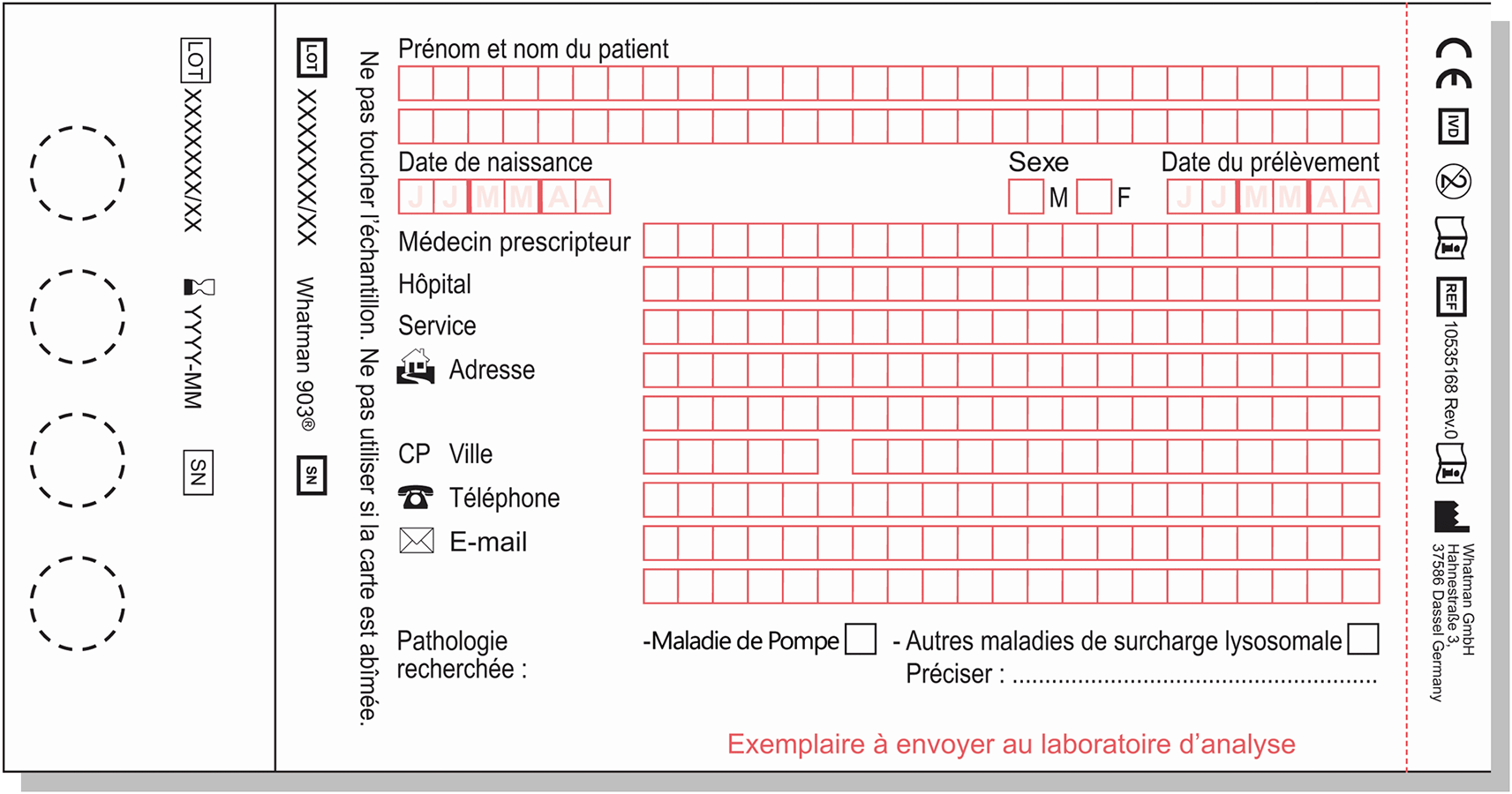
Blood samples of all the patients were collected by applying a blood drops, drawn by lancet from the finger, on a special dry paper (Sigma-Aldrich/USA, Cat. No.: 8.294.0004A) (Figure 1). Then, it was sent to the advanced genetic clinical research lab of Univ.-Prof. Dr. Berthold Streubel in Vienna, Austria and to the Laboratory of Metabolism, University Medical Center Hamburg, Germany by DHL from Iraq. Dried blood spot (DBS) test was conducted for the screening of Fabry disease. In this test, four blood spots were placed on a paper and leave dried at room temperature.
The α-GAL A activity was measured using tandem mass spectrometry, where values between 200–2000 pmol/spot*20 h were considered normal. This diagnosis is confirmed if there is a decrease or absence of α-GALA activity in the dry blood leukocytes. A lysosomal enzyme activity assay <200 pmol/spot*20h is suggestive of Fabry disease with clinical correlation. Any patient with α-GAL A activity <100 pmol/spot*20h was sent for genetic testing for confirmation of the diagnosis.
Molecular testing include several approaches include single-gene testing, multi-gene testing, or comprehensive genomic testing. In this study, multi-gene testing was preferable to use because of multifactorial aetiology of disease. First, the GLA sequence is analyzed; if no pathogenic variant is found, then gene-targeted determined. We invited all patient from different centers to proceed to did dry blood spot paper testing. The information about testing include (Official symbol: GLA; Gene ID: 2717; and Reference sequence: NM_000169.2 (ENST00000218516)). The DNA extraction from Dried Blood Spot; PCR amplification and sequencing of all coding exons and flanking intronic regions were done automated by (PCR thermocycler model Verriti, thermofisher company, USA). All samples were sent to ARCHIMEDlife for PCR testing.
Targeted analysis for the p.Ala143Pro and IVS4+919G>A pathogenic variants were performed.
Mentioned in the test ARCHIMEDlife used a NM_000169.2 for GLA gene ID 2717 as reference sequence for comparison or more scientific term (Alignment) with Human genome HGVS 37. Variants are described according to den Dunnen et al. (2016).18 Common benign variants in the gene may have been identified but have not been included in this report. Mosaicism cannot be excluded. Because of their complexity and their potential implications for other family members, all genetic tests should be accompanied by genetic counseling in accordance with local legislation.
Written informed consent was obtained from all participants. The Medical Ethical Committee at the College of Medicine, University of Basrah, approved this study (1st on 11/06/2018; ID: 2018-00287712 and 2nd on 04/03/2021; ID: 2021-03040818 because the first approval got lost in transport.
Data were coded before entering them on the computer. The data is presented as the total number as well as in percentage. SPSS software version 20 (RRID:SCR_019096) was used to perform statistical analysis. Pearson’s chi-squared test was used for determining the statistically significant differences among variables, which was indicated by a p-value < 0.05.
A total of 1148 patients were enrolled for this multi-center, cross-sectional study conducted across various regions in Iraq.22 After clinical investigation, it was confirmed that 17 of the patients had α-GAL A deficiency (Fabry disease), with each presenting varied clinical symptoms.
Table 1 demonstrates the distribution of Fabry disease according to the gender of the patients. Of the confirmed cases, 16 were male (94.1%) and only one female (5.9%). Comparison of the control group (having normal α-GAL A enzyme level) with the patients showed no statistical difference (p=0.5; Pearson’s chi-square value=0.38).
| Gender | Total | ||||
|---|---|---|---|---|---|
| Male | Female | ||||
| Enzyme | Normal enzyme level | Count | 1012 | 119 | 1131 |
| % of Total | 88.2% | 10.4% | 98.5% | ||
| Deficient enzyme level | Count | 16 | 1 | 17 | |
| % of Total | 1.4% | 0.1% | 1.5% | ||
| Total | Count | 1028 | 120 | 1148 | |
| % of Total | 89.5% | 10.5% | 100.0% | ||
Table 2 shows the distribution of patients with Fabry disease according to age. It was observed that the maximum number of patients belonged to the 10–30 years age group (12 patients; 70.6%), followed by 3 patients in the 31–50 years age group (17.6%). The <10 years and >50 years age groups consisted of only one patient (5.9% of the total Fabry cases each). Patients with enzyme deficiency and the patients with normal enzyme level showed a statistically significant difference (p=0.003; Pearson’s chi-square value=13.8).
Table 3 shows the distribution of patients based on their clinical presentations after the genetic confirmation of the Fabry disease. As expected, renal dysfunction (proteinuria with or without renal insufficiency) was the most common clinical presentation, with 14 out of the 17 confirmed cases (82.3%) showing renal involvement. Ten cases had chronic kidney disease (stage 1–4;eGFR >15 ml/min/1.73 m2) while the other 4 were undergoing hemodialysis (eGFR <15 ml/min/1.73 m2). This was followed by neurological involvement, where 6 patients (35.3%) showed peripheral neuropathy. The other clinical manifestations included angiokeratoma (5 patients; 29.4%), left ventricular hypertrophy (3 patients; 17.6%), and corneal verticillate (4 patients; 23.5%).
Next, we compared the distribution of the various clinical presentations with the control group. As shown in Table 4, renal involvement was found in 1120 patients, of which 1106 patients had normal α-GAL A level, while 14 patients had enzyme deficiency, and hence confirmed Fabry disease. All the confirmed cases had proteinuria with or without renal insufficiency. The confirmed cases showed a statistically significant difference with the control group (p=0.0001; Pearson’s chi-square value=16.7).
In contrast to renal involvement where 1120 patients out of total 1148 patients turned out to be positive for renal dysfunction, cardiac involvement was found in only 37 patients. The number of patients with normal enzyme level was 34 while only 3 patients were confirmed to have Fabry disease (Table 5). These patients suffered from left ventricular hypertrophy. The patients with left ventricular hypertrophy and the control group exhibited statistically significant difference (p=0.001; Pearson’s chi-square test=11.5).
Then, the distribution of neurological involvement was studied among the patients with and without α-GAL A enzyme deficiency (Table 6). Neurological involvement was not found in many patients; there were 7 positive and 1141 negative cases. Out of the total 7 positive cases with peripheral neuropathy, 6 patients had confirmed Fabry disease. The confirmed cases and the control group showed statistically significant difference (p=0.0001; Pearson’s chi-square value=324.5).
Finally, as shown in Table 7, the distribution of dermatological involvement in Fabry disease was studied. In this case, a small number of patients were found to be positive for angiokeratoma, and all those patients had confirmed Fabry disease. The difference between the confirmed cases and the control group was found to be statistically significant (p=0.0001; Pearson’s chi-square value=267).
| Angiokeratomas | Total | ||||
|---|---|---|---|---|---|
| Negative | Positive | ||||
| Enzyme | Normal enzyme level | Count | 1131 | 0 | 1131 |
| % of Total | 98.5% | 0.0% | 98.5% | ||
| Deficient enzyme level | Count | 12 | 5 | 17 | |
| % of Total | 1.0% | 0.4% | 1.5% | ||
| Total | Count | 1143 | 5 | 1148 | |
| % of Total | 99.6% | 0.4% | 100.0% | ||
In addition to the comparison of the clinical presentation between the confirmed Fabry disease group and the control group, we also compared the prevalence of this disease in various regions of Iraq, namely north, middle, and south Iraq. As shown in Table 8, although the maximum number of recruited patients (975) were from middle of Iraq, the maximum number of confirmed cases were from the northern region of Iraq (8 cases; 47%), followed by 6 cases from the middle region of Iraq (35.3%), and 3 cases form south of Iraq (17.7%). A statistically significant difference was observed between the confirmed cases from north of Iraq and the control group (p=0.0001; Pearson’s chi-square value=44.3).
A total of 1148 patients with variable presentations were enrolled for a multi-center, cross-sectional study to realize the prevalence of Fabry disease in Iraq. Fabry disease results due to the deficiency or absence of the lysosomal enzyme α-GAL, whose function is to breakdown glycolipids, which leads to the continuous build-up of Gb3, and consequently, cell abnormalities and organ dysfunction. In this study, lysosomal α-GAL A activity was determined by tandem mass spectrometry, and any patient with α-GAL A activity <100 pmol/spot*20h was sent for genetic testing for the confirmation of the diagnosis.
Interestingly, in our study, only 17 cases were confirmed to have Fabry disease, out of which 16 were male (94.1%) and only one female (5.9%). This skewed gender ratio is explained by Fabry disease being inherited X-linked disorder.1 Men inherit only one X-chromosome, and if the GLA mutation is present on that X-chromosome, they will be affected with the disorder. On the other hand, women have two X-chromosomes. Therefore, the expression of the mutated gene may be reduced or masked by the normal gene on the other X-chromosome.1
Since this was a multi-center study, the regions were distributed as north, middle, south Iraq. The regional distribution of patients with Fabry disease included 8 cases from the north of Iraq (47%), 6 cases from the middle region (35.3%), and 3 cases from south Iraq (17.7%). There was a statistically significant difference between the confirmed cases from the north of Iraq and the control group (p=0.0001).
The clinical manifestations of Fabry disease in the confirmed cases included angiokeratoma (5; 29.4%), peripheral neuropathy (6; 35.3%), left ventricular hypertrophy (3; 17.6%), proteinuria with and without renal insufficiency (14; 82.3%), and corneal verticillate (4; 23.5%). Furthermore, the age distribution of patients with Fabry disease was as follows: one patient was below 10 years of age (5.9%), 12 patients belonged to the 10–30 years age group (70.6%), 3 patients belonged to 31–50 years age group (17.6%), and only one was >50 years of age (5.9%). Considering the age distribution, a statistically significant difference was observed between the confirmed cases with α-GAL A deficiency and the control cases without enzyme deficiency (p=0.003). The distribution of renal involvement (proteinuria with or without renal insufficiency) in patients with Fabry disease demonstrated a statistically significant difference with the control group (p=0.003). This could be explained by the striking glomerular changes that occur in the glomerular visceral epithelial cells. These cells become enlarged and become packed with small, clear vacuoles enclosing the glycosphingolipid.6,10 A progressive decrease in renal function is due to the progressive accumulation of Gb3 in the kidneys, particularly in the endothelial cells, smooth muscle cells, and podocytes, leading to segmental and global glomerulosclerosis.6,10
Cardiac diseases are common in Fabry patients owing to the deposition of Gb3 in all cardiac tissues. Heart diseases like heart enlargement typically left ventricular hypertrophy leads to arrhythmia, hypertrophic cardiomyopathy, and heart failure. Similar to renal dysfunction, a statistically significant difference was observed between patients with cardiac involvement (left ventricular hypertrophy) and the control group (p=0.001). This is consistent with the results of the study by Baptista et al.19 It should be mentioned that the etiology of left ventricular hypertrophy is difficult to distinguish from the other heart diseases using the common cardiac imaging methods, especially echocardiography.19
Another common clinical manifestation of Fabry disease includes peripheral neuropathy (neurological involvement) that may present as neuropathic pain, reduced warm and cold sensation, and gastrointestinal disturbances. In our study, 41.2% of the patients with α-GAL A deficiency suffered from peripheral neuropathy. There was a statistically significant difference between the confirmed cases and the control groups (p=0.0001). A progressive lysosomal accumulation of glycolipids in the neural cells affects the small, unmyelinated nerve fibers, which manifests as neural myopathy, mostly at a young age.
Dermatological involvement was also observed in few of the patients with α-GAL A deficiency. In our study, 29.4% of the confirmed cases had angiokeratoma, which was found to be statistically significant compared to the control group (p=0.0001). In the early stages, reddish to dark blue skin rashes/lesions appear, mostly in the area between the hips and the knees. Angiokeratomas are benign cutaneous vascular lesions characterized by dilated thin-walled blood vessels lying in the upper dermis, associated with an epidermal reaction like hyperkeratosis.20
Although ophthalmological manifestations do not generally result in significant visual impairment, they are important as some manifestations serve as disease markers and have prognostic and diagnostic implications.21 In our study, 23.5% of the patients presented with corneal verticillate, which is the most typical ocular symptom in Fabry disease.
The strength of this study are firstly this is firstly time multi-centers study for rare disease in Iraq and give special recognition for doctors to screening for this genetic disease.
The limitations are small sample size, and the genetic testing done only for female with highly suspected Fabry disease and male with deficient alpha GAL A enzyme.
This study suggested the screening of all patients with a positive or suspected family history of renal disease, suspected angiokeratoma, unexplained peripheral neuropathy, unexplained proteinuria, unexplained cardiovascular disease, as well as children and young hemodialysis patients. It has been established that the early diagnosis of Fabry disease is the cornerstone for the prevention of multiorgan complications, especially renal and cardiac complications. There is a need for effective treatment beyond the palliative and symptomatic care usually provided by the clinicians. Since multiorgan dysfunction is involved, the patients require an individually tailored comprehensive and multidisciplinary treatment approach that includes specific therapies targeting abnormal substrate accumulation and adjuvant therapies that address end-organ damage. Thus, the initiation of enzyme replacement therapy using recombinant human α-GAL A (agalsidase) is of utmost importance for the treatment of Fabry disease. Hitherto, there has been no comparable substitute to enzyme replacement therapy for this damaging, progressive disease till date. It is crucial for future research to focus on the development of protocols for the early diagnosis of Fabry disease. Moreover, effective enzyme protocols are required for the reversal, maintenance, and prevention of the fundamental pathology of the disorder, especially for children and patients with cardiac diseases and compromised renal function.
Zenodo: FABRY disease. https://doi.org/10.5281/zenodo.6879922.22
Data are available under the terms of the Creative Commons Attribution 4.0 International license (CC-BY 4.0).
| Views | Downloads | |
|---|---|---|
| F1000Research | - | - |
|
PubMed Central
Data from PMC are received and updated monthly.
|
- | - |
Provide sufficient details of any financial or non-financial competing interests to enable users to assess whether your comments might lead a reasonable person to question your impartiality. Consider the following examples, but note that this is not an exhaustive list:
Sign up for content alerts and receive a weekly or monthly email with all newly published articles
Already registered? Sign in
The email address should be the one you originally registered with F1000.
You registered with F1000 via Google, so we cannot reset your password.
To sign in, please click here.
If you still need help with your Google account password, please click here.
You registered with F1000 via Facebook, so we cannot reset your password.
To sign in, please click here.
If you still need help with your Facebook account password, please click here.
If your email address is registered with us, we will email you instructions to reset your password.
If you think you should have received this email but it has not arrived, please check your spam filters and/or contact for further assistance.
Comments on this article Comments (0)