Keywords
Acute pulmonary embolism, transesophageal echocardiography, transthoracic echocardiography, CT chest angiography
Acute pulmonary embolism, transesophageal echocardiography, transthoracic echocardiography, CT chest angiography
Pulmonary embolism (PE) can be difficult to diagnose especially in critical ill patients who are hemodynamically unstable notably if the classic symptoms of PE are absent1. However, many cases of PE are diagnosed in an emergency context2. Echocardiography could be considered a useful technique at the bedside in critical care settings for the diagnosis of PE, especially as it is difficult to diagnose using other techniques2. That is why, we present this case of massive pulmonary embolism diagnosed by the combined use of transthoracic echocardiography (TTE) and transesophageal echocardiography (TEE) due to the poor transthoracic window. TEE was useful in ruling out differential diagnoses and finding signs in favor of the diagnosis of PE, which allowed the initiation of adequate treatment without further delay. The aim of this case report was to highlight the pivotal role of TEE in the diagnosis of PE in a hemodynamically unstable patient especially when his mobilization to the radiology department was difficult to achieve.
A 47-year-old north-African man working as an engineer, with no relevant medical history, previous treatments or toxic habits (tobacco, alcohol) was admitted to hospital with a diagnosis of isolated closed fracture of his right leg due to a road accident (he was struck by a motor vehicle). At the time of admission, he was conscious, without any neurological alterations, or hemodynamic and respiratory disorders.
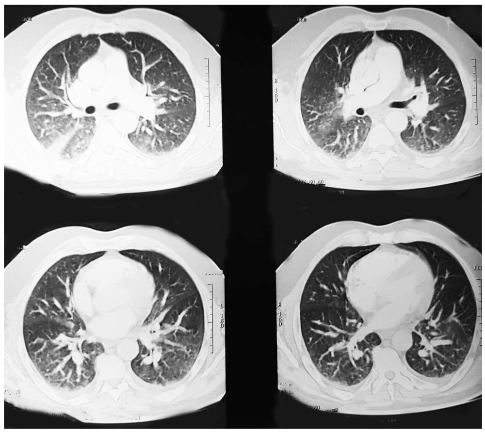
24 hours after admission, the patient suddenly presented a change in his level of consciousness (confusion with Glasgow come scale of 12). He was tachypneic (30 breaths/min) with an oxygen saturation of 94% with a non-rebreather mask. Lung auscultation showed conserved vesicular murmur with bilateral basal crackles. He was tachycardic (heart rate 120 beats/min) and presented a hypotension (blood pressure was 80/40 mmHg). He was not febrile and did not present any cutaneous sign. A 12-lead electrocardiogram showed only a sinusal tachycardia without other signs of acute coronary syndrome or right heart strain. The patient was immediately treated with crystalloid fluid infusion and bolus of epinephrine. After that, a brain scan was done which did not show signs of post traumatic abnormalities, in addition to thoracic CT angiography which did not show any sign of acute pulmonary embolism (Figure 1).

Therefore, he was transferred in emergency to the intensive care unit (ICU) and due to his bad evolution, he was intubated and required mechanical ventilation. Arterial acid-base balance at that time showed fraction of inspired oxygen 100%, pH 7.15, partial pressure of oxygen 86 mmHg, partial pressure of carbon dioxide 52 mmHg, bicarbonates 24 mmol/L, base excess -15, lactic acid 2.5 mmol/L and oxygen saturation 93%. Laboratory findings showed hemoglobin 10g/dl, leukocytes 6.103/mm3, lymphopenia, creatinine 1.5 mg/dl, troponin T 34 µg/L, pro-BnP 400 pg/ml and procalcitonin < 0.05. His respiratory status failed to respond to high-dose of vasopressor and ventilatory support so nitric oxide was introduced in addition to continued infusion of cisatracurium. Chest radiography showed bilateral infiltrate (Figure 2).
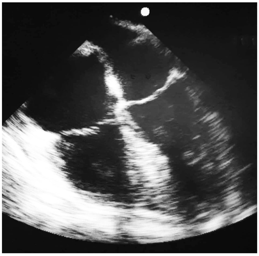
In order to determine the real cause of this instability, TTE was performed however we obtained poor quality images, so it was necessary to do a TEE which was performed by an experiment anesthesiologist. TEE demonstrated a dilated and dysfunctional right ventricle (RV) with a hypertrophic dysfunctional left ventricle (LV). The right atrium (RA) was also severely dilated with a patent foramen oval and septum bowing (Figure 3). The RV end-diastolic diameter to LV end-diastolic diameter ratio was 1.2 suggesting RV pressure overload. RV dilatation led to functional tricuspid regurgitation as the tricuspid annulus enlarged. There was a pulmonary arterial hypertension with a pulmonary artery systolic pressure of 70–80 mmHg. Initially, there was no evidence of a thrombus either in the pulmonary arteries or on the right side of the heart. Due to global heart failure and the low-cardiac-output state, dobutamine was used with the doses of 3–5 µg/kg/min. However, after 24 hours, a control TEE showed an evident thrombus in the right pulmonary artery which was dilated (Figure 4). Massive pulmonary embolism was suspected but we could not confirm it by other complementary test because the unfavorable hemodynamic situation of the patient prevented his transfer. Anticoagulant therapy (non-fractioned heparin) was administrated immediately achieving a favorable clinical outcome with rapid withdrawal of dobutamine, nitric oxide and cisatracurium. No follow up information about the patient was available.

This case highlights the crucial role of echocardiography in ICU for patients with severe shock due to massive pulmonary embolism associated to an unfavorable hemodynamic situation. In addition, like another similar case published in literature, it illustrates the value of TEE over TTE for those who have poor transthoracic window secondary to some clinical situation (supine position or mechanical ventilation)1.
Pulmonary embolism can be difficult to diagnose particularly for patients in ICU who are sedated or on mechanical ventilation because key symptoms are absent (dyspnea, chest pain and syncope). For the diagnosis of PE, pulmonary angiography and spiral CT is the gold standard with a sensitivity of 83% and a specificity of 96% according to the PIOPED II trial3. However, in our scenario, the CT angiography performed initially did not show any sign of acute pulmonary embolism despite the high probability of PE and this could be explained by the occurrence of artifacts or secondary migration of subsegmental thrombosis. So echocardiography was useful in order to rule out some differential diagnoses which caused this hemodynamic instability (tamponade, aortic dissection, hypovolemia…) according to the guidelines of European Society of Cardiology4.
Vignon et al. showed that TEE helped in 98% of clinical decisions in a critical care population so it has higher impact on patient care than TTE which provided adequate images in only 38% of cases5. Concerning the confirmation of PE, TEE has 70% sensitivity and 81% specificity6. In the context of PE, TEE usually shows indirect signs like RV dilatation (RV end-diastolic diameter/LV end-diastolic diameter ratio > 0.9) and exclude other causes7. In addition, serial assessment of RV size, determination of RV systolic pressure and inferior vena cava assessment could be performed in patients with massive PE. Although, thrombus may be seen in some cases. According to Pruszczyk et al.8, the central pulmonary arteries including the proximal lobar branches on both sides could be precisely visualized by biplane TEE. Only the proximal left pulmonary artery is difficult to assess because it is shielded by the left main bronchus. But a perimural artifact may be potentially misinterpreted as thrombus especially when it is present in the right pulmonary artery9.
Significant hemodynamic instability is present in 8% of patient with acute pulmonary embolism. The main cause is acute right ventricular failure which increases mortality from 15% to 42%10. That is why, TEE could be useful for analyzing response to medical interventions such as fluid and drug therapy. It could also be helpful for monitoring RV function and pulmonary artery systolic pressure especially if thrombolytics or anticoagulant were administrated11.
We reported this case in order to show the fundamental role of TEE in ICU especially when the transthoracic window is poor. TEE allows the initiation of adequate treatment without further delay, by avoiding unnecessary mobilization of an unstable patient to perform CT chest angiography and can lead to a better clinical outcome. Although, TEE has some limitations like the cost of the equipment or the inability to place a probe (esophagectomy, esophageal diverticula or varices), complication rates from TEE use are fairly low at 0.2%12. In addition, it has been demonstrated to have a steep learning curve and that physicians could successfully perform focused TEE assessments with a high retention rate after 6 weeks of 4-hour simulation workshop13.
All data underlying the results are available as part of the article and no additional source data are required.
Written informed consent for publication of their clinical details and clinical images was obtained from the patient.
An earlier version of this article can be found on Authorea (doi: 10.22541/au.161467284.43760460/v1).
| Views | Downloads | |
|---|---|---|
| F1000Research | - | - |
|
PubMed Central
Data from PMC are received and updated monthly.
|
- | - |
Is the background of the case’s history and progression described in sufficient detail?
Partly
Are enough details provided of any physical examination and diagnostic tests, treatment given and outcomes?
Partly
Is sufficient discussion included of the importance of the findings and their relevance to future understanding of disease processes, diagnosis or treatment?
Partly
Is the case presented with sufficient detail to be useful for other practitioners?
Yes
References
1. Falster C, Hellfritzsch M, Gaist TA, Brabrand M, et al.: Comparison of international guideline recommendations for the diagnosis of pulmonary embolism.Lancet Haematol. 2023; 10 (11): e922-e935 PubMed Abstract | Publisher Full TextCompeting Interests: No competing interests were disclosed.
Reviewer Expertise: Ultrasound and venous thromboembolism.
Is the background of the case’s history and progression described in sufficient detail?
Partly
Are enough details provided of any physical examination and diagnostic tests, treatment given and outcomes?
No
Is sufficient discussion included of the importance of the findings and their relevance to future understanding of disease processes, diagnosis or treatment?
No
Is the case presented with sufficient detail to be useful for other practitioners?
Partly
Competing Interests: No competing interests were disclosed.
Reviewer Expertise: Echocardiography (transthoracic and transesophageal), appropriate use criteria, critical care cardiology, pulmonary embolism and right heart
Alongside their report, reviewers assign a status to the article:
| Invited Reviewers | ||
|---|---|---|
| 1 | 2 | |
|
Version 1 02 Aug 22 |
read | read |
Provide sufficient details of any financial or non-financial competing interests to enable users to assess whether your comments might lead a reasonable person to question your impartiality. Consider the following examples, but note that this is not an exhaustive list:
Sign up for content alerts and receive a weekly or monthly email with all newly published articles
Already registered? Sign in
The email address should be the one you originally registered with F1000.
You registered with F1000 via Google, so we cannot reset your password.
To sign in, please click here.
If you still need help with your Google account password, please click here.
You registered with F1000 via Facebook, so we cannot reset your password.
To sign in, please click here.
If you still need help with your Facebook account password, please click here.
If your email address is registered with us, we will email you instructions to reset your password.
If you think you should have received this email but it has not arrived, please check your spam filters and/or contact for further assistance.
Comments on this article Comments (0)