Keywords
orbital cellulitis, panophthalmitis, endophthalmitis, pregnancy, bangladesh, epistaxis
orbital cellulitis, panophthalmitis, endophthalmitis, pregnancy, bangladesh, epistaxis
In the updated version, we included the name of the broad-spectrum antibiotic that was injected initially by the otolaryngologist and highlighted the reason why we initially diagnosed this case as orbital cellulitis with endogenous endophthalmitis in the discussion section. We also mentioned the possible source of infection in this case in the discussion section.
See the authors' detailed response to the review by Hidehiro Oku
See the authors' detailed response to the review by Pradeep Kumar Panigrahi
Orbital cellulitis with panophthalmitis is a rare ocular condition, typically trauma-related or endogenous. Panophthalmitis is a severe form of inflammation of all eye coverings and intraocular contents from an internal or external source.1 There are no reports regarding orbital cellulitis with panophthalmitis other than endogenous endophthalmitis (EE). However, EE during pregnancy is extremely rare.2 It’s critical to distinguish endophthalmitis from panophthalmitis. Though both endophthalmitis and panophthalmitis are on the same variety of ocular infection, panophthalmitis is more complicated and happens when worsening endophthalmitis covers the all layers of eyeball, including the orbital contents.
EE is a potentially harmful eye disease that occurs after an organism from the bloodstream reaches the eye, and 2–8% of cases of endophthalmitis are endogenous bacterial endophthalmitis (EBE). Recent research on EE from India reported no systemic infective foci in 67.6% of incidents. The infectious agents retrieved were 48% gram-positive bacteria, 37% gram-negative bacteria, and 15% fungi.3
Specific measures, e.g., surgical procedures or an intravenous line insertion, are established risk factors for endogenous endophthalmitis and panophthalmitis. Moreover, pregnancy, a subtle immunosuppression state in which the body minimizes its natural proinflammatory host responses to protect the developing fetus.4
EE is a rebellious condition since the systemic infectious foci and the ocular inflammation must be managed immediately and appropriately. The patient requires a thorough systemic evaluation to identify the septic foci and treat that with suitable germicides for an extended period. Regardless, the visual outcome can be poor.3 In this case, the management of EE patients presents distinct problems because of the violent nature of EE and concerns about the newborn safety of the systemic and intravitreal medication.2
Despite extensive treatment, panophthalmitis due to S. aureus has poor visual outcomes. All previously described cases of simultaneous orbital cellulitis and panophthalmitis required evisceration or enucleation, with a poor prognosis for globe salvage.5
To our knowledge, orbital cellulitis with endogenous panophthalmitis in pregnant women has never been recorded in Bangladesh or any other South Asian nation. We discussed this unusual presentation of such due to Staphylococcus aureus (SA) in a 33-week pregnant lady with a history of left-sided spontaneous epistaxis to raise awareness of this deadly, vision-threatening complication during pregnancy.
A 22-year-old primigravida, in her third trimester (33 weeks) of gestation, came to us with abrupt, painful vision loss in the left eye coupled with eyelid chemosis, forward bulging of the eye, and purulent discharge for two days.
She reported having several episodes of left-sided spontaneous epistaxis, a 102°F fever, and generalized weakness eight days before she started experiencing visual symptoms. Her obstetrician was consulted about these symptoms, and she was admitted to the hospital for additional assessment and treatment.
She had unremarkable medical history with no personal or familial bleeding tendencies. She was not taking any medications and had a normal range of blood pressure. She had never had any episodes of epistaxis before.
At that time, her blood reports showed mild anemia (normocytic normochromic) with septicemia: 7.2 g/dL of hemoglobin (normal: 10–14 g/dL) and neutrophilic leukocytosis with a total white blood cell count of 12,300/L (normal: 4,000–11,000/L) with 82% neutrophils (normal: 40–75%) and a high serum IgE level of 369.81 KIU/L (normal: 100–150 KIU/L).
The coagulation profile showed no abnormalities. Human immunodeficiency virus (HIV), hepatitis B and C viruses, TPHA, VDRL, QuantiFERON-TB Gold, and Toxoplasmosis IgM and IgG were negative. Routine microscopic urine examinations and renal and hepatic function tests were within normal physiological limits. Consecutive blood and urine cultures showed negative results as well. There were no foci of active infection on systemic examination.
During her hospital stay, she was treated by an otolaryngologist, who applied a nasal pack soaked with a topical vasoconstrictor under the guidance of a nasoendoscope, which halted the bleeding. The nasal pack was removed after 7 hours without any further history of epistaxis. She also received two doses of intravenous dextrose infusion, one bag of B-positive fresh blood (450 ml) transfusion for mild anemia (7.2 gm/dl), and two doses of broad-spectrum injectable antibiotic (ceftraixone 2 gm).
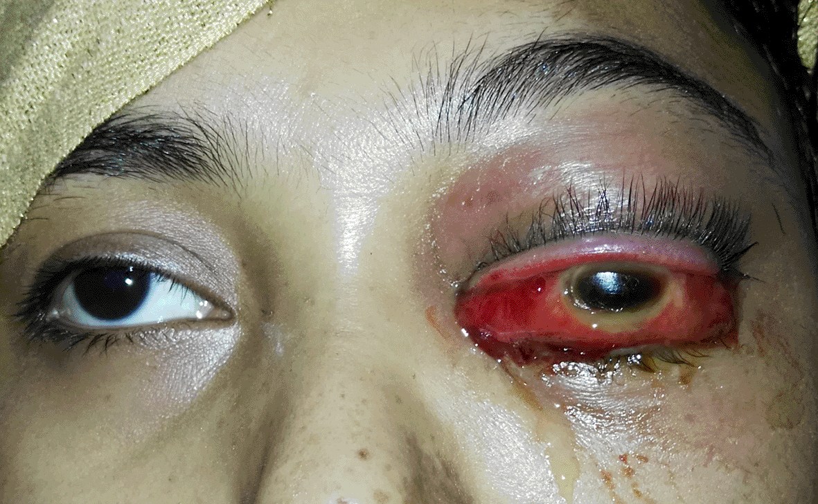
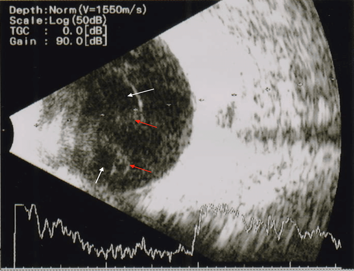
On ocular examination, she only perceived light with an inaccurate ray’s projection in the left eye and 6/6 in the right eye. There were tense lid edema and proptosis ( Figure 1) with severely restricted eye movement in all gazes. There were severely chemosed conjunctiva, circumcorneal congestion and edematous cornea, and no further visibility of the anterior and posterior segments. A B-scan ultrasonogram (USG) showed moderate hyperechogenic shadows with cavitations in the vitreous cavity, which disappeared at low gain with attached retina and increased choroidal thickness in the left eye ( Figure 2). So diagnosed as a case of left-sided orbital cellulitis with endogenous endophthalmitis (EE).
Treatment included intravenous (IV) injection of amoxicillin-clavulanic acid (1.2 g, two times daily) for five days, topical 0.5% moxifloxacin every hour, prednisolone acetate 1% eye drop every two hours, and 1% atropine sulfate eye drop every eight hours.
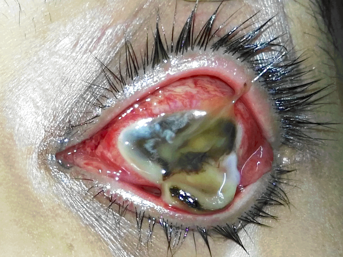
Following the therapy, however, the patient’s condition worsened. She presented with a melted cornea with frank purulent discharge and uveal tissue the following day ( Figure 3). While examining the left eye, there was no light’s perception; no notable pathology in the right eye with a 6/6, N5 visual acuity. The injection site looked healthy, with no signs of infection when checking for the source of infection. So, a diagnosis of orbital cellulitis with endogenous panophthalmitis was made in the left eye.
At that time, her blood investigations came out as a mild anemia with septicemia: 9.6 g/dL hemoglobin, leukocytosis with counts of 13,200/μL, and thrombocytopenia with counts of 2,94,000/μL. The coagulation profile was within normal limits except for a raised activated partial thromboplastin time (APTT) of 45 seconds (control: 30.0 seconds).
After counselling the patient and her husband, evisceration of the left eye was advised. Intraoperatively, the sclera-corneal melting was evident, and completely removed all intraocular contents. All removed ocular contents were sent for microbiological analysis, and significant growth of Staphylococcus aureus (SA) was reported. We continued the previous treatment regimen during the immediate postoperative period.
As her condition improved clinically, we discharged her on the third postoperative day with oral levofloxacin tablets of 750 mg once a day for seven days, moxifloxacin eye drops every four hours for a month, and prednisolone phosphate 1% eye drops every two hours for three days, followed by six times for seven days, four times for seven days, thrice for seven days, and twice for another seven days. She had been on regular follow-ups for the last two months with no further ocular or obstetric complications.
Meanwhile, routine scans for fetus at regular intervals were done to confirm the fetus’s viability. In contrast, the last ultrasonogram of the pregnancy profile showed about 34+ weeks of a single live pregnancy with cephalic presentation. The patient delivered a healthy male infant at 36 weeks without complications.
Pregnancy is a unique immunological condition characterized by several physiological modifications in the maternal immune system. The aggressiveness of infectious uveitis during pregnancy and early postpartum may be due to the relative immune suppression during pregnancy.6 At 33 weeks of pregnancy, our 22-year-old primigravida patient presented with abrupt, severe hazy vision in her left and eyelid chemosis, proptosis, and purulent discharge.
During an eye examination, she perceived light with an inaccurate projection of rays in the left eye, while the right eye had 6/6 vision. There was moderate proptosis, eyelid and conjunctival chemosis, ciliary congestion with corneal edema, and no other view of both segments of the left eye ( Figure 1). As the radiological imaging was not possible during the pregnency state, clinical features, hematological and B-scan USG findings were evaluated to reach the diagnosis of orbital cellulitis with endogenous endophthalmitis (EE) ( Figure 2). Uncontrolled diabetes mellitus, use of catheters, low neutrophil count, immunosupressed patients, and patients on chemotherapy were predisposed to EE. Despite reports of anecdotal cases, pregnancy is not associated with higher EE rates.2 These cases had a history of either septic abortion, handling of an intrauterine device, or IV fluid infusion.2 Our patient received intravenous injections of dextrose infusion, fresh blood transfusion, and broad-spectrum injectable antibiotic as her initial blood reports showed 7.2 g/dL Hb (normal: 10–14 g/dL) and 12,300/μL total white blood cells count (normal: 4,000–11,000/μL) with 82% neutrophil (normal: 40–75%), which her gynecologist prescribed. Her other hematological and urine analysis, including blood and urine cultures, were negative for any organisms, and there were no systemic foci of infections.
While the treatment of endophthalmitis is the similar regardless of the cause, the endogenous endophthalmitis in pregnancy presents unique difficulties, particularly regarding the welfare of intravitreal and systemic medications. Additionally, the safety of any interventional procedure for a pregnant patient is a concern.2 As penicillin, cephalosporins, and erythromycin are the best option in pregnancy,2 we initiated the treatment with IV injection of amoxicillin-clavulanic acid (1.2 g, two times daily) for five days, topical 0.5% moxifloxacin every hour, prednisolone acetate 1% eye drop every two hourly and 1% atropine sulfate eye drop eight hourly. However, the patient’s condition worsened by the next day, and she presented with the features of orbital cellulitis with panophthalmitis in the left eye ( Figure 3). Her blood reports reflected the anemia and sepsis even after blood transfusion and broad-spectrum antibiotics.
Although endophthalmitis and panophthalmitis are distinct kinds of ocular infections, severity of panophthalmitis is more, developing once endophthalmitis deteriorates and spreads to the whole globe and all layers of the eyeball. Panophthalmitis with concurrent orbital cellulitis is rare and has a low likelihood of saving globe, with all earlier documented cases necessitating evisceration or enucleation.1 Under the prior treatment regimen, we chose to eviscerate the left eye and send the ocular samples for microbiological examination, which came back positive for MSSA despite a positive blood culture. 2-8% of all cases of EE belongs to the endogenous bacterial endophthalmitis (EBE).7
Additionally, some forms of immunosuppression were reported in all cases, which adds to the highly violent course, with mostly having cellulitis shortly after symptoms begin.
Despite intensive treatment, panophthalmitis with S. aureus has been shown to have poorer result. Surgical procedures, or any IV insertions, are known to increase the risk of EE and panophthalmitis.4
Before the visual complaints, she experienced bouts of left-sided epistaxis, for which the otolaryngologist used an anterior nasal pack soaked with a vasoconstrictor to successfully control the bleeding. Epistaxis is prevalent during pregnancy, although most episodes do not need medical treatment. Several circumstances increase the risk of epistaxis during pregnancy. Elevated estrogen levels, in particular, promote the nasal mucosal vascularity, and the incidence is 20.3% in pregnant women where 6.2% in nonpregnant women.8 We ruled out epistaxis caused by nasal lesions such as granuloma gravidarum and nasal polyps, as well as clotting problems.
Her blood IgE level was 369.81 KIU/L (normal: 100–150 KIU/L), although she had no rhinosinusitis or allergy symptoms. Pregnancy and allergic rhinitis frequently coexist. Attention deficit hyperactivity disorder (ADHD) is highly linked to total IgE levels in the mother’s prenatal blood but not to levels in her early life or childhood.9 We advised the patient to get the newborn infant checked regularly.
We believe that the combination of immunodepression, presence of unrecognized chronic sinusitis from the begining, and, in conjunction with it, anemia may have contributed to our patient’s ophthalmic situation rapidly progressing from early feature of hazy vision and eye ache to total blindness and proptosis with restricted ocular motility in less than 24 hours. According to Chaudhry et al.,10 the following are possible explanations for orbital cellulitis’ rapid blindness loss:
1. Optic neuritis as a result of a neighboring or contiguous infection,
2. Thrombophlebitis-induced ischemia in the valveless orbital veins, or
3. Ischemia caused by compression or pressure may result in central artery blockage.
After improvement of the patient’s condition post-operatively, she was discharged and followed-up regularly. We ensured her fetal well-being throughout the pregnancy period and later delivered a healthy male baby at her 36-weeks of pregnancy without any complication.
This sort of case scenario requires multidisciplinary team including infectious disease specialist or an internist along with the ophthalmologist and gynecologist and obstetrician to manage promptly and safely.
Pregnancy poses a significant immunocompromised state when any infection, particularly with the women in impoverished countries like Bangladesh, should be identified as soon as possible to prevent further infection, particularly ocular and periocular regions. Orbital cellulitis with panophthalmitis has a disastrous prognosis and nearly invariably requires eye removal. It is critical to intervene quickly to eliminate the infective focus and treat the incidence of anemia in pregnancy. Because of its intricacy, it is a condition that warrants collaborative care by infectious disease, ophthalmology, and perinatal specialists.
| Views | Downloads | |
|---|---|---|
| F1000Research | - | - |
|
PubMed Central
Data from PMC are received and updated monthly.
|
- | - |
Provide sufficient details of any financial or non-financial competing interests to enable users to assess whether your comments might lead a reasonable person to question your impartiality. Consider the following examples, but note that this is not an exhaustive list:
Sign up for content alerts and receive a weekly or monthly email with all newly published articles
Already registered? Sign in
The email address should be the one you originally registered with F1000.
You registered with F1000 via Google, so we cannot reset your password.
To sign in, please click here.
If you still need help with your Google account password, please click here.
You registered with F1000 via Facebook, so we cannot reset your password.
To sign in, please click here.
If you still need help with your Facebook account password, please click here.
If your email address is registered with us, we will email you instructions to reset your password.
If you think you should have received this email but it has not arrived, please check your spam filters and/or contact for further assistance.
Comments on this article Comments (0)