Keywords
Endodontic surgery, Retrograde filling material, Sealing ability, Retrograde Endodontics
This article is included in the Datta Meghe Institute of Higher Education and Research collection.
Endodontic surgery, Retrograde filling material, Sealing ability, Retrograde Endodontics
A successful endodontic therapy is determined by accurate diagnosis, efficient chemo-mechanical preparation of the root canal (RC), and creation of a hermetic barrier which blocks every path of communication between the canal space as well as periapical tissues.1 The reaction of the host to pathogenic bacteria inhabiting the RC system is called apical periodontitis. The essential objective of traditional endodontic therapy is preventing and/or eliminating the state of apical periodontitis.2,3 The success rate of endodontic therapy, which is widely used for treating irreversible pulpitis or necrosis of the RC content, is currently between 85% and 95%.4 But chances of successful treatment are quite slim when infection permeates the periapical tissues.2
Disinfecting a root canal thoroughly is challenging due to its complexity, predominantly in the apical area. The apical third of the root constitutes of the lateral & accessory canals, apical delta, isthmus, ramifications. The lateral and accessory canals, which are situated a few millimeters within the root apex, create an apical delta, which forms channels that allow communication between the canal and the periodontal apparatus.5 Despite endodontic therapy, nutrient supply to bacteria in such ramifications as well as apical deltas will be unchanged, resulting in a persistent presence of bacteria along with remnants of necrotic tissue in the apical end.5 Apical periodontitis and periapical lesions are caused by these root canal irritants egressing into the periradicular tissues.6
The biofilm-resistant microorganisms, poor medicament/irrigant penetration, low irrigant concentration, short exposure period & low volume of the irrigant, and poor irrigant exchange in the apical part continue to pose challenges in nonsurgical endodontic therapy. Additionally, extraradicular biofilms that are connected to the apical root surface, are thought to be a potential reason of post-treatment apical periodontitis, leading to root canal therapy failure.7
When nonsurgical retreatment fails, is impractical, or is unlikely to improve the initial endodontic therapy, endodontic microsurgery is frequently the last resort.8 The primary indications for periradicular surgery include failed non-surgical root canal therapy, the need for surgical drainage of periodontal/periapical abscess, calcific metamorphosis of the pulpal space, procedural errors, anatomic variations, biopsy, corrective surgery, and replacement surgery.8 Particularly, cases with a persistent lesion whose origin is connected to complex canal anatomy, extra-radicular infection, foreign body reacting material, and/or cystic tissue may only be resolved through surgical intervention.4
Harty et al. (1970)9 quantified that most of the non-surgical root canal therapies which failed were due to inadequate apical seal. In order to ensure sufficient apical sealing, periapical surgery facilitates thorough root canal debridement & the introduction of a retrograde filling. For accomplishing the aforementioned objectives, the surgical technique entails a series of consecutive procedures: The RC system is sealed by performing (a) apicoectomy, (b) root-end cavity preparation, & (c) root-end filling with a bioactive & biocompatible material.8,10
Since 98% of apical ramifications, lateral canals, & accessory canals are located in the apical region, the surgical treatment entails resection of 3mm of root.5 This is then followed by preparation of the retrograde cavity & restoration using root-end filling material. The apical resection should be performed in a plane perpendicular to the long axis of the tooth, resulting in a shallow bevel angle (0°–10°), which decreases the exposure of the dentinal tubules thereby reducing microleakage.11 The purpose of endodontic surgery is to provide a sufficient apical seal while also seemingly eliminating the persisting microbes in the apical third by restricting them source of nutrients.12
The retrograde filling material provides an apical seal to an otherwise unobturated root canal and perhaps even enhances the seal of already existing root canal filling material & is biocompatible with periapical tissues because majority of endodontic failures occurs from the outflow of irritants from pathologically involved canals.13 For periapical surgery to be successful, the quality of the apical seal established by the retrograde filling material is reflected to be vital.14
There have been numerous attempts to determine the best methods, tools, and equipment for apical resection and cavity preparation. Numerous investigations examined the possibility of crack formation affecting the apical seal when root end resections were carried out using a variety of equipments, including diamond burs, Lindemann burs, multipurpose burs, Erbium doped yttrium-aluminium-garnet (Er: YAG) laser, carbide fissure tungsten burs, and diamond-coated ultrasonic tips.15–17 Traditionally, high speed micro handpieces have been used for preparing the root end cavity using micro, rounded, and inverted conical burs. This method may result in a number of issues, including lingual perforation of the root and nonparallel cavity walls.18
In comparison to traditional rotary burs, ultrasonic tips offer a better solution and exhibit numerous benefits when used for retro cavity preparation. Because of the availability of tips in variety of shapes along with angulations that are carefully chosen in accordance with the root features and positioning, the introduction of ultrasonic tips significantly improved retrograde preparation.19 Additionally, usage of ultrasonic tips has added benefits, such as the ability to create conservative osteotomy site and to obtain root-end resection with negligibly small or non-existent bevel angles,20 thereby minimizing the number of exposed dentinal tubules and mitigating the risk of microleakage.10 Additionally, these techniques allow in removing isthmus tissue that is situated in between two canals in the same root9 and has a minimal potential for injuring the neighbouring soft tissues during surgery.11 The ultimate effects of ultrasonic preparation include smaller, cleaner, and more retentive root-end cavities that are also more centrally positioned and aligned along with the usual root canal’s orientation.21 Moreover, the possibility of microcracks formation apically after the retrograde cavity preparation using ultrasonic tips has been noted which could have an impact on the apical seal.22 Recently, some efforts have been undertaken to enhance the performance and usability of ultrasonic equipment. The development of new zirconium- and diamond-coated retro preparation tips signify a key subject in this arena.23
The advancement of dental materials is fundamental for raising diagnostic evidence in endodontic surgery. The incorporation of these new materials into clinical settings, as well as developments in equipment technology, procedures, and therapeutic expertise, can, in the author’s opinion, offer treatment possibilities which would have been otherwise unfathomable to foresee. The ideal retrograde filling material should be biocompatible, insoluble in oral tissue fluids, dimensionally stable, and unaffected by moisture. It should also adhere to and adapt to the dentinal walls of the retrograde cavity preparation, preventing the leakage of microbes & their byproducts into the periapical tissues.24,25 With the optimal healing response from periradicular tissues, Mineral Trioxide Aggregate (MTA) is an ideal retrograde filling material for periapical surgery.26 As a result of its capacity to facilitate the biomineralization process, MTA has been demonstrated to be bioactive.27
But nevertheless, MTA has received criticism for failing to meet the necessities of the ideal retrograde filling material in two areas: handling challenges and a delayed setting reaction,28 which may be a factor in leakage,29 surface disintegration, marginal adaptation loss, and continuity of the material, among other issues.30–32
The chemical constitution of MTA (Angelus), according to the manufacturer, includes tricalcium silicate, dicalcium silicate, tricalcium aluminate, calcium oxide, calcium tungstate and certain insoluble residues. According to Duarte et al. (2003),33 Pro-Root MTA is an analogous product to MTA-Angelus, which was developed in Brazil. MTA Angelus offers the same desired qualities as regular MTA but has a shorter setting time & is provided in containers that allow for added precise dispensing.
Bone cement made of polymethylmethacrylate (PMMA), one of the novel materials, may possess the qualities needed for a root-end filling. Orthopedic surgery has frequently used bone cement.34 In the 1970s, the US Food and Drug Administration authorized the usage of bone cement for prosthetic fixation in the hip and knee.34 Since then, bone cement has been used extensively to secure prostheses to living bone, but usage trends have varied.34 Bone cement offers a number of qualities that could make them an excellent choice for a repair material for a number of endodontic procedures. Good handling and working characteristics, superior strength and load bearing capacity,34,35 quicker setting times of about 15 minutes,36,37 and good operating properties. It possesses high tolerance to moisture in the environment and strong marginal adaptability to function as a retrograde filling material,36 and also has low cytotoxicity that is comparable to MTA.37 Furthermore, bone cement lacks bioactivity, a crucial characteristic of reparative materials. Extensive research has demonstrated that incorporating a bioactive substance into bone cement, such as amorphous calcium phosphate, hydroxyapatite, tetra calcium phosphate, or bioactive glass, can aid in the induction of bioactivity.38 The polymer/powder component of bone cement has been modified with MTA in this in vitro study in an attempt to create a bioactive bone cement while retaining all of the material’s beneficial characteristics and avoiding any potential pitfalls of MTA.
In the 1970s, glass ionomers cement was introduced. These cements have superior adhesive qualities because they chemically interact with dentin and are based on the reaction of ion-leachable, acid-soluble calcium fluoro aluminosilicate glass particles with polyalkenoic acid.39 They cause a severe inflammatory reaction, which subsides and is further replaced by bone.39 Novel formulations of Glass Ionomer Cement (GIC) have been introduced, which are original materials, such as “Zirconomer and zirconomer improved”40 that comprises of ceramic & zirconia reinforced GIC and helped combatting the drawbacks of silver amalgam along with tooth-coloured restorative materials exhibiting adequate antibacterial effect. The greater strength of amalgam is demonstrated with Zirconomer, and it preserves the ability of GIC to release fluoride as well as chemical adherence to the surrounding dentine, which can be investigated as well for retrograde filling material for its sealing ability.
Sealability of retrograde filling materials has been gauged via various approaches including dye penetration,41 bacterial leakage,42 fluid filtration,43 capillary flow porometery44 and glucose leakage.45 Optical magnification with or without the use of dyes,46 histological sections,46 stereomicroscopy,47 scanning electron microscopy (SEM),48 & fluorescence confocal microscopy49 are frequently used techniques for analysing the sealability of root-end materials of which the fluorescence confocal microscopy is the most reliable.
This is attributed to the fact that clear images with appropriate shapes of samples can be achieved by exclusion of the light which is not produced by focal plane of microscope. Additionally, images that are more contrasted and less hazy than those produced by conventional microscopes can be obtained.
This study aimed to compare the sealing ability of MTA, Bioactive bone cement, and Zirconomer as root-end filling materials by assessing the degree of microleakage through fluorescence confocal microscopy.
The protocol of this research has been published and the same methodology is being followed in this study.50
The research was performed in the Department of Conservative Dentistry and Endodontics at Sharad Pawar Dental College Sawangi, Wardha.
For better understanding of the study, materials and methods have been discussed under the following headings: Study design, ethical approval, materials, and assessment of outcome parameters.
The study was in vitro experimental study. The present study was performed within a span of two (2020-2022) years.
Ethical approval was obtained from Institutional Ethical Committee DMIMS (deemed to be university) (Ref No – DMIMS (DU)/IEC/2020-21/9386). Written consent was taken from the participants for using the extracted teeth samples for the study. The teeth extracted were due to periodontal and orthodontic reasons.
Source of specimens
Thirty-six extracted single-rooted maxillary incisors and canines were collected from the Department of Oral and Maxillofacial Surgery, Sharad Pawar Dental College, DMIMS, Wardha.
The recommendations and guidelines of the Occupational Safety and Health Administration (OSHA) and the Center for Disease Control and Prevention (CDC) were followed for collection, storage, sterilization, and handling of extracted teeth. Gloves, a mask, and safety glasses were always worn when handling teeth. Any visible blood and gross debris were removed from teeth samples. Wide opening plastic jars were utilised for the initial collection of teeth in distilled water. After being submerged in 10% formalin for 7 days, the teeth were removed from the solution & placed in separate jars with distilled water. The first collection jars, their lids, and the used gloves were disposed of in biohazard disposal containers. With cotton pliers as needed, the teeth were taken out of the jars and cleaned under running water.
Teeth with fully developed and anatomically sound roots, devoid of caries & root canal fillings and with a Single patent canal.
Teeth with fractured roots, open apices, calcified root canals, internal and external resorption, cracks/fractures on examination and multirooted teeth.
Primary Variable:- Sealing ability (Dye penetration)
Mean Difference between for sealing ability between Resin modified glass Ionomer and MTA: 1.60-0.40=1.20 (as per reference article48)
(Estimated difference between two groups)
Pooled Standard Deviation Estimated= (0.516 + 0.966)/2 = 0.741
Total samples required = 9 per group.
To remove the residual debris and tissues, teeth samples were submerged in 2.5% NaOCl solution for 10 minutes. Using hand scalers, mechanical removal of calculus from the surface of the root was done. Fresh distilled water was used to store the teeth until usage.
For this research, 36 maxillary anterior teeth were selected for sample preparation. Crowns of the samples were resected, and length of the teeth was standardized to 16 mm (from apex of root to coronal reference point) by sectioning with a double faced diamond disc perpendicular to the long axis of the root. Access opening was done with high-speed diamond burs along with copious water cooling. A 10 K file was inserted into the root canal until it reached the apical foramen. This measurement was then subtracted by 1 mm to get the working length, which was then radiographically confirmed. A reproducible glide path was established using a 15 K file. Biomechanical preparation of the canals was done with ProTaper Universal file system until F3 size using X-Smart Endomotor and handpiece. For maintaining the apical patency between rotary file insertions, size 10 K files were used. After change of each file, irrigation of root canals with 2 ml of 1% hypochlorite was carried out during instrumentation. For removal of the smear layer, root canals were dried & irrigated with 17% ethylenediaminetetraacetic acid with pH 7.2 for a duration of three minutes.
The canals were dried with paper points, and then sealed with gutta-percha using the single cone obturation technique. The prepared root canals of each tooth sample were fitted with F3 gutta-percha points and sealer (AH Plus) to provide sufficient tug back and successful obturation. The excess gutta-percha was sheared off. Temporary cement was used to seal the access cavities.
The specimens were then stored at 37° Celsius in 100% humidity for 48 hours thus allowing the sealer to completely set.
In all samples, root resection was performed by resecting 3 mm from the apical third of root with a “diamond disc” under continuous irrigation with normal saline solution at 90° angle with respect to the long axis of tooth.
With the usage of ultrasonic Satelac Retrotips (S12 90ND) in a Satelac NSK ultrasonic unit, preparation of retrograde cavities was carried out in all teeth samples measuring 3 mm in depth and 1mm in width. This class I cavity was made parallel to the long axis of the tooth using ultrasonic retro tip with light pressure in a brushing motion. A graduated periodontal probe (API) was utilized for gauging the size of the root end cavity.
Except for the resected apical portion, all specimens were painted with two layers of clear nail varnish for sealing all possible portals of communication with the root canals.
36 teeth specimens were allotted in three groups (n = 12) at random & retrograde filling was carried out as follows (Table 1).
Group I: The retrocavities were filled with MTA
To create a thick putty-like consistency, MTA powder was mixed with liquid provided by the manufacturer in a “3:1 powder to liquid ratio”. MTA carrier was used to transfer MTA in order to fill the retro gaps. Following which it was condensed using “hand pluggers & burnished with a ball burnisher” to remove any material in excess thereby improving marginal adaptation.
Group II: The retrocavities were filled with Zirconomer (Conventional GIC, SHOFU, Japan)
Mixing of this material was done manually as per the manufacturer’s instructions on a paper pad at a “powder to liquid ratio of 2:1” before placing it into the retrograde cavity & is further condensed with micro hand pluggers.
Group III: The retrocavities were filled with Bioactive bone cement (Surgical Simplex P, Stryker)
Preparation of Bioactive bone cement: “In preliminary investigations, the optimal amount of MTA and silane coupling agent that is needed to modify bone cement without altering its handling properties is identified”.51
Powder modification: “Until all of the MTA particles were miscible in the polymer powder, 0.4 mg MTA was mixed with 0.6 mg bone cement”.
Liquid modification: “One mL of monomer liquid was combined with one drop of the silane coupling agent (Monobond S). The generated powder and liquid of the modified bone cement were combined in a 2:1 ratio under ambient conditions at room temperature. Material was compressed into the specified cavity using micro hand pluggers.
Following this the specimens were kept in incubator at 37° Celsius at 100% humidity for 24 hours.
Preparation of reagent
In 10 ml of distilled water, 50 mg of acridine orange was made as a stock solution and stored in a fridge. 50 mL of distilled water was added to “1 mL of acridine orange stock solution and 0.5 mL of glacial acetic acid” to formulate a working solution.
Principle of the fluorescent dye
Acridine orange (AO), a metachromatic stain, intercalates with nucleic acid, altering the dye’s optical properties and causing it to fluoresce bright orange red under ultraviolet light. All cells containing nucleic acids will fluoresce orange. Against a green-fluorescing or dark background, the empty voids and spaces will fluoresce bright orange. It is an intercalating dye capable of penetrating both living and dead cells. To generate green fluorescence, AO will stain all nucleated cells.
Its maximum excitation/emission pair is 502 nm/525 nm in the DNA bound state & 460 nm/650 nm in the RNA bound state. It can also cross acidic compartments such as lysosomes, where the cationic dye is protonated. Acridine Orange is excited by light in the blue spectrum in this acidic environment, whereas emission is strongest in the orange region.
The samples were submerged in Acridine orange dye 24 hours. Following this, the samples were retrieved from the dye reservoir and rinsed using running water for 15 minutes to remove any excess dye.
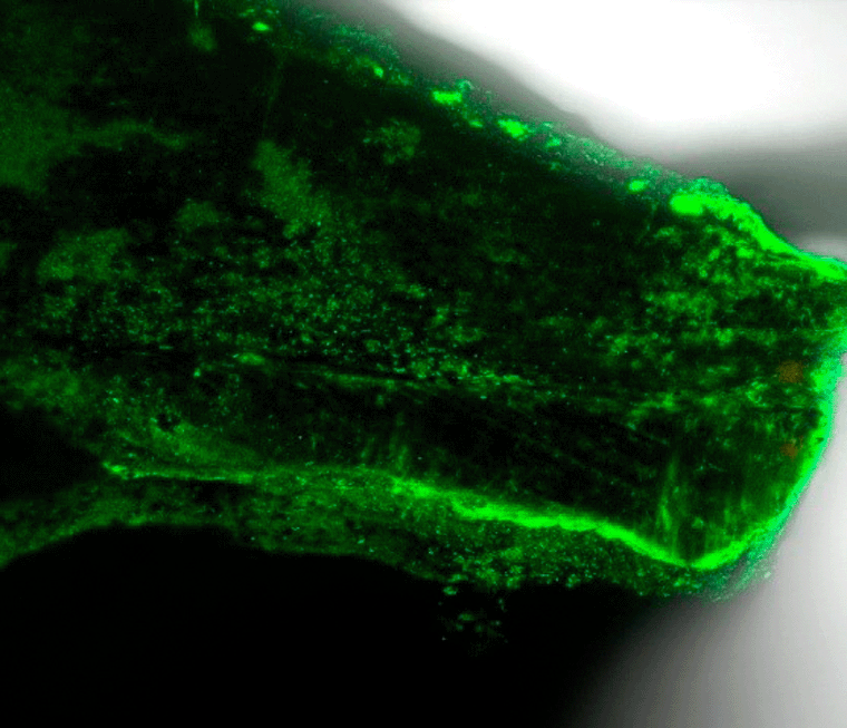
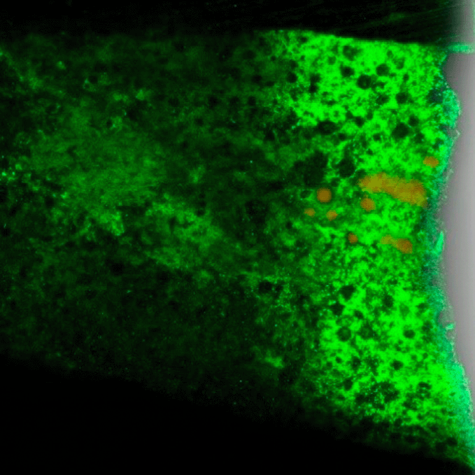
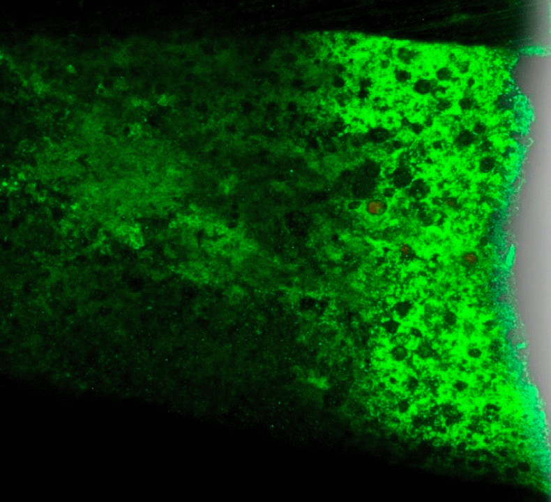
Each root was sectioned longitudinally into two halves with the usage of a diamond disc. Using a Confocal Fluorescent microscope (ZEISS LSM 800) at 10x magnification and a green filter, each sliced specimen was examined for marginal adaptation of the retrograde filling material to the walls of the prepared cavity, the presence and absence of gaps/voids, & the extent of penetration of the dye (wavelength 546 nm). Digimizer Image Analysis Software 6.3.0 was employed in assessing the amount of penetration of dye in three groups.
Scoring criteria for dye penetration for checking sealing ability.49
0-No dye penetration; 1 - Dye penetration into apical one third of retrograde filling material; 2 - Dye penetration into apical middle third of retrograde filling material; 3 - Dye penetration into full length of retrograde filling material; 4 - Dye penetration beyond retrograde filling material”.
For this study, descriptive and analytical statistics were used. The Shapiro-Wilk test was used to determine whether the data were normal.52 The data were analysed using parametric tests because they had a normal distribution.
The mean differences between the groups were examined using the one-way analysis of variance (ANOVA) test. Tukey’s HSD test was used in the post hoc analysis.
The software used was SPSS (Statistical Package for Social Sciences) Version 22.0 (IBM Corporation, Chicago, USA).
Group II (Zirconomer group) had the maximum mean (4.1 ± 0.57) followed by Group I (MTA group) (1.74 ± 0.65) and lastly Group III (Bioactive Bone Cement group) (1.4 ± 0.51) demonstrating highest microleakage due to increased dye penetration in Group II Zirconomer and least microleakage in Group III bioactive bone cement, thus indicative of highest sealing ability of Bioactive bone cement and least sealing ability with Zirconomer (Figures 1-3). There was statistically discernible dissimilarity in the amount of sealing ability admist the three groups of retrograde filling material with the p value of <0.001 (Table 2).


| Parameters | Group I: MTA | Group II: Zirconomer | Group III: Bioactive bone cement |
|---|---|---|---|
| Mean | 2.3 | 4.1 | 1.4 |
| Standard deviation | 0.65 | 0.57 | 0.51 |
| F Value | 69.01 | ||
| p-Value | <0.001 | ||
On intergroup comparison (Table 3), it was seen that the mean difference observed between Group I (MTA) and Group II (Zirconomer) of 1.8 is a statistically significant difference (i.e. p = 0.02) followed by the mean difference observed between Group I (MTA) and Group III (Bioactive Bone cement) of 1.1 which is a statistically significant difference (i.e. p = 0.03). Although, the Mean difference observed between Group III (Bioactive Bone cement) and Group II (Zirocnomer) of 3.3 is a highly statistically significant difference (i.e. p = 0.001).
Most endodontic tribulations can be resolved by utilising a standard conventional therapeutic approach.1 The fundamental goal of an endodontic therapy is to establish a three-dimensional hermetic seal between the periodontium and the root canal. For prevention of leakage and the entry of oral fluids along with organisms that could contribute to reinfection of the tooth, this seal should be established apically as well as coronally.53 It is essential to reduce the microbial load in these conditions to address the main etiological cause.54 Another cause of endodontic failure is complicated root morphology with an apical delta and lateral canals. Most of the endodontic failures arise as a consequence of irritants and bacteria seeping into the periapical region from infected root canals.1 According to dental estimates, 10 to 38% of root canal reintervention attempts have been shown to fail.55 The presence of microorganisms aggregated as biofilm in the root canal is a primary factor cited in those failed cases.54 Reduced microbial load is achieved in part by mechanically debriding out the length and diameter of the infected root canal.56 The retrograde filling serves as a second line of defense against persistent bacteria entering periapical tissues, hence the chosen material’s ability for sealing is crucial. Additionally, under specific circumstances, such as those that involve significant periapical lesions, instruments that are separated in the canals, apical variations, inadequate obturations, canal calcification, and dilacerated roots, etc., necessitates surgical intermediation as cleaning and shaping of the apical third becomes very challenging.57–59
Introducing the paradigm shift to endodontic surgery which is an approach employed in cases of periapical pathology that remains persistent or resistant to healing following nonsurgical retreatment60 and is cantered on the usage of equipment, materials, and techniques that integrate biological principles with tested clinical outcomes for healing of endodontic lesions.59
Resection of the root end, retrograde cavity preparation, and root-end filling are the measures taken to accomplish the goal of securing the root canal at the cut root end against microbes.10 Periradicular surgery followed by retrograde filling not only removes the infected periapical tissues & root apex, but also reseals the root canal. Creating a hermetic seal at the root apex with a root-end filling will inhibit bacteria and their byproducts to enter or exit the canal.1
A strong apical seal established with a retrograde filling material that is properly adapted is essential for the success of periradicular surgery. Improper marginal seal of the root ends, characterised by insufficient contact between the restorative material & the tooth surface, is typically the cause of apical surgery failure.61 Interstitial fluid may drive bacteria into the root canal, therefore apical closure should limit such leakage into the canal. Retrograde preparations and apicectomy increases the likelihood of leakage from the residual root, which emphasizes the need for retrofilling.62 So, a good apical seal must be provided by a retrograde filling substance that adheres well to the canal wall. Additionally, it should be biocompatible as well as capable of exhibiting osteoinductive/osteoconductive properties that will hasten healing in the periapical area and lower the risk of failures. The purpose of introduction of a retrograde filling is to create a fluid-tight apical seal, which prevents the leaking of residual irritants from the canal into the perirapical tissue & vice versa.63 Therefore, in the present study, the apical sealing ability of three root end filling materials to the root dentine was assessed.
Gilheany et al. (1994)11 stated that root resection at angles other than 90 degree and <3 mm from the apex exposes a greater number of dentinal tubules and leads to ineffective removal of root ramifications. Therefore, in the present investigation, root end resections were performed perpendicular to the long axis of the tooth and 3 mm from the apex. According to a research by Mjör et al. (2001),64 reduction in 98% of apical ramifications and 93% of lateral canals while decreasing the amount of exposed dentinal tubules was achieved by removing at least 3 mm of the root end.
Root-end resection can be carried out in three different planes to the tooth’s long axis: 30°, 45°, and 90°. Large number of exposed dentinal tubules, increased mechanical loads, and loss of dentine-cementum bone that arise from 30° & 45° resection angles impede healing after periapical surgery.65 The most preferred of them is 90° because it has the least impact on adaptability of retrograde material and reduces the possibility of leakage via the dentinal tubules that have been severed.
The performance of retrograde filling material and the outcome of surgical endodontic therapy are both influenced by a diverse range of variables. The strategy employed to prepare the root end cavity is one of them. Burs were traditionally employed as the tools to prepare the root end cavities. In comparison to cavities made with diamond-coated ultrasonic tips, it has been found that cavities prepared utilizing rotary instruments resulted in increased production of debris and smear layer.66 These remnants are permeable67 and permit microleakage, which prevents filling material from making complete contact with cavity walls.
Ergo, in the present era of dentistry, piezoelectric devices demonstrate a revolutionary method to accomplish the same goal while conserving soft tissue in the surgical field and selectively cutting mineralized tissue.68 With walls that are “parallel to & coincide with the anatomic outline of the root canal space”, the optimal root-end preparation should be at least 3mm into the root dentine.59 The apical cavities conform considerably more easily, safely, and precisely; they are smaller and more centrally positioned, which lowers the possibility of root perforation; and finally, improved cavity disinfection, which lowers the amount of dentinal remnants.69–71 Diamond-coated tips aid in preventing the propagation of dentinal cracks.72 Therefore, in the current investigation, S12 90ND Retrotips (NSK, Satelac) with diamond coating were used to prepare retrograde cavities at the optimal depth of 3 mm.
Different techniques have been devised to evaluate the sealing ability of root canal filling materials. These techniques are typically based on the same concept, which is to analyse the tracer’s capacity to penetrate along the obturated canal of an extracted tooth. For assessment of microleakage and subsequently the sealing ability, number of tracers including dye, radioisotope, bacteria and their products have been employed.73 Dye penetration technique is most often utilised for microleakage research since the dyes are affordable, secure, widely accessible, reasonably simple to store & use, and most significantly, their penetration can be analysed quantitatively.74
Indicators of potential leakage, such as dye penetration, should be taken into consideration. This is due to the fact that a filler material that can withstand the entry of small compounds like dyes may also be able to withstand the penetration of larger bacteria and their byproducts, according to Torabinejad et al. (1994).75 As a result, the dye penetration method was used in this investigation since it provides reliable information.
For dye leakage experiments, several different dyes are employed, such as India Ink, erythrosine B solution, aqueous fuchsin solution, fluorescent solution, silver nitrate, Methylene blue, rhodamine B, acridine orange, and others.76 Dye leakage studies depend on a number of factors, including the length of time the specimens are immersed in the marker, the time of immersion, whether negative pressure (vacuum) is used or not to release air trapped within filling gaps, whether the specimens are immersed completely in the dye, or only partially, the type of seal used, the quantity of specimens, the size of the marker, the position of the specimens during immersion, and, most importantly, the kind of material used.77 Methylene blue is frequently utilised in various studies of dye leakage. However, it has drawbacks, such as discoloration caused when methylene blue comes in proximity with an alkaline filling material. This occurs as a consequence of the ‘hydrolysis of methylene blue’, which produces a clear molecule named thioxine.78 MTA exhibits a high pH (12.5) & contains calcium oxide, which when in contact with water forms calcium hydroxide and is stained by methylene blue. Therefore, dye solutions that do not adversely affect the alkalinity of their marking capability are employed in evaluating the sealing capability of MTA.78
In the present investigation, acridine orange dye was utilised in place of methylene blue since it has additional benefits, such as smaller particle size, greater penetration, water solubility, diffusability, and hard tissue non-reactivity. The water-soluble fluorescent dye acridine orange, is a metachromatic vital stain, is not harmful to the substrate or material in touch, is readily identifiable even at low concentrations, flows freely along the interface, has low toxicity, and is stable in an aqueous environment. It diffuses more effectively on human dentin in comparison to methylene blue. When excited by blue light, the fluorescent dye helps in differential staining where the voids or spaces fluoresce bright orange against a green-fluorescing dark background.
A linear or spectrophotometric measurement of dye penetration is achievable. However, in a study comparing longitudinal splitting, cross sectioning, and cleaning of the specimens, it was shown that longitudinally split samples exhibited improved dye penetration.77 Hence, after longitudinally splitting the samples with a diamond disc, the linear measurement of dye penetration is assessed in the current investigation. This study employed Confocal Fluorescent Microscopy (ZEISS LSM 800) at 10× magnification in conjunction with a green filter (wavelength 546 nm) to assess the sealing ability. This method provides advantages over other traditional techniques such as “scanning electron microscopy or transmission electron microscopy. The advantages of this technique include the elimination of out-of-focus blur in images as well as the ability to create three-dimensional images that disclose more precise information than two-dimensional images.79 The confocal fluorescent microscope does not require a specific sectioning method, hence the incidence of artifacts is lower when compared to a scanning electron microscope.80
The retrograde filling material groups were as follows: Group I (MTA), Group II (Zirconomer) and Group III (Bioactive Bone cement). The sealing ability values were as given below: Group I (MTA): 2.3 ± 0.65; Group II (Zirconomer): 4.1 ± 0.57 and Group III (Bioactive Bone cement): 1.4 ± 0.51. Group II (Zirconomer group) had the highest mean (4.1 ± 0.57) followed by Group I (MTA group) (2.3 ± 0.65) and lastly Group III (Bioactive Bone cement group) (1.4 ± 0.51).
When Group I (MTA) (2.3 ± 0.65) was compared with Group II (Zirconomer) (4.1 ± 0.57), the difference of 1.8 ± 0.80 was found. There was statistically discernible dissimilarity found amid Group I and II with the p value <0.001.
The study’s findings revealed that every material examined demonstrated microleakage. The highest microleakage values were produced by Group II Zirconomer which is attributed to the high solubility possessed by this material. Compromised sealing produced by Zirconomer could be described by the presence of zirconia ceramic filler particles that make up Zirconomer’s chemical makeup. It is probable that the zirconia fillers would interfere with the chelating process between the calcium ions (Ca2+) in tooth apatite and the carboxylic group (-COOH) of polyacrylic acid.81 This was in congruence with a study done by Asafarlal S et al. (2017)82 which showed inferior adaptation of Zirconomer at the tooth restoration interface owing to larger size of zirconia filler particles.
MTA showed better sealing ability when compared to Group II zirconomer. Good sealing ability & marginal adaptation of MTA is caused by the deposition of hydroxyapatite crystals & the formation of a hybrid layer between dentine and calcium silicate–based materials. The formed hydroxyapatite crystals gradually grow and nucleate in order to fill the microscopic gaps between calcium silicate based cements & the dentinal wall.83 The sealing ability of MTA may have improved as a result of Ca ions bioavailability inside these materials which stimulates the production of bone-associated proteins through Ca channels and large amounts of Ca ions could eventually activate adenosine triphosphate, which is important in the mineralization process.84 Additionally, the good sealing ability of MTA may be due to dihydrogen monoxide absorption during the powder’s hydration, which causes expansion during setting. The positioning of the cementum and periodontal ligament strands adjacent to MTA is another intriguing feature.85
Due to the MTA Angelus’s reduced particle size and higher specific surface area of its particles, the wetting volume, water binding capacity, and hydration rate are all increased. Additionally, because MTA is hydrophilic and expands during setting expansion when cured in a moist environment, moisture does not alter its setting or its qualities during periapical surgery.86 This finding is reinforced by a study by Xavier et al. (2005),41 which found that MTA Angelus had better marginal adaptability and sealing capability than super EBA and Vitremer.
Conversely, studies by Ahirwar et al. (2020),87 Bolbolian et al. (2020)88 and Fatinder Jeet Singh et al. (2022)89 were in contradiction to this present study as they concluded that the production of tag-like structures made of crystalline deposits rich in calcium or phosphate at the junction of tooth and retrograde filling materials and the shorter setting period, i.e., 12 minutes would be the reason for the greatest sealing ability of Biodentine over MTA.
An invitro study by Dhivya et al. (2022)90 was in contradiction to the present study as it concluded that Zirconomer had enhanced sealing ability due to “incorporation of zirconia fillers which is an uneven compound and hence deviations in its phase from monoclinic to tetragonal and then to cubic and vice versa lead to increase in volume thereby counteracting the volumetric shrinkage expressed during polymerization”.91
When Group I (MTA) (2.3 ± 0.65) was compared with Group III (Bioactive Bone cement group) (1.4 ± 0.51), the difference of 1.1 ± 0.14 was found. There was statistically discernible dissimilarity seen amid Group I and III with the p value <0.001.
The current study revealed that Group I MTA had inferior sealing ability when compared to Bioactive bone cement. This can be attributed to MTA’s extended maturation phase, which is linked to the creation of a passivating trisulfate coating over hydrated crystals, simulating a terrible clinical condition” in congruence to a study conducted by Chaudhari et al. (2022).92
In comparison to MTA & Zirconomer, the acquired results showed that group III bioactive bone cement had superior adaptation to the root dentine in the longitudinal sections. These findings are consistent with those of the earlier dye diffusion investigation conducted by High and Russell et al. (1989),93 who demonstrated a successful marginal seal when bone cements (CMW and Palacos bone cement with gentamicin) were used as retrograde filling materials. Additionally, Holt and Dumsha’s (2000)94 leakage research demonstrated that Simplex P bone cement offered a reliable retrofill seal. The results of earlier studies by Charnley et al. (1970),95 who used a fluid displacement model and observed that the volume of cement increases to a maximum during polymerization before shrinking slightly, but not to its initial volume, may help to explain how bone cement is able to adhere to the dentin wall properly despite the well-known polymerization shrinkage of acrylics.
According to Miyazaki et al.96 (2003), Si-OH groups produced on the material’s surface can act as a catalyst to promote the formation of apatite by releasing Ca2 + ions into the body fluid. The addition of silane to the liquid component results in the formation of a Si-OH group due to the hydrolysis of alkoxysilane following exposure to the body environment, which causes heterogeneous HA nucleation. In contrast, the addition of MTA powder to bone cement results in the formation of Ca ions. The addition of silane to bone cement improves its mechanical properties because chemical bonding can be achieved with PMMA. Additionally, any PMMA polymerization shrinkage is offset by the slight MTA expansion that occurs during setting, increasing the sealing and strength of modified bone cement. This could account for the better apical sealing capabilities of Bioactive bone cement.
Additionally, the exothermic reaction released during the setting of bone cement did not seem to have any negative effects due to very small amount needed in root end fillings as opposed to quantities required in orthopaedics.97 Less quantities needed would result in a considerably lower exothermic reaction & a reduced quantity of free monomer as stated by Badr et al (2010).25 Bone cement could thus be used in both dentistry and medicinal applications. There are other bone cements available with antibiotics incorporated.
An in vitro study performed by Chordiya et al. (2022)98 & Akbulut et al. (2018)99 are in contradiction to the present study. They claimed that the cell reduction caused by the bone cement was comparable to that caused by the Micro-Mega mineral trioxide aggregate (MM-MTA), Smart Dentin Replacement (SDR) and ProRoot MTA (PMTA). These results are in accordance with those of Badr et al. (2010),25 who observed that PMMA bone cement & MTA have similar cytotoxic effects on fibroblasts. Although, the poor biocompatibility of PMMA bone cement may be caused by residual methylmethacrylate monomer.100
When Group II (Zirconomer) (4.1 ± 0.57) was compared with Group III (Bioactive Bone cement group) (1.4 ± 0.51), the difference of 3.3 ± 0.06 was found. There was statistically discernible dissimilarity found amid Group II and III with the p value <0.001.
This study proved bioactive bone cement to have highest sealing ability. This can be attributed to its excellent interlocking property. With superior handling capabilities that make it possible to mould it into a dough form and place it in the root end cavity area, the bone cement could also endure a damp environment and is unaffected by blood contamination. This is in congruence to a study performed by Ann Saji et al. (2022).101
Within the limitations of this investigation, it can be concluded that the seal created by bioactive bone cement in retrograde filling was superior to Ziconomer and comparable to MTA. It offered more potent operating qualities that could outweigh any prospective MTA drawbacks. In light of this, bioactive bone cement appears to be an advantageous and promising material for retrograde filling. It also has the potential to be employed as a repair material in a variety of other clinical settings, including furcation repair and resorptive defects.
Advancements in dental materials will help broaden the perspective to have a holistic approach with better marginal adaptability and sealing ability in retrograde preparations preventing microleakage. More recent materials for root-end filling can be evaluated and compared. The teeth with multiple canals can be evaluated that might change the outcome of the study. Further in vivo studies should be carried out to provide more accurate results.
Zenodo: Comparison of Root End Sealing Ability of three Retrograde Filling Materials in Teeth with Root Apices Resected at 900 using dye penetration method under fluorescent microscope. https://doi.org/10.5281/zenodo.7857676.
This project contains the following underlying data:
Zenodo: Strobe checklist for the study. https://doi.org/10.5281/zenodo.7861151.
This project contains the following extended data:
Data are available under the terms of the Creative Commons Attribution 4.0 International license (CC-BY 4.0).
| Views | Downloads | |
|---|---|---|
| F1000Research | - | - |
|
PubMed Central
Data from PMC are received and updated monthly.
|
- | - |
Is the work clearly and accurately presented and does it cite the current literature?
Yes
Is the study design appropriate and is the work technically sound?
Yes
Are sufficient details of methods and analysis provided to allow replication by others?
Partly
If applicable, is the statistical analysis and its interpretation appropriate?
Yes
Are all the source data underlying the results available to ensure full reproducibility?
Yes
Are the conclusions drawn adequately supported by the results?
Yes
Competing Interests: No competing interests were disclosed.
Reviewer Expertise: Dental biomaterials, restorative dentistry, prosthodontics, dental education, mechanical properties
Is the work clearly and accurately presented and does it cite the current literature?
Partly
Is the study design appropriate and is the work technically sound?
No
Are sufficient details of methods and analysis provided to allow replication by others?
Partly
If applicable, is the statistical analysis and its interpretation appropriate?
Yes
Are all the source data underlying the results available to ensure full reproducibility?
No
Are the conclusions drawn adequately supported by the results?
No
References
1. Chaudhari P, Chandak M, Ramdas A, Bhagat P: Comparison of Root End Sealing Ability of Three Retrograde Filling Materials in Teeth with Root Apices Resected at 90° using Dye Penetration Method under Fluorescent Microscope: An In-vitro Study. Journal of Pharmaceutical Research International. 2021. 319-325 Publisher Full TextCompeting Interests: No competing interests were disclosed.
Reviewer Expertise: Endodontics
Alongside their report, reviewers assign a status to the article:
| Invited Reviewers | ||
|---|---|---|
| 1 | 2 | |
|
Version 1 29 Aug 23 |
read | read |
Provide sufficient details of any financial or non-financial competing interests to enable users to assess whether your comments might lead a reasonable person to question your impartiality. Consider the following examples, but note that this is not an exhaustive list:
Sign up for content alerts and receive a weekly or monthly email with all newly published articles
Already registered? Sign in
The email address should be the one you originally registered with F1000.
You registered with F1000 via Google, so we cannot reset your password.
To sign in, please click here.
If you still need help with your Google account password, please click here.
You registered with F1000 via Facebook, so we cannot reset your password.
To sign in, please click here.
If you still need help with your Facebook account password, please click here.
If your email address is registered with us, we will email you instructions to reset your password.
If you think you should have received this email but it has not arrived, please check your spam filters and/or contact for further assistance.
Comments on this article Comments (0)