Keywords
Crohn, Emergency, Surgery, Laparoscopy, Inaugural, Small bowel, Peritonitis, Appendicitis,
Crohn, Emergency, Surgery, Laparoscopy, Inaugural, Small bowel, Peritonitis, Appendicitis,
Crohn’s disease (CD) is a chronic transmural inflammation that can affect the gastrointestinal tract from the mouth to the anal margin.1 Inaugural spontaneous perforation with generalized peritonitis is infrequent and potentially life-threatening occurrence.2 Herein, we present a case study of acute generalized peritonitis caused by the perforation of a distal ileal loop, leading to the diagnosis of Crohn's disease.
A 43-year old man presented to the emergency department with a one-day history of abdominal pain and vomiting. There were no signs of bowel obstruction or fever. The patient had no medical or surgical history, and was a non-smoker. He reports paroxysmal abdominal pain and diarrhea for the last year.
On examination, his temperature was 37.4 °C and generalized abdominal tenderness were noticed. The maximum pain was at the level of the right iliac fossa. Hemodynamic state remained stable, and the respiratory rate was normal.
Laboratory data showed elevated white blood cell count (18500/mm3) and C-reactive protein level (140 mg/l). The results of the serum electrolyte and kidney function tests were normal.
Acute appendicitis complicated with peritonitis were considered and the patient underwent urgent laparoscopic exploration, without morphologic exploration.
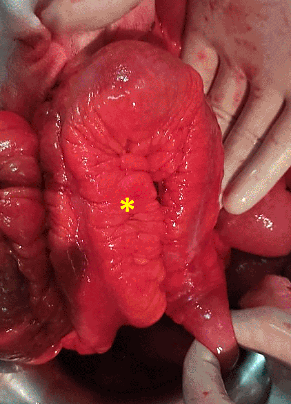
The peroperative exploration of the abdominal cavity found a free purulent fluid caused by a perforation in the anti-mesenteric border at the distal ileum (Figure 1), 40 cm from the ileocecal valve. Furthermore, we observed the presence of an inflammatory-looking conglomerate of small bowel loops associated with sclerolipomatosis (Figure 2 and 3). Terminal ileal loop, caecum and appendix were grossly normal. Due to poor exposure conditions, we decided to convert to a midline procedure.
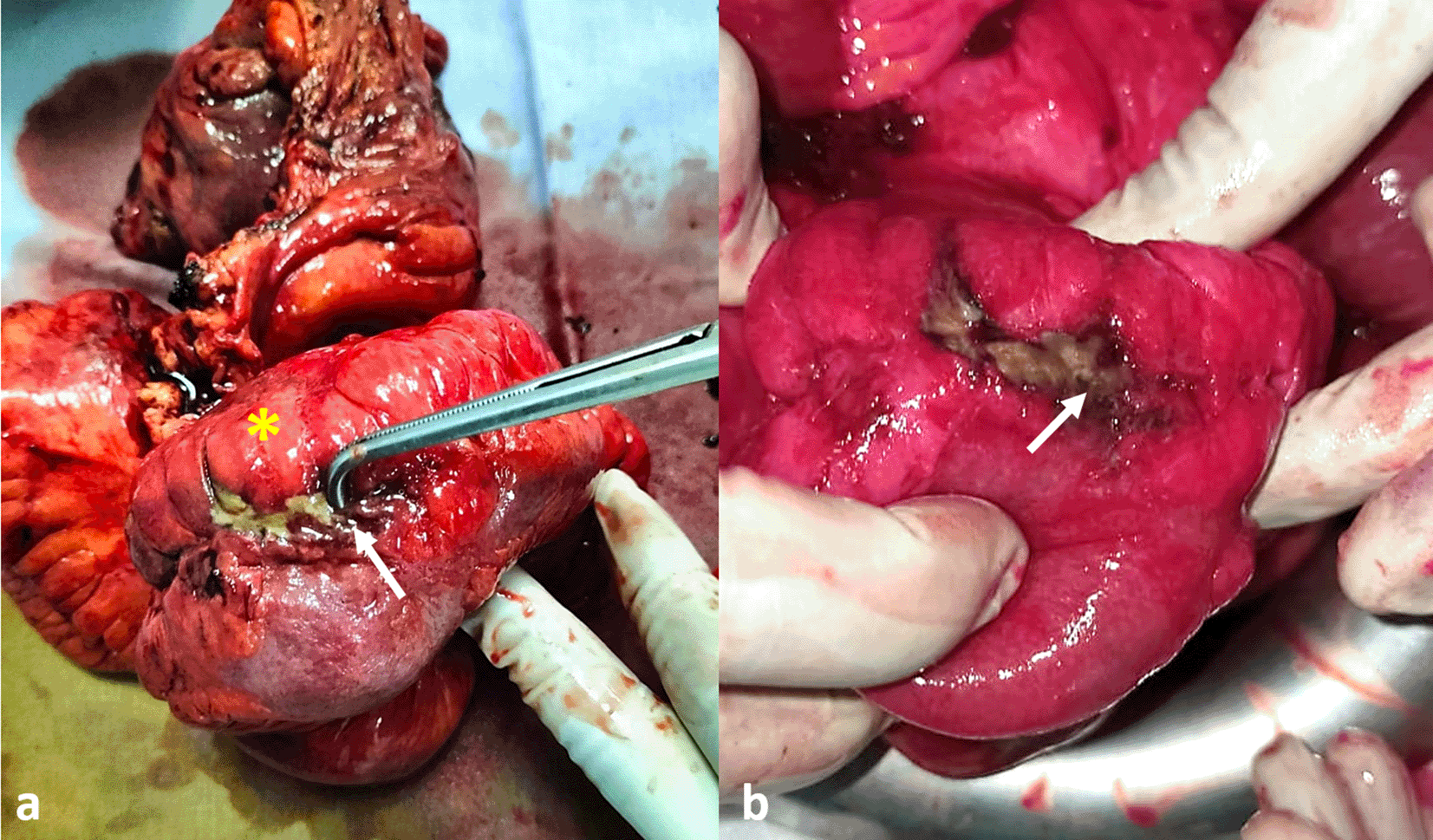
a: Specimen after resection, (the yellow asterisk shows the sclerolipomatosis), b: Appearance of the perforation before resection.

An abundant peritoneal toilet with serum salin and ileocaecal resection carrying out 50 cm of bowel were performed. Remaining bowel length was estimated at 4.5 meter. An ileo-colostomy in the right iliac fossa was performed.
The post-operative course was uneventful. First oral intake was allowed since the first post-operative day. The patient was discharged on the fifth postoperative day.
The histopathological analysis of the specimen revealed a chronic granulomatous ileitis lesions, which are indicative of Crohn's disease.
Crohn's disease is a chronic inflammatory disease of unknown origin that has the potential to impact the entire gastrointestinal tract from the mouth to the anus.2 Although, the terminal ileum and colon are the most frequently involved areas.3 The transmural characteristic of Crohn's disease (CD) can lead to localized perforation, which has the potential to progress rapidly into generalized peritonitis.1 Free perforation of the small intestine in the peritoneal cavity is a rare and serious complication in Crohn's disease (CD).4
The originality of our case lies in the fact that peritonitis secondary to ileal perforation was inaugural, leading to the diagnosis of Crohn's disease.
The diagnosis of acute peritonitis can be considered on physical examination.4 However, the challenge of conducting etiological investigations is particularly pronounced, especially given the absence of supportive findings in the medical history for Crohn's disease.5 In our case, paroxysmal abdominal pain and diarrhea suggest Crohn's disease and a CT scan could be performed to better support the diagnosis. However, the surgery was obviously required, regardless of the scan features. For this reason, the patient underwent a laparoscopic exploration without any morphological investigations.
Perforative peritonitis as a primary manifestation of Crohn's disease is very rare, and occurs in 1 to 3 % in general population.2,4 However, The CONNECT retrospective cohort Korean study reported a higher incidence of 6,5% which is similar to a Japanese meta-analysis result.2,3,6
Among the reported cases of perforation in various literature, the most frequently site affected is ileum, particularly the terminal ileum (80%).2,4 Less frequently involved locations consist of other segments of the ileum, the colon, and the jejunum.2,4
The precise mechanism behind the occurrence of free perforation in Crohn's disease remains unidentified. However, many risk factors may be incriminated such as anti TNF therapy or significant distension upstream of a stenosis.3,4
Surgical treatment involves performing a limited resection of the affected segment of the bowel, followed by either a primary anastomosis or a temporary stoma.3 In this particular case, considering the peritoneal contamination, an ileocecal resection with a stoma was performed.
Although Crohn’s free perforation in the peritoneal cavity is infrequent, it should be kept in mind especially in front of suggestive features.4 Timely diagnosis and prompt treatment significantly enhance the prognosis.5 Surgery is obviously required for acute peritonitis, with limited resection with or without anastomosis.
| Views | Downloads | |
|---|---|---|
| F1000Research | - | - |
|
PubMed Central
Data from PMC are received and updated monthly.
|
- | - |
Provide sufficient details of any financial or non-financial competing interests to enable users to assess whether your comments might lead a reasonable person to question your impartiality. Consider the following examples, but note that this is not an exhaustive list:
Sign up for content alerts and receive a weekly or monthly email with all newly published articles
Already registered? Sign in
The email address should be the one you originally registered with F1000.
You registered with F1000 via Google, so we cannot reset your password.
To sign in, please click here.
If you still need help with your Google account password, please click here.
You registered with F1000 via Facebook, so we cannot reset your password.
To sign in, please click here.
If you still need help with your Facebook account password, please click here.
If your email address is registered with us, we will email you instructions to reset your password.
If you think you should have received this email but it has not arrived, please check your spam filters and/or contact for further assistance.
Comments on this article Comments (0)