Keywords
Epithelial-mesenchymal transition, Histopathological marker, Histological evaluation, Invasive tumor front, Lymph node metastases, Oral squamous cell carcinoma, Prognosis, Tumor budding
This article is included in the Oncology gateway.
This article is included in the Datta Meghe Institute of Higher Education and Research collection.
Epithelial-mesenchymal transition, Histopathological marker, Histological evaluation, Invasive tumor front, Lymph node metastases, Oral squamous cell carcinoma, Prognosis, Tumor budding
In the abstract, there was a repetition of the words - lymph node metastasis, removed in the new version.
In the histological features, images are replaced and correlated with grading parameters that are - High-intensity tumor buds are >5 clusters of tumor cells and Low-intensity tumor buds are <5 clusters of tumor cells.
To read any peer review reports and author responses for this article, follow the "read" links in the Open Peer Review table.
In India, oral cancer contributes to two-thirds of cancer cases. Oral squamous cell carcinoma (OSCC) has been categorized as the third most common malignancy and is considered a major risk factor because of increased consumption of tobacco and areca nut.1 OSCC has a low survival rate due to an early tendency to metastasize and recurrent recurrences. Therefore, early detection, correct diagnosis, and timely treatment are necessary to reduce morbidity and mortality. The invasive tumor front is the region where the most advanced layers of tumor reside, this gives the detailed prognosis of OSCC cases.2 OSCC cases are usually managed by surgery followed by postoperative radiation therapy. Although diagnostic techniques and treatment methods have improved, the mortality rates of patients with OSCC are still inadequately low with a poor survival rate of five years, challenging the current approaches of prognostic assessment. The aggressive clinical history of OSCC, which includes frequent locoregional relapses and lymph node metastasis (LNM) in more than 60% of cases, makes the prognosis uncertain.3,4 Tumor budding (TB) is the gold standard and reliable histomorphological parameter that has been studied in many cancers. TB is characterized by a dispersed invasive pattern in which epithelial tumor cells are visible at the invasive front as small clusters (up to five cells) spread throughout the stroma at different distances.5 In the 1950s, Imai et al.,6 originally described TB as “sprouting”, Gabbert et al.,7 termed this feature as “tumor dedifferentiation”. Later Morodomi et al.,8 and Hase et al.,9 coined the term “budding” as they observed the budding of undifferentiated cells and nests from larger tumor islands.1 TB is thought to represent the epithelial-mesenchymal transition (EMT), i.e., the important mechanism for the progression of epithelial cancers.10 As a result, we aimed to evaluate the importance of TB in the prognosis of OSCC in the current study.
This study aimed to perform an evaluation of TB, determine its association with various histopathological parameters, and thereby the prognosis in OSCC.
The objectives of this study were as follows: i) to assess TB in OSCC; ii) to evaluate TNM staging in OSCC; iii) to determine the association between TB with LNM in OSCC; iv) to determine the association between TB and the five year survival rate in OSCC; and v) to determine the association between TB with histopathological grade.
The procedure that is employed the most frequently is the manual assessment of TB on hematoxylin and eosin (H&E) stained slides. For the classification of TB intensity, several authors have proposed a variety of techniques with a range of criteria. However, the technique by Wang et al.,5 from 2011, is one that is the most frequently acknowledged and used in OSCC. According to this approach, TB refers to isolated single cells or clusters of five tumor cells found at the invasive front. To locate the fields with the highest density of TB, a section is initially scanned at low power. Following this, the tumor buds are counted in a single field at a resolution of ×200 or ×20 objective. The samples are then divided into two groups: high-intensity TB (five or more buds) and low-intensity TB (five buds or fewer) or absence TB. Each section in the present study was reviewed by three observers, then by consensus to remove bias. The cases underwent follow-ups at intervals of two months. This study followed the STROBE guidelines.11
Daniel formula for sample size estimation:
Where,
Z2 - The level of significance at 5%
i.e. 95% confidence interval =1.96
P = Prevalence of OSCC in India = 80% = 0.80
d = Desired error of margin = 10% = 0.10
Formula Reference: Daniel et al.12
The present study was conducted from November 2022 at the Department of Oral Pathology and Microbiology, Sharad Pawar Dental College and Hospital, after receiving approval (dated, 05/10/2022) from the Institutional Ethical Committee [DMIMS (DU)/IEC/2022/292] of Datta Meghe Institute of Higher Education and Research, Sawangi (M), Wardha, Maharashtra, India. A total of 30 surgically operated cases of OSCC from the year 2010-2015 at this institute were retrieved from the archival of the department. There were 21 cases that had evaluated TB in OSCC, which included eight cases of well-differentiated squamous cell carcinoma (WDSCC), 12 cases of moderately differentiated squamous cell carcinoma (MDSCC), and one case of poorly differentiated squamous cell carcinoma (PDSCC) with their average age between 30-70 years. In order to assess the survival data, we had telephone conversations with patients or with their relatives. Informed written and verbal consent was taken from all participants involved in the study.
The histopathologically diagnosed and surgically operated cases of OSCC with various histopathological grades were retrieved from the departmental archives. The staging of the patients has been done according to the “American Joint Committee of Cancer” (AJCC) staging system.13 The cases who had undergone presurgical radiotherapy or chemotherapy, history of second primary or local and distant recurrence, and patients with any other systemic diseases and with coexisting malignancy were excluded from the study. The detailed information of the study population, including the clinical staging of the disease, comprehensive history of pertinent habits with their duration, and histopathological features such as grades of OSCC and lymph node metastasis had been noted.
A chi-squared test was used to test the possible relationship between various clinicopathological variables. The statistical analysis was conducted using IBM SPSS Statistics (RRID: SCR_016479) v27.0 software, and p<0.05 is thought to be the threshold of significance. A chi-squared test, descriptive statistics, and inferential statistics were all used.
Each clinical and histological feature and TB are cross-tabulated sequentially. Stromal TB was seen in each of the 21 cases. Nine out of the 21 cases (less than five tumor buds) had low-intensity TB, while 11 had high-intensity TB (more than five tumor buds) (Table 1).25 Out of 30 patients in the current study, 20 cases demonstrated an association between TB and lymph node metastases, while the other 10 cases did not (Table 2). We discovered that out of 20 cases that showed the presence of TB, 12 patients are dead and eight are alive, indicating that there is no connection between TB status and the likelihood of a patient surviving (Table 3). When compared to cases of WDSCC and PDSCC, cases of MDSCC demonstrated the presence of TB (Table 4). This finding suggests that TB can be used as a reliable histopathological parameter for early detection and diagnosis and to determine the prognosis of OSCC cases.
| TB | HI/LI | Total | χ2-value | ||
|---|---|---|---|---|---|
| HI | LI | No findings | |||
| Present, n (%) | 11 (55%) | 9 (45%) | 0 (0%) | 20 (66.67%) | 25.95 P=0.0001, S |
| Absent, n (%) | 0 (0%) | 1 (10%) | 9 (90%) | 10 (33.33%) | |
| Total, n (%) | 11 (36.67%) | 10 (33.33%) | 9 (30%) | 30 (100%) | |
| TB | LNM | Total | χ2-value | |
|---|---|---|---|---|
| Yes | No | |||
| Present, n (%) | 11 (55%) | 9 (45%) | 20 (66.67%) | 8.68 P=0.003, S |
| Absent, n (%) | 0 (0%) | 10 (100%) | 10 (33.33%) | |
| Total, n (%) | 11 (36.67%) | 19 (63.33%) | 30 (100%) | |
| TB | Survival | Total | χ2-value | |
|---|---|---|---|---|
| Dead | Alive | |||
| Present, n (%) | 8 (40%) | 12 (60%) | 20 (66.67%) | 0.28 P=0.59, NS |
| Absent, n (%) | 3 (30%) | 7 (70%) | 10 (33.33%) | |
| Total, n (%) | 11 (36.67%) | 19 (63.33%) | 30 (100%) | |
The H&E stained section of OSCC shows the presence of TB as indicated by the presence of cell nests containing single-cell infiltration. The invasive front of OSCC is shown by a histological investigation to have tumor buds that are spreading into the underlying connective tissue. High-intensity TB is the presence of >5 cell clusters at the invasive tumor front of the lesion (Figure 1, marked by black arrow), and low-intensity TB is the presence of <5 cell clusters at the invasive tumor front of the lesion (Figure 2, marked by black arrow).
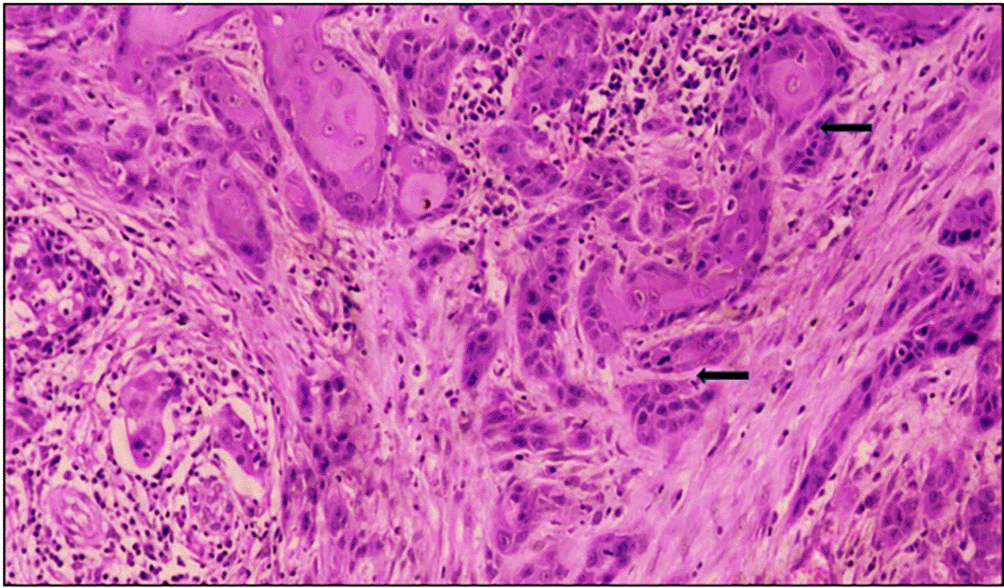
Under high power view (magnification, ×40). TB, tumor budding.
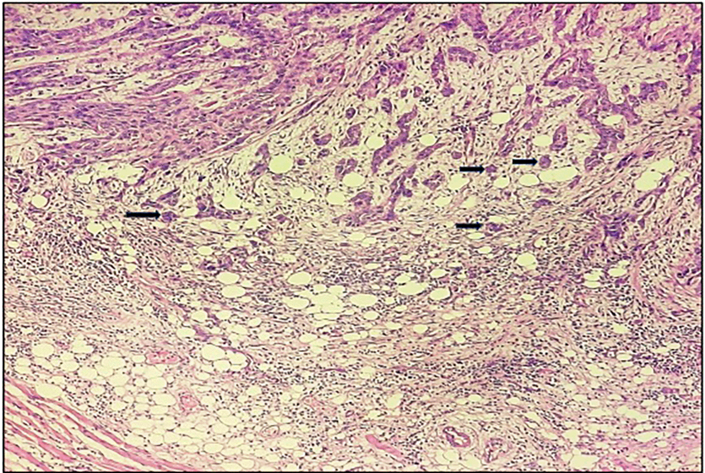
Under low power view (magnification, ×10). TB, tumor budding.
OSCC contributes to 95% of oral cancers and predominantly metastasizes to the lymph nodes of the neck. Metastasis of cancer cells to lymph nodes i.e., positive lymph nodes (LN) is an important adverse prognostic factor for the survival of OSCC cases.14 In several studies of OSCC, TB has been recorded. Some of these studies have shown that TB is an important prognosticator of LNM, distant metastatic disease and local recurrence.15–18 Strong evidence linking TB and LNM has been established, and multivariate analysis shows that TB is associated with aggressive tumor activity and a bad prognosis in instances, suggesting that TB count can be used to gauge the risk of LNM.1,19 TB count does not require the use of any other expensive techniques or equipment because it is a simple procedure used on a daily basis with the help of H&E stained sections.15,20 TB is not a static histological feature, but it is a series of various processes occurring in an aggressive tumor with the potential to disseminate and metastasize. This biological phenomenon is a dynamic process carried out by a tumor with the potential to invade other tissues, spread and metastasize.16
TB might be a sign of cells going through EMT, a process in which highly polarized epithelial cells become mobile mesenchymal cells. Therefore, EMT can be regarded as an indicating factor for invasion and subsequent metastasis.21 Metastasis can be evaluated from the increased distance of the tumor bud and reduced tumor bud size. Significantly more metastases were linked to decreased tumor bud size and increased tumor bud distance. The extent of EMT increased with the distance the tumor buds progressed. Thus, EMT has been related to poor prognostic outcomes in OSCC.22 Treatment plans and prognosis could be enhanced with precise histological grading and scoring of particular features. Sundar et al.,23 evaluated the prognostic value of TB in OSCC in 2019. They evaluated TB at the invasive tumor front in all OSCC cases. They discovered that advanced malignancies with tumor sizes larger than 4 cm exhibited high-intensity TB. They came to the conclusion that TB always manifested as an invasive front with cell infiltration in cords or groups. The fact that TB occurs before lymph node metastasis suggests that it is a reliable prognostic marker. They found a significant relationship between LNM and TB. Their research highlighted the significance of TB in the treatment of OSCC cases.23 The worst pattern of invasion (WPOI) and TB were two histological markers that were evaluated by Chatterjee et al., in 2019.17 The risk of LNM, recurrence, and death in early-stage OSCC were found to be best predicted by early detection of these characteristics. The treatment plan and outlook for OSCC are based on the tumor stage and LNM status. During the course of four and a half years, they examined the histological characteristics in 126 samples, including the histological grade, WPOI, TB, lymphovascular emboli, perineural invasion, depth of invasion, and host lymphocyte response. The findings of the study resulted in the recommendation that WPOI and TB be included in the guidelines for histopathological reporting as important risk factors for predicting LNM in all stages of OSCC and reliable prognostic markers.17 Almangush et al. (2014),15 investigated the effect of TB on prognosis in OSCC. They examined 16 studies to test this in a meta-analysis. For a better prognosis, they advised using the Reporting Recommendations for Tumor Marker Prognostic Studies (REMARK) criterion in histology reporting. They assessed the prognostic significance of TB in OSCC. Their research demonstrated a link between lymph node metastasis and TB. TB, which can be seen easily, is a valid prognostic indicator for OSCC and may help with individualized treatment, according to their findings.15 When compared to lymph nodes without metastasis, TB is significantly (P=0.003) associated with the presence of lymph node metastasis in our study (Tables 1 and 2), which is in accordance with previous studies by Sundar et al. (2019),23 Chatterjee et al. (2019),17 Almangush et al. (2014).15
TB was examined by Joshi et al. (2020),16 as a potential predictive histopathological feature in oral squamous cell carcinoma. Overall, 30 OSCC cases from January 2018 to August 2019 were investigated. While analyzing invasive tumors, they considered both high-intensity tumor bud clusters of cancer cells and low-intensity tumor bud clusters of cancer cells. There found no evidence of a significant relationship between TB and the survival rate.16 Candanedo-Gonzalez et al. (2020),18 investigated incidences of OSCC in the tongue (TSCC), which had a susceptibility to early local spread and recurrence. They reported that TSCC at an early stage (T1/T2N0M0) is not necessarily associated with a favorable prognosis. It is essential to identify prognostic indicators that can be assessed using biopsies. They identified that the EMT, specifically in TSCC, is histologically represented by TB. As a result, they determined that TB in the TSCC was a biological marker that predicted a poor prognosis.18 The findings of our study (P = 0.59, not significant) (Table 3), demonstrated that there is no relationship between the presence or absence of TB and patient survival rates, which is in accordance with previous studies by Joshi et al. (2020)16 and Fernando Candanedo-Gonzalez et al. (2020).18
Nandita et al. (2016),24 investigated the contribution of TB to the diagnosis of OSCC. For this investigation, 30 histological samples of OSCC with a biopsy-proven diagnosis were chosen. They found tumor cells at the invasive front of the tumor. The number of tumor buds in which TB intensity was at its highest in the histological fields is counted. They used an independent t-test to compare prognostic variables with TB. As a result, they came to the conclusion that the relationship between TB and poor tumor differentiation, as well as its presence, could be crucial indicators to forecast the prognosis of patients with OSCC.22 Our research is consistent with that of Nandita et al. (2016).24 In the current study, we came to the conclusion that tumor buds are present in WDSCC and MDSCC at the invasive tumor front as compared to PDSCC (Table 4), suggesting the relevance of TB in early diagnosis of oral squamous cell carcinoma after assessing the existence of TB in various histological grading.
To conclude, tumour budding is an essential factor for determining the behaviour of tumours. TB refers to clusters of neoplastic cells at the invasive tumour front than the neoplastic cells in the primary tumour mass. TB is a frequent, repeatable, and easy-to-identify histological factor in OSCC. This study highlights the relevance of TB in prognosis as well as the significance of its evaluation and integration into the routine management of OSCC.
| Views | Downloads | |
|---|---|---|
| F1000Research | - | - |
|
PubMed Central
Data from PMC are received and updated monthly.
|
- | - |
Provide sufficient details of any financial or non-financial competing interests to enable users to assess whether your comments might lead a reasonable person to question your impartiality. Consider the following examples, but note that this is not an exhaustive list:
Sign up for content alerts and receive a weekly or monthly email with all newly published articles
Already registered? Sign in
The email address should be the one you originally registered with F1000.
You registered with F1000 via Google, so we cannot reset your password.
To sign in, please click here.
If you still need help with your Google account password, please click here.
You registered with F1000 via Facebook, so we cannot reset your password.
To sign in, please click here.
If you still need help with your Facebook account password, please click here.
If your email address is registered with us, we will email you instructions to reset your password.
If you think you should have received this email but it has not arrived, please check your spam filters and/or contact for further assistance.
Comments on this article Comments (0)