Keywords
Keywords- Gingival pigmentation, Scalpel technique, BlueM gel, Active oxygen species, Coe-pack, wound healing, Gingival hyperpigmentation, Esthetics.
This article is included in the Datta Meghe Institute of Higher Education and Research collection.
Keywords- Gingival pigmentation, Scalpel technique, BlueM gel, Active oxygen species, Coe-pack, wound healing, Gingival hyperpigmentation, Esthetics.
The smile is a significant element of social relationships and has an immense effect on making a positive first impression because it conveys emotions and confidence. Dark-coloured gingiva looks aesthetically unpleasant and is a matter of concern that should be taken into account, especially with a gummy smile or high lip line. Dark pigmentation is frequently caused by an excessive accumulation of melanin in the gingival epithelium; a brown pigment that can range in colour from pale pink to dark brown. In every ethnic group, gingiva have some degree of melanin pigmentation, which is a physiological process, and its presence is not indicative of risk.1
Periodontal plastic surgery called gingival depigmentation uses a range of techniques to eliminate or to reduce hyperpigmentation. The primary rationale for depigmentation in patients is a desire for better aesthetics.2 One of the earliest and most widely used methods used is the scalpel technique which requires removal of undesirable pigmentation of epithelium of gingiva and underneath connective tissue layer with surgical scalpel. Thus, the connective tissue denudation healing occurs by secondary intention. It is a conventional, simple, non-invasive, and economical technique.3 But also, it results in an open wound where bleeding tissues are visible, and it heals by secondary intention. Thus, to reduce undesirable bleeding, pain, and the possibility of postoperative infection, a periodontal dressing must be required.4
Dr. A.W. Ward pioneered the use of periodontal dressings in 1923 and recommended using them after periodontal surgery. One of the key advantages of using periodontal dressings is preventing mechanical damage to the wound and maintaining stability at the surgical site throughout the healing process after surgery. Other advantages includes decrease in tooth hypersensitivity in the early postoperative hours, providing additional comfort as the surgical site heals, providing support in free gingival grafts, preventing post-operative haemorrhage or infection, protecting the clot from pressures generated by speech or mastication, preventing detachment of gingiva from the surface of the root, preventing coronal flap displacement in apically repositioned flaps, and preserving and protecting denuded bone during the process of healing.5
Over the years, composition of the periodontal dressing has been modified. There are three distinct categories of materials: those that contain both eugenol and zinc oxide (Ward’s Wonderpak), those that contain zinc oxide only (Coe-Pak, PeriPac, Vocopac, SeptoPack, Periocarea), and those that don’t have zinc oxide and eugenol (Resopac, Mucotect, Barricaid).5
The cytotoxic effects of Coe-Pak may become more pronounced with continued use and cause oral allergenic reactions and allow plaque to build up underneath them thus, slowing the healing process.5
BlueM gel has components that release oxygen in addition to antibacterial elements which provides therapeutic effects such as neovascularization, eliminating toxins, aids in the stimulation and synthesis of new blood cells, increases stem cell proliferation which promotes faster healing, and has antihistamine and antimycotic effects.1
Therefore, the current study will be implemented to overcome the limitations of the periodontal dressings as there is the scarcity of the literature regarding newer material. BlueM gel will therefore present novel perspectives on the proper management of a periodontal dressing.
To compare the effectiveness of BlueM active oxygen gel with Coe-pack dressing on post-surgery healing of gingival depigmentation in a split-mouth trial.
1. To evaluate the effectiveness of BlueM oxygen gel on pain, healing of the gingival tissue and reepithelization acceleration on post-operative gingival depigmentation healing.
2. To evaluate the effectiveness of Coe-pack dressing on pain, healing of the gingival tissue and reepithelization acceleration on post-operative gingival depigmentation healing.
3. To compare the effectiveness of BlueM active oxygen gel and Coe-pack dressing on pain, healing of the gingival tissue and reepithelization acceleration on post-operative gingival depigmentation healing.
Comparative split-mouth randomised parallel group trial will be performed at the Sharad Pawar Dental College, Datta Meghe Institute of Higher Education and Research, Sawangi, Meghe, from the outpatient department of periodontics. 20 systemically healthy patients with maxillary gingival hyperpigmentation will be selected using following criteria.
Inclusion criteria
• Patients with maxillary gingival hyperpigmentation
• Age between 18 and 45 years.
• Patient with scoring criteria for Dummet Oral Pigmentation (DOP) index >1
Exclusion criteria
• Recent periodontal surgery within the last three months
• Gingival hyperpigmentation linked to many disorders and lesions
• Plaque and gingivitis indices scores more than 1 and
• DOP index scores lower than 1 were excluded.
• Smokers
• Pregnant and lactating women
• Patients with medically compromised health
Coe-Pack dressing
The well-known non-eugenol periodontal dressing, Coe-Pack, is composed of two pastes: a base paste and a catalyst paste to physically protect the surgical sites. The Coe-Pack dressing will be applied to the denuded sites in the control group during surgery, and it will be removed out at the initial recall appointment after one week.1
BlueM oxygen gel
Ingredients like sodium perborate and honey enzymes are used in an active oxygen formula (Blue M gel). In the affected tissues, it is capable of releasing oxygen at a therapeutic concentration. BlueM gel contains additional antibacterial components along with its oxygen-releasing elements.1
Initial therapy
The objectives will be discussed and explained to the patients. Before enrolling them, informed consent will be acquired orally and in writing by the primary investigator. After proper oral examination and diagnosis, initial treatment will be carried out including scaling until a plaque score of 1 or less is achieved plaque control instructions will be reinforced. Oral hygiene instructions will be given. Ethical committee of Datta Meghe Institute of Higher Education and Research will first provide its approval to the study protocol before it may proceed further.
At baseline, clinical parameters will be assessed and again at the first, second, third, and fourth weeks, respectively, prior to the surgical procedures. These are:
1. Patient with scoring criteria for Dummet Oral Pigmentation (DOP) index >1
Score 0: Pink tissue (No clinical pigmentation)
Score 1: Mild light brown color (Mild clinical pigmentation)
Score 2: Medium brown or blue -black tissue (Heavy clinical pigmentation)
Score 3: Deep brown or blue -black tissue (Heavy clinical pigmentation)6
2. Plaque index at first week, sixth weeks and three months post-surgery.
3. Papillary bleeding index will be assessed at six weeks and three months after surgery.
4. Pain index i.e severity of pain experienced after 2 hours (as soon as the effect of anesthesia gets resolved), and after 24 hours, 48 hours, after 72 hours will be marked post-operatively and assessed by Visual analogue scale (VAS).
All the patients will be asked to mark the level of pain (0–10) experienced on each side based on the numeric pain rating scale (Visual Analog Scale, VAS) where scores will suggest the level of pain from “no pain” to “severe pain” [0 = no pain; 1–3 = mild pain; 4–6 = moderate pain; 7–10 = severe pain].7
5. Healing index- Early healing index [Wachtel et al.] or postoperative healing will be assessed after 1- and 2-weeks post-surgery by EHI differentiating into five different closure types.8
a. Complete flap closure – no fibrin line in the interproximal area
b. Complete flap closure – fine fibrin line in the interproximal area
c. Complete flap closure – fibrin clot in the interproximal area
d. Incomplete flap closure – partial necrosis of the interproximal tissue
e. Incomplete flap closure – complete necrosis of the interproximal tissue
6. Reepithelization index- All surgically depigmented sites on test and control group will be stained with toluidine blue at one-week, two-week, three-week and four-week follow-up postoperatively.
20 systemically healthy patients will be selected which will be considered suitable for the study after initial therapy. Prior to surgery, randomly 20 defects from the test and control groups will be assigned to each group by a coin flip in keeping with a randomised parallel design and sequentially numbered.
Following all the aseptic precautions, two symmetrical halves of the maxillary gingival pigmentation between the two second premolars will be divided. Each half will undergo the required steps: Using a no. 15 surgical blade, the entire gingival epithelium’s pigmented layer and layer of connective tissue underlying will be removed after topical anaesthesia with 2% lidocaine has been given. The depigmentation procedure will be carried out, and procedure will be continued till the removal of pigmented layer. Direct pressure will be applied with a sterile gauze to control haemorrhage during surgery.
Each half will undergo surgical depigmentation using a conventional scalpel, with a one-week interval to assess pain indices and reepithelization indices accordingly. As a postoperative dressing, Coe-Pack or Blue gel will next be distributed at random to the split sections. After the procedure, periodontal dressing will be applied over the open wound. In the test group. The depigmented area will be covered with BlueM oxygen gel, whereas the control group will be provided with Coe-Pack periodontal dressing and will ask patient to apply BlueM gel three times per day for one week at home. Patients will be recalled for follow up visit after 24 hours and 1 week post-operatively for the removal of Coe-pack dressing.
There will be no special criteria for discontinuing or modifying allocated interventions.
There will be follow ups involved in the study and outcomes will be checked at the specific time points for each outcome.
There is no concomitant care permitted or prohibited during the trial as this is a surgical procedure carried out for the purpose of improving aesthetics.
1. Assessment of pain based on the numerical scale for grading pain will be done by Visual analogue scale (VAS) i.e severity of pain experienced post-operatively. Patients will be addressed to rate their degree of pain (0–10) each side after two hours (immediately after the anaesthetic effect subsided), after twenty-four hours, after two days, and after three days, respectively.7
2. Reepithelization index study
Toluidine blue staining will be performed on all surgically depigmented sites in the respective test groups and control groups at the postoperative follow-up intervals of one, two, three, and four weeks. 1% Toluidine Blue solution is used in accordance with the application protocol to measure re-epithelialization. The patient is instructed to rinse for 20 seconds with water to get rid of food particles, then to rinse for 20 seconds with 1% acetic acid to eliminate salivary components. Then, using a piece of cotton, 1% toluidine blue is applied for 20 seconds. In addition, participants rinse for again 20 seconds with 1% acetic acid. After that, sterile water is used to rinse for 20 seconds with a syringe. The amount of stain that a given area absorbs reflects the inflammatory concentration beneath it. The presence of inflammatory cells in the operated area is well demonstrated by the toluidine blue score. Connective tissues bind to toluidine blue dye, which gives them a blue colour. Therefore, if dye is not retained it suggests that clinically, the wound is healed which indicates the formation of keratinized epithelium. The interpretation depends upon the colour. When no colour is visible, it is considered as a negative stain otherwise a heavy blue stain is viewed as positive, a light blue stain as questionable.9
The results will be recorded on the 7th, 14th, 21st, 28th day post-surgery by putting the cellophane sheet over the surgical site. The cellophane sheet’s dark blue color’s surface area is calculated. The overall wound area is subtracted from the dark blue area to determine the re-epithelialization surface area. At one week, two-week, three-week, and four-week follow-ups, complete reepithelization of gingiva in the BlueM group and Coe-Pack group will be assessed, respectively.10
Clinical measurements to record will be plaque index, papillary bleeding index, pain index, healing index and reepithelization index. For three days following surgery, analgesics like Ibuprofen 400 mg or Paracetamol 500 mg twice a day will be prescribed. Patients will be asked to rinse with 0.2% chlorhexidine gluconate twice daily for a period of six weeks. One week after surgery, the periodontal dressing will be taken off. For six weeks after treatment, chewing or mechanical oral hygiene procedures are prohibited, and given cleaning instructions for an extra two to three weeks after treatment using cotton pellets soaked in 0.12% chlorhexidine gluconate and later using a soft toothbrush. Following this period, patients will be instructed to resume mechanical oral hygiene practices, such as gentle brushing with a soft toothbrush, using an interdental brush to clean between teeth, and discontinue chlorhexidine usage.
Patient recalls will take place at 1st, 2nd, 3rd day and 4th day for assessment of pain and after 1st, 2nd, 3rd, and 4th week post operatively for assessment of healing and re-epithelization.
At follow-up appointments, a thorough post-operative evaluation will be carried out. The clinical parameters will all be assessed.
20 patients in all will be a part of this study. After one, two, three, four, and five days of using BlueM gel, statistically significant differences are anticipated to appear in the pain index and healing index. After one, two, three, and four weeks, the reepithelization index is anticipated to show a higher significant difference in favour of the BlueM gel group.
Human Post-operative pain is reduced with use of periodontal dressing.11
In comparison to the control group, the percentage of sites with PPD 5 mm or more was considerably lower in the periodontal dressing group. There may be fewer periodontal dressing group sites that require surgery.12
By preventing the coagulum from detaching, periodontal dressings can promote healthy healing and improve periodontal indices. The test group experienced a higher PD reduction, measuring 2.4 0.6 mm on average. The improvement in parameters following scaling and root planning procedure is evident from the difference in PAL, which was 2.5 0.4 mm.13
Pain score was significantly lower in periodontal flap surgical sites with periodontal dressing.14
BlueM oral gel is a newer formula developed for dental professionals as a local drug delivery system. Wound healing improves significantly due to intensified levels of oxygen which can be delivered to periodontal pockets and oral wounds.15
According to Juliana et al. 2022, 50% of patients in the BlueM group had no pain or just mild to moderate discomfort after surgery, but in the Coe-Pack group, after two hours, 30% had no pain, and 70% had just slight pain. After 24 hours, BlueM group experienced 50% no pain and 50% mild pain, compared to 20% no pain, 50% mild pain, and 30% moderate pain in the Coe-Pack group. After two days, 90% of the patients in the BlueM group reported no pain, while 10% reported mild pain, whereas 30% in the Coe-Pack group reported no pain, 40% reported mild pain, and 30% reported moderate pain. After three days, BlueM group had no pain, compared to 20% of the patients in the Coe-Pack group who had no pain, 80% had mild pain, and 20% had moderate pain.1
The participant timeline is as follows: 01 September 2023 to 01 March 2024.
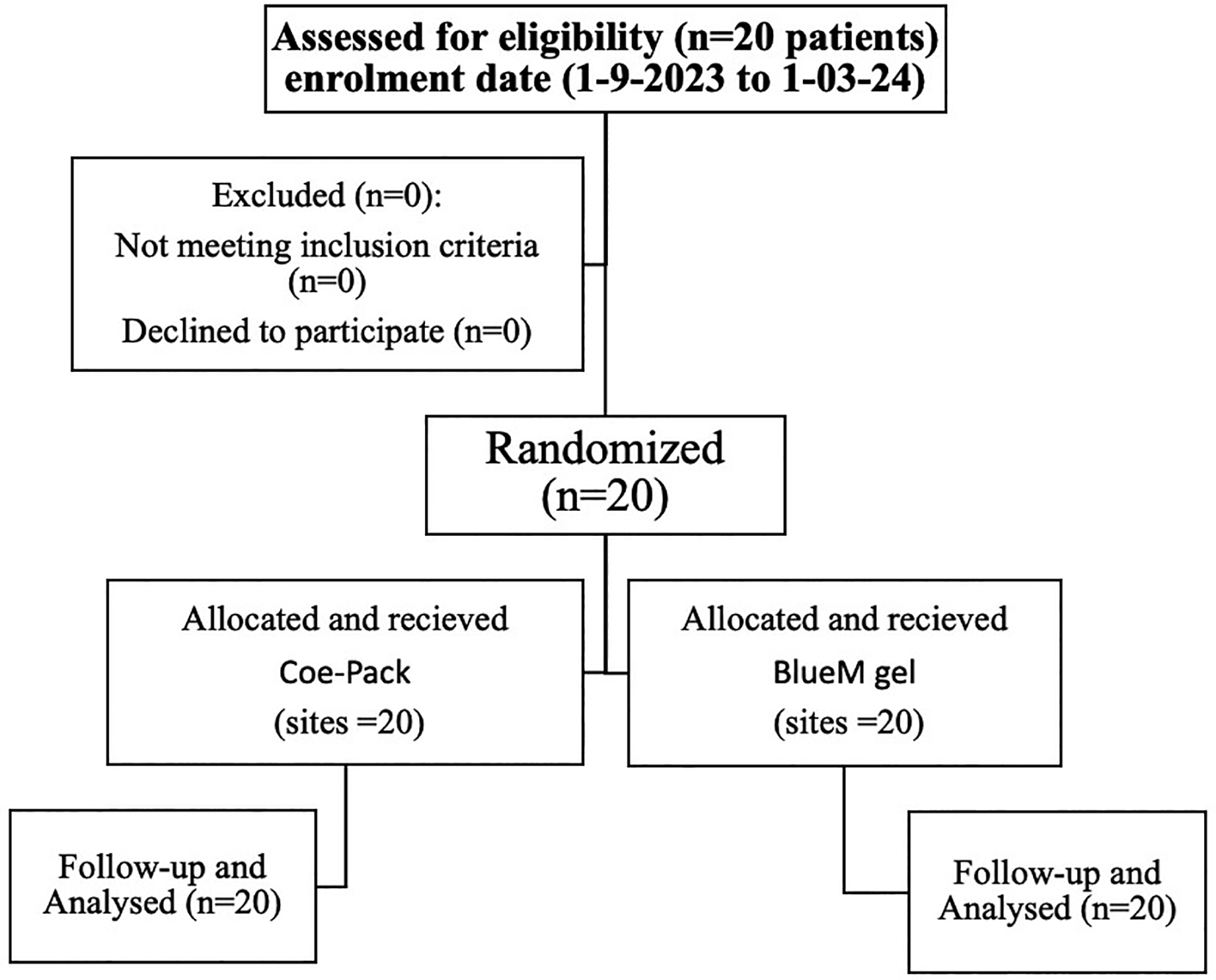
Figure 1 shows the Flow diagram for patient recruitment.
Sample size will be calculated using the reference from the previous study1 using OpenEpi (version 1), open-source calculator – SS Mean (calculation shown at the end).
Ratio of sample size (Group 1/Group 2) = 1:1
Sample size in Group 1 is 20
Sample size in Group 2 is 20
Sample size depends on the true mean difference, d, standard deviations for the two groups, and a level of significance α (type|error), and the power. The total sample size n=n1+n0 is minimized when r = σ1/σ0.
With the calculation above, a sample size of 20 was determined. To accommodate for dropouts, an estimated sample size of 20 samples per group was used. 20 is the total sample size required to achieve a 90% confidence level.
Principal investigator of the department will enrol participants and will assign participants to interventions by flipping the coin and thus sequentially numbered. Trial participants will be blinded.
Patients will be evaluated for primary and secondary outcomes at baseline, one month and three months. The data collected from participants will be entered on case history proforma which also includes the personal information of the participants.
Electronic data management will be done by entering the data from patients into the database by principal investigator.
For the pain index and re-epithelization index, the mean and standard deviation (Mean SD) values will be determined. Data from the baseline up to four weeks for each treatment group will be correlated using a paired t-test of the students. Comparisons between treatment groups will be made at baseline, 24 hours, 48 hours, 36 hours, and 48 hours as well as at the first, second, third, and fourth weeks using the student’s unpaired t test. The observed difference will be considered significant if the probability value (p) is less than 0.05 and non-significant if it is greater than 0.05.
The study will be monitored till completion by DMC which includes PG Guide, head of the department, research convener and chief advisor of university research cell.
Principal investigator (PI) will have access to these interim results and make the final decision to terminate the trial.
Data will be collected, assessed, and spontaneously reported during adverse events and other unintended effects of trial interventions or trial conduct.
The project management group meet will review the trial conducted every month. The trial steering group and the independent data monitoring and ethics committee meet will review and conduct the trial period till the trial is complete.
Procedures will be performed in keeping with the ethical standards of the institutional and/or national research committee. This protocol is permitted by the Institutional Ethics Committee of Datta Meghe Institute of Higher Education and Research, Sawangi, Meghe, Wardha [Ref.No. DMIHER (DU)/IEC/2023/1085].
In order to protect confidentiality personal information about potential and enrolled participants will be managed by keeping the records in the computer and protected by password.
Only principal investigator, co-principal investigator and institutional ethics committee will have access to the final trial dataset.
Model consent form and other related documentations will be given to the participants and authorised surrogates.
| Views | Downloads | |
|---|---|---|
| F1000Research | - | - |
|
PubMed Central
Data from PMC are received and updated monthly.
|
- | - |
Provide sufficient details of any financial or non-financial competing interests to enable users to assess whether your comments might lead a reasonable person to question your impartiality. Consider the following examples, but note that this is not an exhaustive list:
Sign up for content alerts and receive a weekly or monthly email with all newly published articles
Already registered? Sign in
The email address should be the one you originally registered with F1000.
You registered with F1000 via Google, so we cannot reset your password.
To sign in, please click here.
If you still need help with your Google account password, please click here.
You registered with F1000 via Facebook, so we cannot reset your password.
To sign in, please click here.
If you still need help with your Facebook account password, please click here.
If your email address is registered with us, we will email you instructions to reset your password.
If you think you should have received this email but it has not arrived, please check your spam filters and/or contact for further assistance.
Comments on this article Comments (0)