Keywords
Craniomaxillofacial defects, Cranial Prosthesis, Digital Workflow, Conventional Workflow, Hybrid Workflow, Cranioplasty
This article is included in the Datta Meghe Institute of Higher Education and Research collection.
Craniomaxillofacial defects, Cranial Prosthesis, Digital Workflow, Conventional Workflow, Hybrid Workflow, Cranioplasty
Cranioplasty aims to reconstruct skull defects from tumours, congenital anomalies, decompressive craniectomies, and fractures in a cosmetically acceptable manner. In addition to attempting to safeguard underlying brain tissue from cranium defects, cranioplasty also seeks to re-establish normal calvarial shape in a way that is aesthetically appropriate. It can be carried out using either alloplastic devices for repair or osteoplastic reconstruction. Among the materials used for cranial alloplastic implants are metal, acrylic resin, polyethylene, and silicone.1
Several centers now consider methyl methacrylate cranioplasty to be preferable to titanium mesh due to its radiolucency, cheaper cost, ease of shaping, light weight, and lower rates of infection when compared to titanium implants.1 Previous studies have demonstrated the potential to transform a conventional workflow from one that is highly skill-dependent, time-consuming, labour-intensive, costly, and unpleasant for the patients due to discomfort of the procedure, to one that is straightforward and predictable digitally.1
One of the earliest publications on digital maxillofacial prosthesis Penkener et al. in 1999 described a method for creating a patient’s unique, life-sized, three-dimensional ear model using a computer and a CT scan Endoplan (Medical Diagnostic Computing, Zeiss, Germany) with a semiautomatic contouring program for CBCT segmentation of the soft tissue, based on Hounsfield units (HU) thresholding.2
Recently, maxillofacial prosthodontics has adopted digital technology more and more. The studies on the production of maxillofacial prostheses with CAD and CAM (computer-aided design and computer-aided manufacturing) technology that have been published in the last 20 years2 have shown that it is feasible to switch from a traditional workflow that is extremely skill-dependent, laborious, costly, and unpleasant for the patients to a digitized version.
Although there have been advances in digital fabrication for prosthetics, there are still some constraints that prevent a full transition to digital fabrication for the final stages of prosthesis construction. Advanced training and modification of current treatment procedures are required due to the introduction of new technologies and methods in the field of maxillofacial prosthodontics.3
As of right now, only highly skilled dental professionals or CAD engineers may utilise the interface and software used for processing and developing maxillofacial prosthesis as CAD software is expensive and requires a skilled personnel to carry out the fabrication process.4
Digital dentistry currently faces something of a split; industry-driven technological solutions (primarily in the CAD-CAM and imaging sectors) are already realities, whereas AI-technologies concentrating on diagnosis and treatment planning are currently theoretical rather than reliable applications. One important job in the future is to bridge this gap. There is great need for collaborative and interdisciplinary research that combines technology and dental care.
The purpose of this research is to develop a hybrid protocol for fabricating a prosthesis that could be more accurate than the conventional technique in terms of accuracy, precision and fit of the prosthesis, and provide ease for both the patient and the maxillofacial team. This research will help prosthesis providers understand the application of a digital workflow with the addition of a conventional workflow and how they could use this in providing the finest prosthesis.
Ethical approval for this protocol was granted by Datta Meghe Institute of Higher Education and Research, Sawangi, Wardha, with IEC reference number DMIHER (DU)/IEC/2023/853 on 31st March 2023.
Written informed consent will be taken from the participants. In the case of unconscious patients, the relatives including the parents or spouse can provide written informed consent instead. In the case of minors the parents can provide written informed consent.
This research comprises a hospital based observational and analytical study, carried out in the Department of Prosthodontics, Sharad Pawar Dental College, Sawangi (Meghe), Wardha, Datta Meghe Institute of Higher Education and Research (Deemed to be University).
Data will be collected after the IEC approval and this study will be completed in 2 years.
Eligible patients will be those above 10 years of age, who have been referred from the Department of Neurology to the Department of Prosthodontics with a cranial defect, which may be due to a congenital or acquired anomaly. Only patients who have given full consent to participate in the study will be included.
This will be a hospital-based study on patients who report to Department of Prosthodontics, referred from the Department of Neurology for the fabrication of their outer cranial stent for defects due to congenital and acquired anomalies. In prosthodontics, we deal with maxillofacial prostheses which includes fabrication of stents and cranial rehabilitation for patients from the neurosurgery department. The cranial stent will be fabricated from polymethyl methacrylate with four different workflow protocols for the research, for the purpose of checking their accuracy. Each patient will have four stents fabricated. The cranial stent fabricated using CT scan 3D workflow will be taken as gold standard. This stent will be then compared for accuracy using reverse engineering principle using a software analysis.
Initially, the impression of the patient will be taken and the CT scan will be taken of the defect.
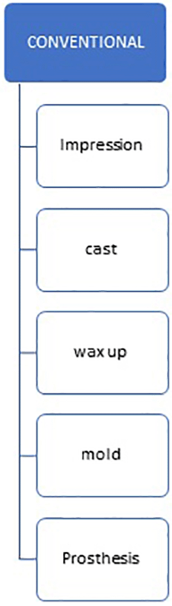
First, the impression of the defect will be created using the traditional method by the primary investigator for the conventional prosthesis, and the prosthesis will be designed with conventional workflow i.e., an impression will be made and then the cast will be fabricated in type 3 dental stone, and over the master cast the wax pattern is fabricated. This wax pattern is flasked and packed with polymethyl methacrylate, and after this finishing and polishing is done (Figure 1).
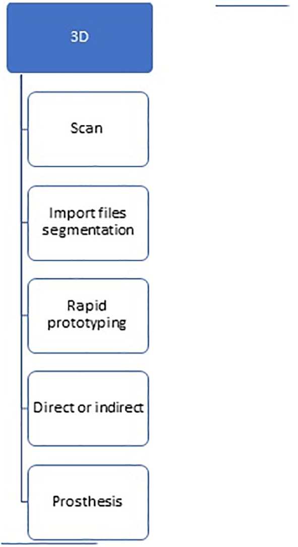
For the 3D prosthesis, a CT scan will be taken of patient in the neurology department, and the prosthesis will be designed in the prosthodontics department with CAD software and, with the help of mirroring the image, the 3D print will be created (Figure 2).
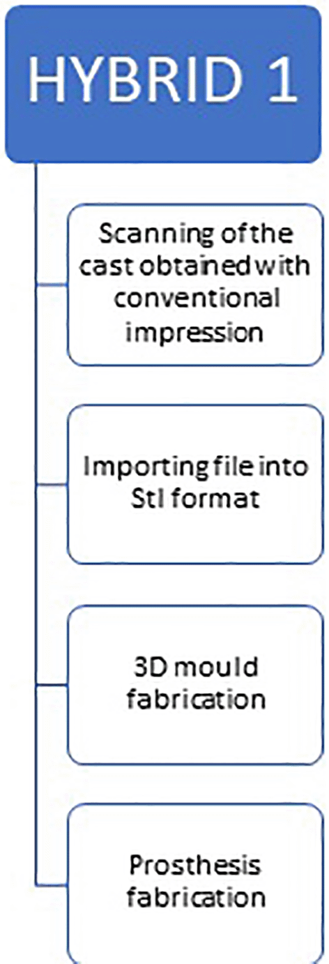
For the first hybrid prosthesis, an impression will be taken of the defect by the primary investigator using conventional alginate, and the cast will be fabricated with the type 3 dental stone. The cast will then be scanned and the mould will be fabricated with rapid prototyping, i.e., we will 3D print it and the prosthesis will be fabricated conventionally from PMMA (Figure 3).
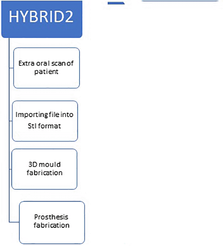
The second hybrid prosthesis will be made from the CT scan of the patient and the stl file that it generates. The prothesis will be fabricated by rapid prototyping, i.e., 3D printing and designed conventionally from PMMA (Figure 4).
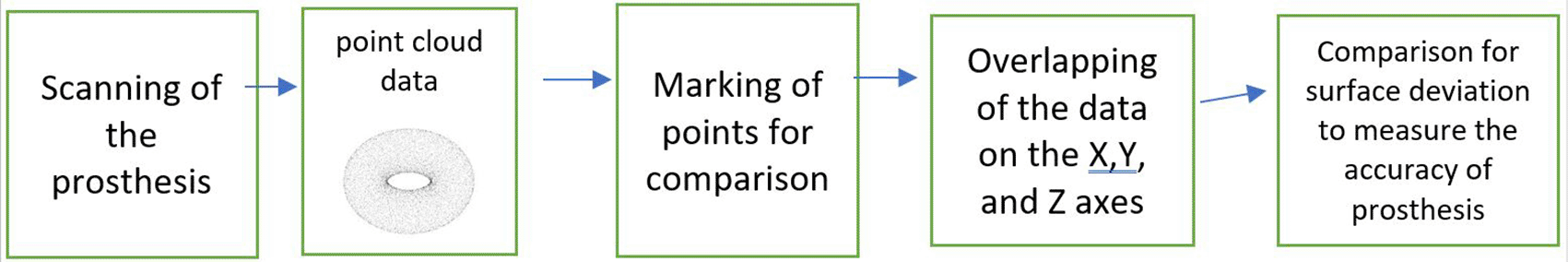
All four prosthesis will be designed using conventional, 3D, and the two hybrid workflows, and will be scanned using an extraoral EinScan Pro 2x 2020 scanner, and will be converted into point cloud data that is the points taken from the four prostheses on a 3D shape in software depicting its coordinates in the X,Y, and Z axis. This data will be compared by overlapping the prosthesis on the control group, i.e. 3D group, and compared for its change in the surface deviation in 3D space. Charts will be prepared for the surface deviation thus depicting the dimensional accuracy of prosthesis (Figure 5).
The study will measure the accuracy of all the prostheses made from the 4 different workflows, which will check the size, shape, and fit of the prostheses. As this study is an in vitro study, the fit cannot be checked on a patient. Since the prosthesis is fabricated with different protocols, the cost will differ for each prosthesis; the best fitting and most accurate prosthesis may be more expensive, so this is a variable that can act as a confounding factor and will be considered in the analysis.
The expected outcome of the study is that the hybrid prosthesis would be a better fit and more accurate than its conventional counterpart. For dental professionals the paradigm shift towards digital technology has many obstructions, such as in infrastructure and costly equipment, so for an accurate prosthesis opting for a hybrid solution is needed so that we can benefit from both technology and the conventional process.
Bias will be minimized by random selection of patients based on inclusion and exclusion criteria.
The calculation of sample size is described in this section. The primary variable is the cranial prosthesis defect measurement.
Conventional method
Max (positive) deviation in conventional prosthesis = 3.5, max (negative) deviation in conventional mould = 1.0
Average deviation for prosthesis mould = (3.5+1)/2 = 2.25
Digital CAD method
Max (positive) deviation in CAD fabricated prosthesis mould = 2.5, max (negative) deviation in CAD mould = 1.0
Average deviation for prosthesis mould = 1.755
Mean difference of deviation between two treatments = 0.5
Estimated standard deviation = (0.4) mean difference [mean difference between the two average deviations of prosthesis mould from conventional and digital method].
Assumption
Minimum sample size required N = 2 *[(1.64+ 0.84)2(0.4)2] / (0.5)2 = 10 per group
All results obtained from the comparison of the prostheses fabricated from the four different workflows will be analyzed using R software version 4.3.2. Data collection from the prostheses fabricated for patients with congenital and acquired cranial defects who are undergoing cranioplasty, made with four different workflows, will be converted into point cloud i.e. the points of all four prostheses on 3 dimensional axis, X,Y, and Z. This data will be fetched from (point cloud) software and will be comparatively analysed for surface deviation in the 3 dimensional axes, defect significance (mean) in size, shape, fit and accuracy, using the one way ANOVA test. A p-value <0.05 will be considered as significant value for providing greater accuracy.
The findings related to this study will be published in indexed journals and presented in scientific tracks/conferences. This study will help us in standardizing a protocol for hybridization of digital technology with conventional methodology to establish a more accurate and precise treatment deliverance of maxillofacial prosthesis.
The craniomaxillofacial prostheses made with different workflows can be checked for their accuracy and cost efficacy, and the best suited one can be further used in patients.
The limitations for this study are that only cranial defects will be considered, but the branch of maxillofacial prosthodontics as a whole deals with all the maxillofacial prosthesis. This is an in vitro study; as the prosthesis is not fitted in a patient its final fit cannot be assessed.
De La Pena et al. conducted a case study of a 3D-designed PMMA cranial prosthesis in 2018 and found that the prosthesis conformed precisely to the osseous defect. No problems, such as rejection, toxicity, local or systemic infection, were seen at the 6-month follow-up, and the cosmetic improvement was extremely noticeable and pleasing. A customised 3D PMMA prosthesis has benefits in terms of price, cosmetic quality, operation time, and biocompatibility.6
Schon et al. in 2020 in 16 patients demonstrate that systematic in-hospital manufacture of patient-specific implants made of conventional PMMA with 3D printer assistance is viable for severe calvaria deformities and may be simply applied in routine practise. When a patient needs a cranioplasty but does not have access to a bone flap, this manufacturing method may lower treatment expenses.7
Rotaru et al. did a study to assess quantitatively whether a symmetric reconstruction of the calvaria could be achieved using 3-dimensional (3D) custom-made implants and to examine any complications caused by the cranioplasty. He found out that the implants could be highly helpful for treating significant and irregularly shaped cranial abnormalities.8
Beri et al did in year 2022 published a trial to evaluate the impression making process in the craniomaxillofacial defects by traditional method 3D scanning and photogrammetry. In these trial all the impression was assessed by the point cloud data and checked for its accuracy.9
The purpose of this research is to develop a hybrid protocol for fabricating a prosthesis that could be more accurate than the conventional technique in terms of accuracy, precision and fit of the prosthesis, and provide ease for both the patient and the maxillofacial team. This research will help prosthesis providers understand the application of a digital workflow with the addition of a conventional workflow and how they could use this in providing the finest prosthesis.
| Views | Downloads | |
|---|---|---|
| F1000Research | - | - |
|
PubMed Central
Data from PMC are received and updated monthly.
|
- | - |
Provide sufficient details of any financial or non-financial competing interests to enable users to assess whether your comments might lead a reasonable person to question your impartiality. Consider the following examples, but note that this is not an exhaustive list:
Sign up for content alerts and receive a weekly or monthly email with all newly published articles
Already registered? Sign in
The email address should be the one you originally registered with F1000.
You registered with F1000 via Google, so we cannot reset your password.
To sign in, please click here.
If you still need help with your Google account password, please click here.
You registered with F1000 via Facebook, so we cannot reset your password.
To sign in, please click here.
If you still need help with your Facebook account password, please click here.
If your email address is registered with us, we will email you instructions to reset your password.
If you think you should have received this email but it has not arrived, please check your spam filters and/or contact for further assistance.
Comments on this article Comments (0)