Keywords
Tuberculosis, knee, abscess, diagnosis, therapeutic
Tuberculosis, knee, abscess, diagnosis, therapeutic
Osteoarticular tuberculosis remains prevalent in countries where tuberculosis is endemic. It represents 1 to 5% of all forms of the disease. It predominantly affects the spine (40%), hip (25%), and knee (8%).1,2
The symptoms of the extra-spinal form are non-specific, with an insidious clinical picture and inconsistent general signs, which explains the diagnostic difficulties. Early management is necessary to prevent serious complications.3 In the knee, tuberculosis typically presents as joint involvement: synovitis, arthritis, or an infected Baker’s cyst, and rarely as extra-articular involvement.4,5
Currently, antitubercular antibiotics are the standard treatment. Moreover, surgical treatment is limited to cases resistant to medical treatment.4
This is a 17-year-old male, Tunisian high school student, from central-western Tunisia (Sbeïtla, Kasserine) with no notable medical history but with a history of exposure to tuberculosis seven years ago (his brother had pulmonary tuberculosis) and a history of regular consumption of unpasteurized milk. The patient presented with a painful, red, hot swelling in the right popliteal fossa that was resistant to first-line medical treatment with analgesics. Furthermore, he reported a weight loss of 5 kilograms over the past 2 months, as well as fever and night sweats over the past 2 weeks.
On examination, the patient had a limp due to an antalgic stance in the knee flessum. An inflammatory swelling of 10 cm in diameter was found in the right popliteal fossa. It was a painful, soft, and mobile swelling. In addition, there was no joint effusion in the knee and no metaphyseal pain in the femur and tibia. An inguinal lymph node on the same side was found.
Laboratory tests showed a biological inflammatory syndrome with a white blood cell count of 12,040 and a C-reactive protein level of 67.3 mg/L. Chest and knee radiographs were normal (Figures 1 and 2).
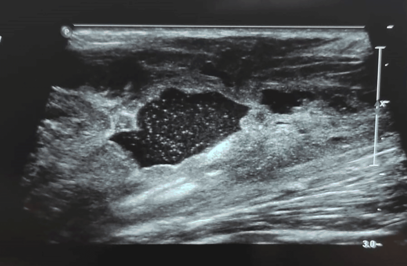
Doppler ultrasound of the lower limb showed a collection of the right popliteal fossa with an echogenic content, a clean wall independent of the popliteal pedicle, and thrombophlebitis of the right saphenous vein (Figure 3). The patient was treated with anticoagulant therapy in the form of low-molecular-weight heparin overlap and Sintrom (acenocumarol).

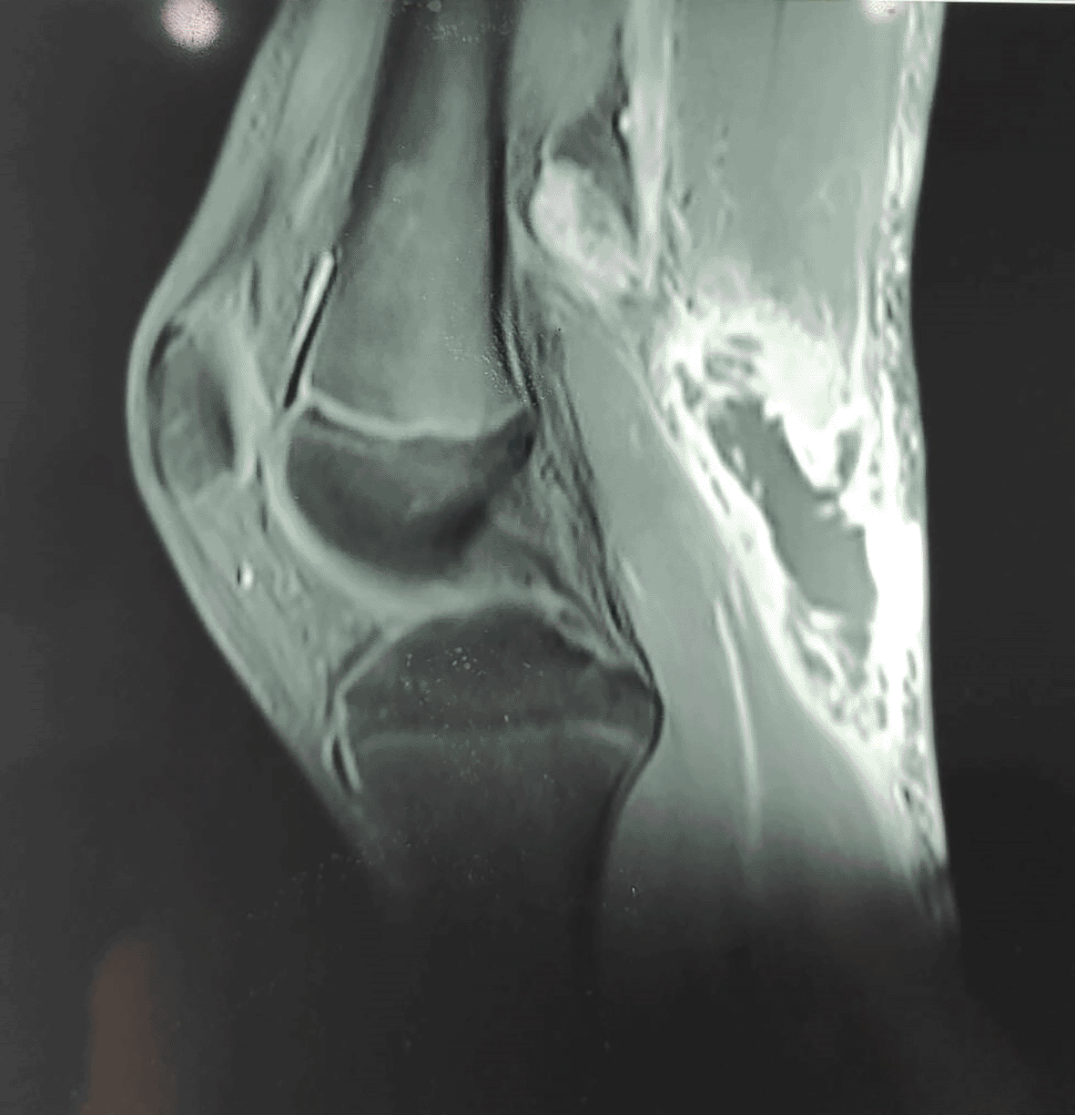
A magnetic resonance image (MRI) showed a well-vascularized mass of 8 cm in diameter that was partly liquefied and developed in contact with the semimembranosus and medial gastrocnemius muscles, popliteal vessels, and posterior tibial nerve, with muscle and adipose edema and popliteal lymphadenopathy but no synovial effusion or thickening. This aspect suggested an infectious etiology (Figure 4).

The etiological investigation showed negative tuberculin skin test, negative direct examination and culture for Koch’s bacillus in urine and sputum, negative Rose Bengal serology and negative Wright serology.
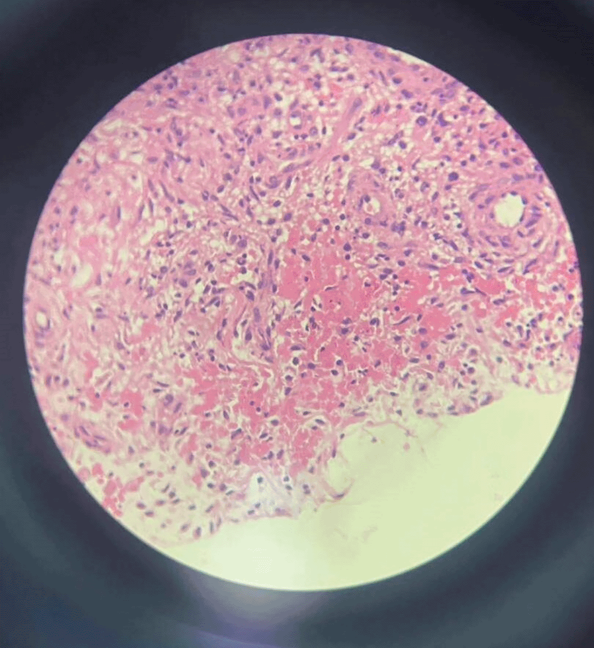
An ultrasound-guided biopsy puncture was done. The bacteriological results were negative. Histopathological examination revealed the presence of inflammatory granulation tissue with acellular eosinophil deposits and epithelioid cells, suggesting a tuberculous origin (Figure 5).

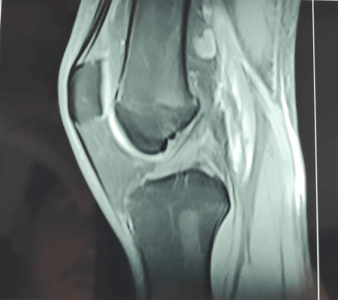
The decision was to start an antituberculosis trial treatment with isoniazid (H), rifampicin (R), pyrazinamide (Z), ethambutol (E) and a pre-treatment assessment. The patient was treated with anti-tubercular drugs for 9 months according to the 2HRZE/7HR regimen with regular monitoring of liver and hematological function. The dosage was 4 HRZE tablets per day associated with 3/4 Sintrom tablets to control the thrombophlebitis of the internal saphenous vein. The patient’s condition improved, and a control MRI was performed in the eighth week of treatment that demonstrated complete disappearance of the previously described mass in the popliteal fossa, with no joint effusion or synovial thickening. Muscular and fatty edema had disappeared, and the bone signal was normal (Figure 6).

With 2 years of follow-up, the clinical and radiological evolution was favorable (Figure 7): clinical examination showed complete disappearance of the swelling and inguinal lymphadenopathy, with no joint effusion and full active mobility of the knee, and radiography revealed no bone lesions.
Soft tissue tuberculosis, defined as involvement of tendons, bursae, muscles, or deep fascia, is a rare form of musculoskeletal tuberculosis. It is usually associated with immunocompromised patients.6 Isolated localization in the popliteal fossa is also exceptional.7
Several authors8–10 have reported in the literature that isolated or primary soft tissue tuberculosis without bone involvement in immunocompetent patients represents a rare form of extrapulmonary tuberculosis and its exact incidence is not known. Its clinical manifestations can mimic malignant diseases or other inflammatory diseases, making diagnosis difficult. The main differential diagnoses are pyogenic abscesses, atypical mycobacterial abscesses, hydatid cysts, and certain tumors.11
It is interesting to note that most cases of soft tissue tuberculosis reported in the literature were located near joints and bursae. The pathophysiology of this isolated involvement remains difficult to understand.12 However, extension from an adjacent joint, bone, or bursa and even direct inoculation have all been reported. In this context, Lupatkin et al.13 reported that tuberculous abscesses resulted from hematogenous, lymphatic, or local contamination from adjacent or other primary infection areas and, in rare cases, direct inoculation.
In our case, there was no clear explanation for the focal origin of the popliteal fossa involvement. Furthermore, this isolated and primary involvement could be attributed to hematogenous spread or extension from neighboring bursae. This hypothesis is most likely due to the presence of nearly 13 bursae around the knee.
Tuberculosis of soft tissues mainly affects the extremities, presenting as localized masses accompanied by local inflammatory signs and pain. General symptoms such as fever and weight loss are inconsistent.14,15 Recent cases of soft tissue tuberculosis published in the last 10 years were identified by conducting a search on PubMed (Table 1). We found 17 reported cases in the literature, including 10 males and seven females. The patients ranged from 7 to 79 years old. 12 cases were reported in Asian countries, four cases in African countries, and one case in America. In 13 patients, the lesions were predominantly in the extremities including the thigh, calf, forearm, and wrist. The other four patients had lesions in the buttocks, back, chest, and iliopsoas. The diagnosis of tuberculosis was made by a combination of bacteriological and pathological examination. The majority of cases improved under medical treatment based on antituberculosis drugs without any associated surgical procedure.
PCR: Polymerase chain reaction, NS: Not specified.
| Reference | Year and country | Age and gender | Predisposing factors | Location | Clinical signs | Diagnosis | Treatment |
|---|---|---|---|---|---|---|---|
| Arora et al.16 | 2012 India | 15/male | NS | Left thigh | Swelling + systemic signs | Bacteriological examination | Medical treatment for 6 months |
| Lee et al.17 | 2013 Korea | 62/male | NS | Right thigh | Swelling | Bacteriological and histological examination | Medical treatment for 6 months |
| Elshafie et al.18 | 2013 Oman | 25/male | History of tuberculosis exposure | Right buttock | Swelling | Bacteriological and histological examination | Drainage followed by 9-month antibiotic |
| Neogi et al.19 | 2013 India | 11/female | NS | Right thigh and calf/Left arm | Swelling | Bacteriological and histological examination | Medical treatment for 6 months |
| Meena et al.20 | 2015 India | 25/male | NS | Right arm | Swelling | BK PCR | Medical treatment |
| Dhakal et al.21 | 2015 Nepal | 9/female | NS | Right forearm and calf | Swelling | Bacteriological and histological examination | Medical treatment |
| Sbai et al.22 | 2016 Tunisia | 45/male | NS | Right wrist | Swelling | Bacteriological and histological examination | Drainage followed by 8-month antibiotic |
| Al-khazraji et al.23 | 2017 USA | 33/female | Lupus nephritis | Right calf | Inflammatory swelling | Bacteriological and histological examination | Drainage followed by antibiotics |
| Alaya et al.24 | 2017 Tunisia | 23/female | History of tuberculosis exposure | Right thigh | Inflammatory swelling + pain | BK PCR + Histological examination | Medical treatment for 12 months |
| Manicketh et al.14 | 2018 India | 55/female | Pulmonary tuberculosis | Left wrist and right calf | Inflammatory swelling with systemic signs | Bacteriological examination | Medical treatment |
| Hashimoto et al.25 | 2018 Japan | 79/male | NS | Left wrist | Inflammatory swelling | Histological examination | Drainage followed by antibiotics |
| Zeng et al.26 | 2019 China | 49/male | Pulmonary tuberculosis, corticosteroid therapy | Both thighs and calves | Inflammatory swelling | BK PCR + Bacteriological and histological examination | Medical treatment |
| Zitouna et al.27 | 2019 Tunisia | 42/female | NS | Right lumbar paraspinal muscles | Swelling | Bacteriological and histological examination | Medical treatment |
| Moyano et al.28 | 2019 Senegal | 29/male | NS | Right hemithorax | Increased volume of the hemithorax | Bacteriological examination + BK PCR | Medical treatment |
| Fahad et al.29 | 2020 Pakistan | 45/Female | NS | Right forearm | Swelling | Histological examination | |
| Murugesh et al.30 | 2020 India | 31/male | Renal transplantation under immunosuppressants | Right calf and foot | Inflammatory swelling | Bacteriological and histological examination | Drainage followed by antibiotics |
| Tone et al.15 | 2021 Japan | 29/male | Tuberculous pleurisy | Right iliopsoas muscle | Pain and fever | Bacteriologial and histological examination + BK PCR | Percutaneous drainage followed by antibiotics |
In the literature,7 to confirm a definitive diagnosis, it is essential to identify the Koch bacillus. However, tuberculous soft tissue disease is paucibacillary. Often, the Ziehl-Nielsen test is negative and it is necessary to wait for the results of Lowenstein culture. Histological examination is more sensitive than microbiological tests and can confirm the diagnosis in 80% of cases.31 A normal chest radiograph, the absence of systemic symptoms, or the absence of detectable tuberculosis focus should not deter from considering this diagnosis.
In our case, faced with a history of tuberculosis exposure and a painful isolated swelling of the popliteal fossa, we performed an ultrasound-guided biopsy puncture of the swelling. The diagnosis was finally established by histopathology, which showed pathognomonic caseating granulomas of tuberculosis.
In the literature,10 the treatment protocol is not clearly defined. The main debated questions concern the duration of medical treatment as well as the need and modalities of any surgical treatment. Some authors32,33 advocated for primary medical treatment with a combination of antituberculous antibiotics. The choice of drugs is generally the same as for pulmonary tuberculosis (isoniazid, rifampicin, pyrazinamide, and ethambutol). This treatment is divided into two phases: an initial phase based on quadritherapy that lasts two months and a secondary phase with a bitherapy that lasts 4 to 7 months or longer in extensive cases. Moreover, surgical drainage is reserved for cases resistant to medical treatment.16 Postoperative outcomes are marked by local recurrence in 50% of cases within a year.25 Indeed, our patient responded well to exclusive medical treatment for 9 months. The evolution was spectacular with complete regression of the mass and no recurrence with a follow-up of 2 years. The patient expressed a high level of satisfaction regarding the clinical outcomes following the treatment. He reported a successful recovery and the ability to resume his normal physical activity level.
Our case study had several strengths: we conducted a meticulous clinical examination, along with radiological investigations using ultrasound and MRI of the knee. Given that we are in an endemic country and taking into account the patient’s history of tuberculosis exposure, the diagnosis of tuberculous abscess should always be considered as a first possibility. Therefore, we decided to perform a biopsy to confirm the etiology before proceeding with surgery. After confirming the diagnosis through histology and reviewing the literature, we opted for antibiotic treatment and monitoring of the progression. Consequently, the lesion regressed totally overtime.
Tuberculosis of soft tissues is a rare form of the disease. It is even rarer for it to present as a primary abscess in the popliteal fossa. The lack of specificity of clinical and radiological signs and the insidious and progressive course make the diagnosis difficult.
This case report emphasizes the importance of considering the diagnosis of tuberculosis in the differential diagnosis of any unexplained soft tissue swelling in endemic areas, despite the absence of systemic and pulmonary symptoms. Thus, a more thorough investigation can prevent delayed diagnosis and its devastating complications.
Written informed consent for the publication of the patient’s clinical details and clinical images was obtained from the patient and his parents.
| Views | Downloads | |
|---|---|---|
| F1000Research | - | - |
|
PubMed Central
Data from PMC are received and updated monthly.
|
- | - |
Is the background of the case’s history and progression described in sufficient detail?
Yes
Are enough details provided of any physical examination and diagnostic tests, treatment given and outcomes?
Yes
Is sufficient discussion included of the importance of the findings and their relevance to future understanding of disease processes, diagnosis or treatment?
Yes
Is the case presented with sufficient detail to be useful for other practitioners?
Yes
Competing Interests: No competing interests were disclosed.
Reviewer Expertise: orthopedic surgery, spine surgery, bone and joint infectious diseases, Extra-pulmonary Tuberculosis research Unit (University of sfax, faculty of medicine)
Is the background of the case’s history and progression described in sufficient detail?
Yes
Are enough details provided of any physical examination and diagnostic tests, treatment given and outcomes?
Yes
Is sufficient discussion included of the importance of the findings and their relevance to future understanding of disease processes, diagnosis or treatment?
Yes
Is the case presented with sufficient detail to be useful for other practitioners?
Yes
Competing Interests: No competing interests were disclosed.
Reviewer Expertise: spine surgery, orthopedic surgery
Is the background of the case’s history and progression described in sufficient detail?
Yes
Are enough details provided of any physical examination and diagnostic tests, treatment given and outcomes?
Yes
Is sufficient discussion included of the importance of the findings and their relevance to future understanding of disease processes, diagnosis or treatment?
Yes
Is the case presented with sufficient detail to be useful for other practitioners?
Yes
Competing Interests: No competing interests were disclosed.
Reviewer Expertise: rheumatology, orthopedics
Alongside their report, reviewers assign a status to the article:
| Invited Reviewers | |||
|---|---|---|---|
| 1 | 2 | 3 | |
|
Version 1 25 Sep 23 |
read | read | read |
Provide sufficient details of any financial or non-financial competing interests to enable users to assess whether your comments might lead a reasonable person to question your impartiality. Consider the following examples, but note that this is not an exhaustive list:
Sign up for content alerts and receive a weekly or monthly email with all newly published articles
Already registered? Sign in
The email address should be the one you originally registered with F1000.
You registered with F1000 via Google, so we cannot reset your password.
To sign in, please click here.
If you still need help with your Google account password, please click here.
You registered with F1000 via Facebook, so we cannot reset your password.
To sign in, please click here.
If you still need help with your Facebook account password, please click here.
If your email address is registered with us, we will email you instructions to reset your password.
If you think you should have received this email but it has not arrived, please check your spam filters and/or contact for further assistance.
Comments on this article Comments (0)