Keywords
Level VI, Level VII, central compartment, metastasis, oral cancer
This article is included in the Datta Meghe Institute of Higher Education and Research collection.
Level VI, Level VII, central compartment, metastasis, oral cancer
Oral cancer is ranked sixth amongst all the cancers.1 India harbours the most amount of oral cancer patients and it has one-third of all the oral cancer cases around the world.1 Metastasis to the cervical group of lymph nodes is a critical prognostic factor in cases with oral squamous cell carcinoma.2 Presence of cervical metastasis in oral cancer is indicative of aggressive behaviour.3 Node-positive (N+) patients have a 50% reduction in survival compared with node-negative (N−) patient.4 About 30% of oral cancer patients have subclinical metastasis to the neck lymph nodes.5 Kalnins et al., quote survival rates of 75% for a true N0 neck, falling to 49% if one node is involved, 30% if two nodes are involved, and 13% if three or more nodes are involved.6
Oral squamous cell carcinoma (OSCC) predominantly captivates the level I, II and III cervical lymph nodes and less commonly it spread to cervical lymph nodes in the level IV and level V groups, which represent 20% and 4% of cases, respectively.7
Involvement of the central compartment secondary to oral squamous cell carcinoma involving the Level VI and VII neck nodes is extremely unconventional which is also ascertained by Likhterov et al. in 2015 who reported two cases with central compartment involvement in recurrent oral squamous cell carcinoma for the first time in English literature.8 We report a certainly rare case of central compartment involvement in a recurrent case of squamous cell carcinoma of tongue.
A 41-year-old Indian daily wage construction site male worker came to our institute with a non-healing growth over the lateral border of tongue of the right side. There was no relevant family or medical history. On examination, there was a proliferative growth over the right lateral border of tongue of size 1.5×1.5 cm. The biopsy of this growth was reported as ‘well differentiated squamous cell carcinoma’. The MRI report was suggestive of a heterogeneously enhancing altered signal intensity lesion seen over the right lateral border of tongue of size 1×0.5×0.5 cm. The patient underwent wide local excision of the lesion, modified radical neck dissection (type III) of right side and primary closure which was done under general anaesthesia.
The post-operative margin report was suggestive of ‘well differentiated squamous cell carcinoma’. All margins were clear, pT size 2×1 cm, lymph nodes level I to level V bilaterally were negative for infiltration by malignant cells. The lesion was pT1N0Mx. He was under observation after that, no adjuvant therapy was given. After 8 months, the patient noticed a nodular swelling over the left submandibular region of size 2×1 cm. All other cervical lymph nodes were clinically non-palpable. Fine needle aspiration cytology from left submandibular gland was done which was suggestive of “metastasis of squamous cell carcinoma with foreign body granulomatous reaction” following which he underwent modified radical neck dissection of left side. The histopathological report of the specimens suggested the level IB node to be positive for infiltration by malignant cells. Radiotherapy was given to bilateral face and neck to a dose of 60 Gy in 30 fractions using the intensity modulated radiation therapy [IMRT] technique with weekly Inj. Cisplatin 50 mg/mL. The patient was on follow up after that.
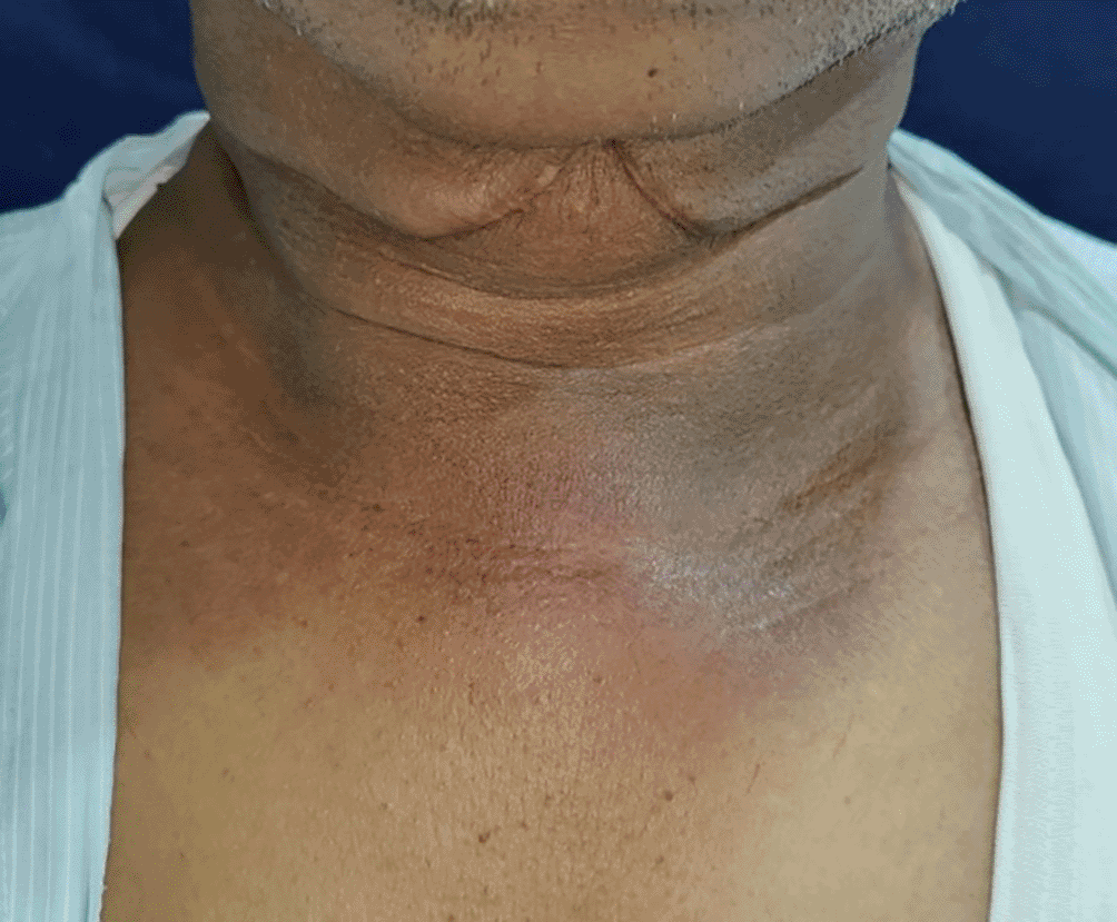
The patient then reported 1 year later, complaining of swelling over suprasternal region [Figure 1] which gradually increased to the size of 3×2.5 cm and was associated with dull aching pain. On oral examination, there was no evidence of any recurrence of lesion or any second primary tumor in the oral cavity.
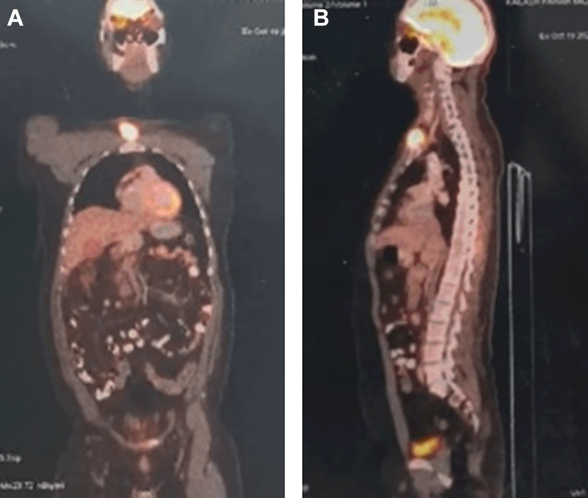
Fine needle aspiration cytology of the swelling over the suprasternal region was suggestive of “deposits of squamous cell carcinoma”. The PET scan showed no evidence of any distant metastasis [Figure 2].
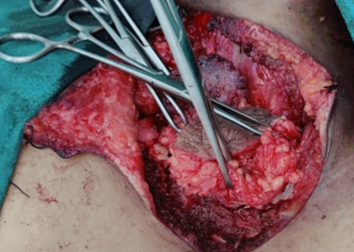
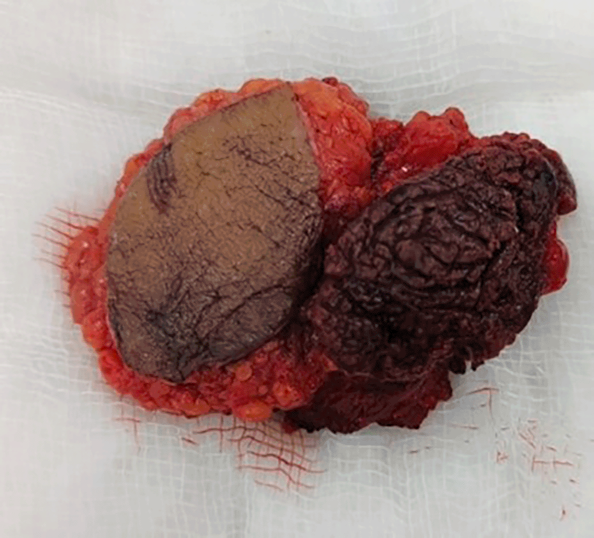
Wide local excision of the lesion over the sternoclavicular joint was carried out with 1 cm of margin [Figure 3]. The tumor was seen encroaching the bone [Figure 4]. Hence, removal of the right sternoclavicular joint was carried out [Figure 5]. Histopathological report of the resected specimen was suggestive of “Moderately differentiated squamous cell carcinoma”. The decalcified bone was positive for infiltration by malignant cells. The patient was on regular follow up. On 6 months clinical follow up, the surgical site was healthy. There was no evidence of recurrent lesion. Nor were there any signs of lymphadenopathy.

The level VI or the tracheoesophageal group of lymph nodes is surrounded superiorly by the hyoid bone, inferiorly by the suprasternal and laterally by the strap muscles.9 The anterior aspect of the neck, pharynx, larynx, oesophagus, thyroid drain lymph into level VI cervical group of lymph nodes. Lymph nodes in the tracheoesophageal groove and the anterior upper mediastinum are considered to be level VII lymph nodes. It reaches the innominate artery from the suprasternal notch.9
Ferlito et al. suggested that the skip metastasis of the oral tongue to clinically negative nodes is seen in 20% to 30% of patients.10 The patients who have undergone surgical ablation of the primary tumor and neck dissection followed by adjuvant radiation therapy, the rates of local, regional, and locoregional recurrence are 16.5%, 5.3%, and 4.9%, respectively.11 The predominant lymphatic drainage of the tongue takes place in Level I, II and III cervical lymph nodes.
Metastasis is a familiar sequela of oral carcinoma. If the metastasis skips any regional lymph node and show dysplastic features into the peripheral lymph node then it is known as ‘skip metastasis’ (SM) or ‘nodal skip metastasis’ (NSM). Byers RM., et al. (1997) concluded that the usual supra-omohyoid neck dissection is inadequate for a complete pathologic evaluation of all the nodes at risk for patients with squamous cell carcinoma of the oral tongue due to skip metastasis to level IV in 15.8% of population.12 Pantvaidya et al. (2014) prospectively studied 583 cases of oral cancer through lymph node mapping and found that in 95.7% the metastasis was seen in Level I to Level IV cervical lymph nodes.13
Cancer of oral cavity metastasizes to the cervical lymph nodes in a predictable fashion based on the primary sites or subsites. Metastasis of tumor cells to level VI and level VII group of lymph nodes is rare. The incidence of this distinctive phenomenon of level VI lymph node involvement in primary oral squamous cell carcinoma was noted by Zhang et al. in 2021 to be 0.69% of incidence for the first time in the literature.7 Level VII lymph node involvement is uncommon but it does exist. Five of the 779 head and neck cancer patients depicted by Probert et al. acquired mediastinum metastasis, of which four cases were found by autopsy.14
There are a few theories mentioned about this odd occurrence. Lymphatic drainage is altered after previous surgical interventions. This theory's most likely interpretation is that the obstruction of the afferent lymphatic vessels causes the lymphatic drainage to be diverted along other pathways.
This is well established in breast cancer studies. Estourgie et al. reported on lymphoscintigraphy studies before and after excisional biopsies of breast masses. Changes in axillary and internal mammary lymph node drainage were noted in 68% of patients.15
An alternative hypothesis is the seeding of malignant cells in the middle aspect of the neck during previous tracheotomy as is mentioned by Likhterov et al.8 In our case, the patient did not undergo tracheostomy or any intervention in the central compartment during the previous surgery. Hence, seeding of tumor cells would not be applicable in this case.
In this case, the lesion was seen encroaching the bone but did not involve the thoracic cavity. In PET scan, there was no evidence of distant metastasis. Hence, wide local excision of the lesion was done. The lesion was fixed to the sternoclavicular joint of the right side. Removal of the right sternoclavicular joint was done which was found to be positive for infiltration by malignant cells. With the surgical approach, the lesion was removed with adequate margins. The patient could resume his work 4 weeks after the surgery and could carry out everyday chores smoothly.
There are no extensive studies in the literature which have assessed the aberrant lymphatic drainage after neck dissection in head and neck cancer surgeries.
Nodal involvement of central compartment in level VI and level VII is a rather very rare finding, but is still possible in cases of oral cavity cancers. This could be expected in cases which have undergone neck dissection previously. Thorough evaluation of the entire neck should be done to rule out metastasis at these aberrant areas of the neck and appropriate management should be carried out.
| Views | Downloads | |
|---|---|---|
| F1000Research | - | - |
|
PubMed Central
Data from PMC are received and updated monthly.
|
- | - |
Provide sufficient details of any financial or non-financial competing interests to enable users to assess whether your comments might lead a reasonable person to question your impartiality. Consider the following examples, but note that this is not an exhaustive list:
Sign up for content alerts and receive a weekly or monthly email with all newly published articles
Already registered? Sign in
The email address should be the one you originally registered with F1000.
You registered with F1000 via Google, so we cannot reset your password.
To sign in, please click here.
If you still need help with your Google account password, please click here.
You registered with F1000 via Facebook, so we cannot reset your password.
To sign in, please click here.
If you still need help with your Facebook account password, please click here.
If your email address is registered with us, we will email you instructions to reset your password.
If you think you should have received this email but it has not arrived, please check your spam filters and/or contact for further assistance.
Comments on this article Comments (0)