Keywords
unexplained ascites, tuberculosis infection, constrictive pericarditis, pericardiectomy
This article is included in the Datta Meghe Institute of Higher Education and Research collection.
unexplained ascites, tuberculosis infection, constrictive pericarditis, pericardiectomy
Constrictive pericarditis is a result of chronic pericardial inflammation characterized by fibrosis, calcification, and thickening of the pericardium leading to impaired cardiac dispensability and diastolic filling leading to right heart failure. Pericarditis can occur following causes like inflammatory conditions, non-inflammatory conditions, idiopathic disease, cardiac surgery, and radiation to the mediastinum for malignancy like Hodgkin’s disease or lymphoma.1 Malignancy is the least common cause of constrictive pericarditis.2 One to two percent of pulmonary tuberculosis (TB) patients and around 1% of all autopsied cases of TB had tuberculous pericarditis caused by Mycobacterium tuberculosis. It is the most common cause of pericarditis in a country like India, in which TB remains a major public health problem.3 Appropriate diagnosis and treatment of tuberculous pericarditis can prevent mortality. The primary pericarditis condition is rare and systemic, frequently a symptom of systemic illness, and can cause severe cardiac compromise that can be fatal.
A six-year-old male child born out of non-consanguineous marriage, second by birth order with normal birth and development history, no co-morbidities with no significant past medical, surgical, or family history presented to us with a complaint of distension of the abdomen for three years, and difficulty in breathing for one year. He was referred to our hospital, Acharya Vinoba Bhave Rural Hospital, in June 2022 before being referred to Jawaharlal Nehru Medical College, Wardha. The patient was apparently normal three years before, but he then developed a fever, which was low grade. The patient had three to four episodes every year and each episode lasted for several days. Later, he developed abdominal distension, which progressively increased but it was not associated with pain in the abdomen. The patient also complained of increased difficulty in breathing, aggravated by lying down but he was comfortable when sitting up. He went through extensive diagnostic workups in multiple hospitals for three years.
The patient had facial edema, a faint Kayser-Fleischer ring in both eyes, pallor, jugular vein distension, but haemodyanamically stable. On abdominal examination gross abdominal distension was observed with an everted umbilicus. The skin over the abdomen was stretched with a visible vein on the abdominal wall. On palpation, the abdomen was tense with mild hepatomegaly. On percussion fluid thrill was present. Cardiovascular examination showed jugular venous pressure (JVP) was raised, and muffled heart sounds were present. Respiratory and central nervous system examinations were normal. Anthropometry-wise was normal meaning weight and height were normal for age.
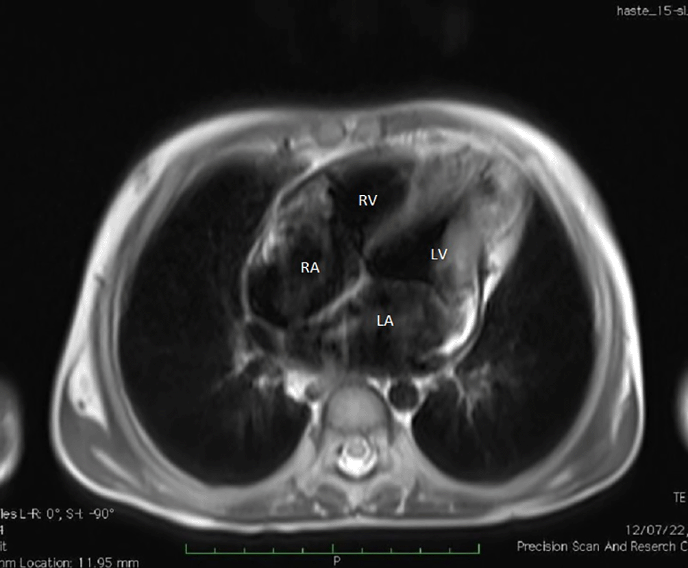
In a routine investigation, haemogram, erythrocyte sedimentation rate, renal and liver function were normal. Autoimmune workup including antinuclear antibody, double stranded DNA, and paraneoplastic markers like Carcinoembryonic antigen and cancer antigen 125 were normal, and serology for human immunodeficiency virus, hepatitis B surface antigen and hepatitis C virus were normal. (Detailed investigations are shown in Table 1). An electrocardiogram showed a low voltage QRS complex. A tuberculin skin test was performed, and the result three days later showed 14 mm induration. Ultrasonography of the abdomen showed increased echotexture, and irregularities suggestive of cirrhosis of the liver. There was no evidence of hepatic and portal vein thrombosis with gross ascites. Contrast-enhanced computed tomography of the abdomen showed gross ascites, but the rest of the study was normal. Ascetic fluid tapping showed an increase in protein 5 mg/dl with serum ascites albumin gradient 1.4 suggestive of transudative fluid. There was no malignant cell, and lymphocytic predominance. Cartridge-based nucleic acid amplification testing of ascetic fluid was negative, sputum was also negative but the Monteux test was strongly positive (3 cm) (Table 2). Based on the clinical findings, laboratory findings, imaging, and positive tuberculin skin test, the possibility of tuberculous peritonitis was considered and we started him on antitubercular therapy: isoniazid 300 mg, rifampicin 600 mg, pyrazinamide 1500 mg and ethambutol 750 mg given once daily, along with prednisolone, aldactone. His 2-D echocardiogram was done which was suggestive of constrictive pericarditis. These findings were confirmed by cardiac magnetic resonance imaging (MRI) (Figures 1–3) which showed diffuse enhancing pericardial thickening with maximum thickness along the lateral wall of left ventricle with small sized left and right ventricle and dilated right, left atrium SVC and IVC. Septal bounce was seen, as well as infective/inflammatory changes in the myocardium.
| PH | 7.4 |
| Protein/albumin (mg/dl) | 4.8/2.5 |
| Sugar mg/dl | 104 |
| Lactate dehydrogenase (LDH) mg/dl | 101 |
| TLC (P/L) | 615(60/40) |
| Culture | No growth |

Because of constrictive pericarditis, a pediatric cardiothoracic surgeon's opinion was requested and pericardiectomy was advised and performed. Histopathological examination also confirmed the diagnosis of tubercular constrictive pericarditis after pericardiectomy. The child clinically and significantly improved and was discharged home in July 2022, on anti-tuberculous medication and prednisolone.
We tapered and stopped prednisolone after four weeks. The patient is reviewed in August 2022 in the pediatric outpatient department. Chest radiography was conducted after six months of anti-tuberculosis treatment in January 2023, and this showed that the lung and heart were normal; no pericardial effusion was seen on a 2D- echocardiogram.
A high index of suspicion is required to diagnose constrictive pericarditis. The incidence of constrictive pericarditis is low in the pediatrics age group because of underdiagnosed or delayed diagnosis. A rare, severe documented consequence of acute pericarditis is constrictive pericarditis.4 Constrictive pericarditis can present with a variety of symptoms, making diagnosis difficult. Consideration of it as a differential diagnosis, careful history taking, and a thorough physical examination are the most crucial components of the diagnosis.5 In India, there are few prospective studies that have assessed the particular risk in the context of various etiologies of constrictive pericarditis.4 This patient commonly presents with chest pain, breathlessness, pedal edema, narrow pulse pressure, pulse paradoxes, increased JVP, hepatomegaly, ascites, pericardial frictional rub and muffled heart sound. The long-lasting symptoms of this patient from refractory ascites led to pericardial involvement and constriction.
Investigations such as ECG show atypical ST-T wave changes and low voltage QRS complex. The chest radiograph showed an enlarged cardiac silhouette like an Erlenmeyer flask/water bottle appearance. A 2-dechocardiography is the choice of investigation as it identifies the distorted anatomical structure of heart like pericardial thickness of more than 4 mm, septal bounce, dynamic respiration variation, and enhanced ventricular interaction.6
It is particularly important to confirm the diagnosis of subacute constrictive pericarditis; however, sometimes this typical finding might not be present in some cases so cardiac magnetic resonance imaging is required. CT angiography is noninvasive, more tolerable and easier to perform. The pericardium may appear thickened and calcified, and the suprahepatic inferior vena cava may look dilated, both of these signs are indicative of constriction. Cardiac MRI is valuable for addressing the difficulties in confirming a diagnosis of constrictive pericarditis because it enables imaging of the thickened pericardium as well as the use of free breathing sequences to look for ventricular septal flattening on inspiration suggesting constrictive physiology.7 Cardiovascular catheterization is the gold standard, but it is an invasive procedure, thus it is only used in circumstances where other noninvasive tests have failed and there is uncertainty. The ratio of right ventricular to left ventricular systolic area during inspiration and expiration is known as the “systolic area index,” which has a 97% sensitivity prediction rate and a 100% positive predictive power when higher than 1.1.8 Tubercle bacilli in the pericardial fluid or a histopathological section of the pericardium are required for a confirmed diagnosis of tuberculous pericarditis.9 Accurately diagnosing and treating conditions like pericardial effusion or constriction is made simpler by integrating new imaging tools. Tissue doppler and colour M-mode echocardiography help with the clinical problem of distinguishing between constrictive pericarditis and restrictive cardiomyopathy.10
Pericarditis is most of the time self-limiting and responds to non-steroidal anti-inflammatory agents in uncomplicated cases. The majority of pericardial effusions may be safely managed using an echo-guided percutaneous approach. Pericardiectomy is still the most effective treatment for constrictive pericarditis and often reduces symptoms. The diseased pericardium is still best removed during surgery to treat constrictive pericarditis. While the exact timing of surgical operation is controversial, many doctors recommend pericardiectomy in non-responders following four to eight weeks of antituberculosis chemotherapy and a trial of medication for noncalcified pericardial constriction.9
After the pericardiectomy in our case, ascites significantly decreased, and the patient was discharged. This case demonstrated chronic severe refractory ascites caused by prolonged undiagnosed constrictive pericarditis. The initial diagnostic test used to assess individuals with suspected constrictive pericarditis is echocardiography; even if the results of the test are nor- mal, constrictive pericarditis should be taken into consideration. For diagnostic certainty to confirm the diagnosis and begin the final course of therapy, the complementary roles of multidetector computed tomography (MDCT) and cardiac MRI are frequently useful.11
The patient’s symptoms were relived after pericardiectomy and treatment with antitubercular drugs and the parents of the patient were satisfied with treatment received.
This case demonstrated chronic constrictive pericarditis as the cause of chronic severe ascites. Pediatricians need to consider constrictive pericarditis when evaluating patients with unexplained ascites. Two-dimensional echocardiography is the first diagnostic test of choice in evaluating patients with suspected constrictive pericarditis, even if echocardiography is reported as being unremarkable, constrictive pericarditis may still be a possibility on the basis of clinical signs and symptoms and to rule out the possible common causes of ascites. The important role of multidetector computed tomography and cardiac magnetic resonance imaging can often provide diagnostic certainty to confirm the diagnosis and initiate treatment. Final or confirmatory diagnosis can be given by histopathology. After giving surgical or medical treatment such as antitubercular drugs the disease outcome can be changed.
Written informed consent for publication of the patient’s clinical details and clinical images was obtained from the parents of the patient.
| Views | Downloads | |
|---|---|---|
| F1000Research | - | - |
|
PubMed Central
Data from PMC are received and updated monthly.
|
- | - |
Is the background of the case’s history and progression described in sufficient detail?
Partly
Are enough details provided of any physical examination and diagnostic tests, treatment given and outcomes?
Partly
Is sufficient discussion included of the importance of the findings and their relevance to future understanding of disease processes, diagnosis or treatment?
Partly
Is the case presented with sufficient detail to be useful for other practitioners?
Yes
Competing Interests: No competing interests were disclosed.
Reviewer Expertise: pericardial diseases, tuberculosis, mycobacterial lung diseases, interstitial lung diseases
Is the background of the case’s history and progression described in sufficient detail?
Yes
Are enough details provided of any physical examination and diagnostic tests, treatment given and outcomes?
Yes
Is sufficient discussion included of the importance of the findings and their relevance to future understanding of disease processes, diagnosis or treatment?
Yes
Is the case presented with sufficient detail to be useful for other practitioners?
Yes
References
1. Mayosi BM, Ntsekhe M, Bosch J, Pandie S, et al.: Prednisolone and Mycobacterium indicus pranii in tuberculous pericarditis.N Engl J Med. 2014; 371 (12): 1121-30 PubMed Abstract | Publisher Full TextCompeting Interests: No competing interests were disclosed.
Reviewer Expertise: Pericardial disease.
Alongside their report, reviewers assign a status to the article:
| Invited Reviewers | ||
|---|---|---|
| 1 | 2 | |
|
Version 1 26 Sep 23 |
read | read |
Provide sufficient details of any financial or non-financial competing interests to enable users to assess whether your comments might lead a reasonable person to question your impartiality. Consider the following examples, but note that this is not an exhaustive list:
Sign up for content alerts and receive a weekly or monthly email with all newly published articles
Already registered? Sign in
The email address should be the one you originally registered with F1000.
You registered with F1000 via Google, so we cannot reset your password.
To sign in, please click here.
If you still need help with your Google account password, please click here.
You registered with F1000 via Facebook, so we cannot reset your password.
To sign in, please click here.
If you still need help with your Facebook account password, please click here.
If your email address is registered with us, we will email you instructions to reset your password.
If you think you should have received this email but it has not arrived, please check your spam filters and/or contact for further assistance.
Comments on this article Comments (0)