Keywords
pemphigus Vulgaris, autoimmune, autoantibodies, lesions, steroids, case report.
This article is included in the Datta Meghe Institute of Higher Education and Research collection.
pemphigus Vulgaris, autoimmune, autoantibodies, lesions, steroids, case report.
Pemphigus vulgaris (PV) is a grave autoimmune disorder characterised by blisters on the skin and mucous membranes. The root cause of this condition lies in the disruption of desmosomes, leading to a loss of adhesion among epidermal keratinocytes above the basal layer, resulting in flaccid blisters and erosions.1 Pemphigus is classified as an autoimmune disease owing to the production of autoantibodies and the development of IgG and IgA reservoirs against desmosome components.2 This condition typically affects individuals between the ages of 50 and 70, with children rarely affected by PV.3
The primary treatment options for PV include high-dose steroids and immunosuppressive medications, with rituximab being approved as an adjuvant medication.4 In India, the commonly used treatments are dexamethasone cyclophosphamide pulse therapy or oral corticosteroids with or without adjuvant immunosuppressants such as azathioprine, cyclophosphamide, mycophenolate mofetil, and cyclosporine. However, these medications can result in severe side effects, and many patients may succumb to treatment-related complications. Furthermore, some patients may not tolerate conventional treatments, which necessitates the use of alternative therapies.
Rituximab, a monoclonal antibody that targets the CD 20 antigen on the cell surface of B lymphocytes, is highly effective in treating pemphigus, particularly in cases of resistant disease or potentially fatal pemphigus.5 However, the high cost of this therapy compared to conventional options limits its use on a large scale in India.
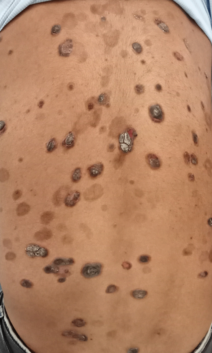
A 28-year-old male presented to the outpatient department complaining of fluid-filled lesions on his forehead and oral cavity, accompanied by burning pain and itching. The patient reported bursting the lesions, resulting in a clear fluid discharge. Over one-month, new fluid-filled lesions developed all over his face, followed by his upper limbs, trunk, and lower limbs. These lesions were associated with mild itching and pain and showed little tendency to heal (Figures 1 and 2).
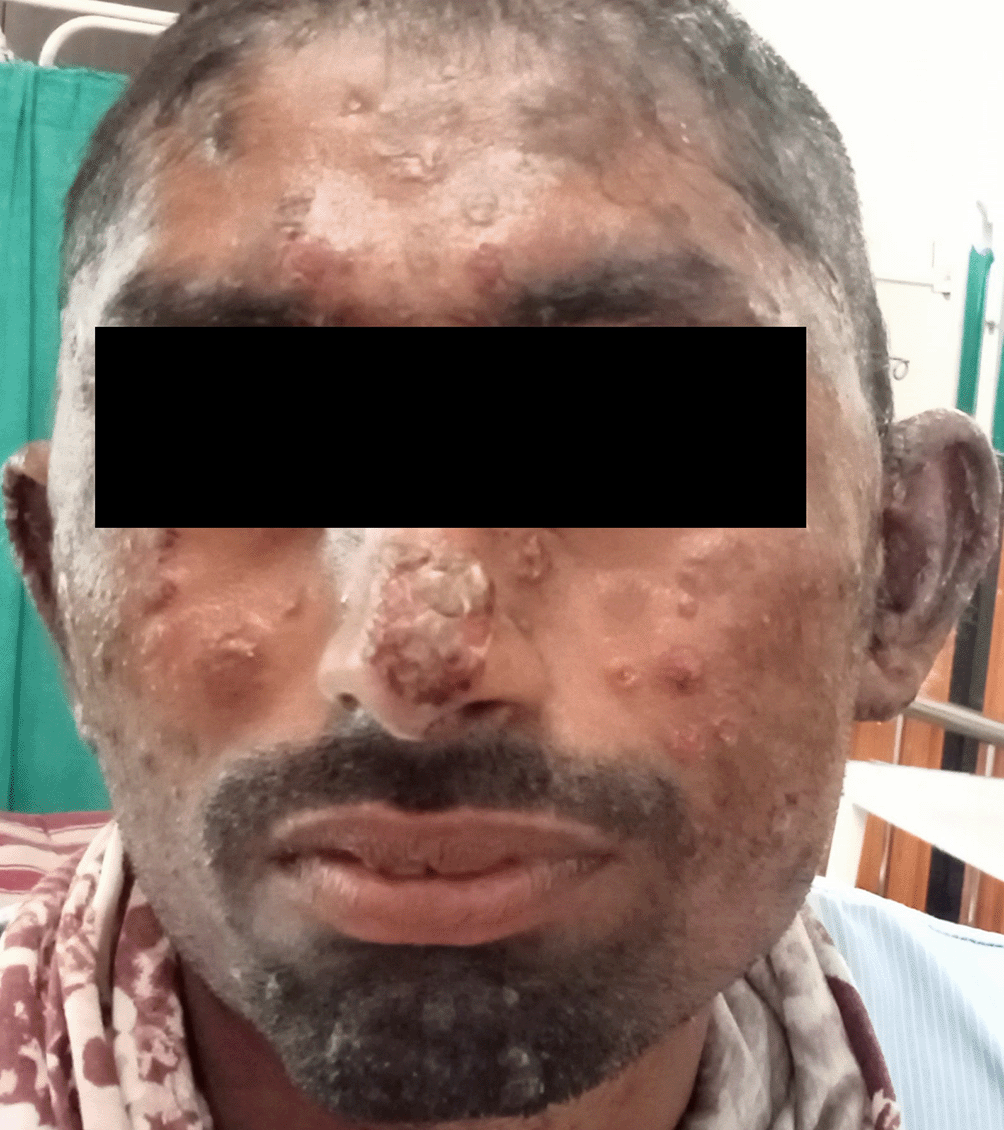
The patient reported no significant medical, family, psychosocial, or genetic history. He is a farmer by occupation. Initially, he developed lesions on his forehead that spread to his cheek and back. He sought treatment from a private hospital, where he was prescribed oral and topical medications. However, after following this regimen for 4–5 months, the patient reported incomplete resolution of his symptoms. He subsequently consulted another private practitioner due to worsening symptoms and the development of ulcers in his oral cavity, making it difficult for him to consume spicy foods.
The practitioner prescribed a course of oral antibiotic minocycline 100 mg twice daily and referred the patient to a tertiary care hospital for further treatment. The underlying cause of the patient’s symptoms must be identified and treated appropriately to prevent further complications.
During his medical evaluation, he underwent a thorough cutaneous examination, which brought to light several hypopigmented patches and plaques with crusting and a few lesions on his trunk bilaterally, upper and lower extremities, scalp, and face. Additionally, upon examination of the buccal mucosa, mucus with a raw base was noted. All in all, it was determined that approximately 25% of his body surface area had been affected by these dermatological conditions.
The patient expressed dissatisfaction with the aggravation of his symptoms, which had persisted for one month. Specifically, he had developed fluid-filled lesions on both his forehead and within his oral cavity, which had caused him significant discomfort and concern.
Additionally, blood tests were conducted, revealing a white blood cell count of 10,300 cubic millimetres. Furthermore, a skin biopsy was taken from a filled lesion, and the outcome indicated rounded-up and separated keratinocytes.
All of the investigations that were conducted have confirmed the diagnosis of pemphigus vulgaris.
The patient began his treatment regimen with oral medication tablets, including Defcort 30 mg and 12 mg twice daily, alternating between doses. He was also prescribed Pantoprazole 40 mg once a day to be taken before meals and Cetirizine 10 mg once a day for allergy symptoms.
In addition to these oral medications, the patient received topical treatments for his body and face lesions. Specifically, he was instructed to apply Clop-G cream twice daily to his body lesions and Momate-F cream twice daily to his face. He also used Mucopain gel twice daily for 20 minutes before meals and Tessoral gel twice daily for 20 minutes after meals. Finally, the patient soaked in saline twice daily to address crusts on his skin.
The patient’s condition stabilised with symptomatic treatment, and a plan was made to administer injected rituximab. After obtaining the patient’s consent, rituximab was administered in 500 cc normal saline over six hours while the patient was observed on the fifth day of treatment. Following the completion of the injection, the patient began taking Azathioprine 50 mg tablets as a maintenance regimen for six weeks.
On the seventh day, the symptoms exhibited by the patient were significantly reduced, leading to a favourable prognosis. Consequently, the medical team overseeing the patient’s care deemed it appropriate to discharge them from the hospital. However, they were advised to schedule a follow-up appointment in two weeks to ensure the patient continues to recover properly and monitor their progress.
PV is a severe and potentially life-threatening autoimmune disease. It can be fatal due to the loss of the epidermal barrier and body fluids, leading to subsequent infections. Therefore, PV must be diagnosed early and treated promptly to prevent fatal consequences. For a clinical diagnosis confirmation, histopathological analysis is required.6
The cornerstone of treatment for PV is systemic steroids, either with or without immunosuppression. However, some individuals may require higher doses of corticosteroids that can be unacceptable and difficult to manage. Unfortunately, we sometimes cannot reduce their high doses of drugs or provide treatment to those suffering from extreme cases.7
There are potential side effects linked with using rituximab. Seven patients had good drug tolerance and no adverse effects.5 Two individuals experienced angioedema, an acute side effect identified two hours after the infusion, and one patient required stopping of infusion. In contrast to earlier reports,5 the effects were seen during the second infusion. The second infusion was mainly uncomplicated, with the exception of one patient who experienced a slight infusion reaction during her initial infusion. Following rituximab infusion, sepsis occurred in two individuals (20%); they received the proper antibiotic treatment. Sepsis was experienced by patient number seven two weeks after beginning therapy. Acinetobacter was isolated from the blood on two separate occasions. Patient number eight developed sepsis after one week of initiating treatment, and the blood culture showed the growth of Staphylococcus aureus. The proper intravenous antibiotics were administered to both patients. Patient number seven made a full recovery. However, patient eight died because of sepsis. We recommended that rituximab be administered under close observation and in a setting with access to resuscitation equipment in case of infusion reactions. Sepsis is the most common side effect in patients with eroding skin, which serves as a portal of entry for organisms, including concurrent immunosuppressive medications. Sepsis is the most frequent cause of mortality in pemphigus.8 Strict clinical attention is necessary to identify sepsis early on, and efficient therapies are essential.9
Rituximab has a manageable adverse reaction, which includes infrequent infusion reactions and a low risk of infection—very few reports of severe adverse reactions to rituximab infusions.10 The injection rituximab received by our patient was well tolerated.11
Pemphigus is a prevalent and potentially lethal organ-specific autoimmune disease arising from the production of autoantibodies targeting keratinocyte proteins. In cases where conventional immunosuppressive medications and corticosteroids fail or when significant adverse effects occur, rituximab may be an effective therapeutic regimen for pemphigus vulgaris (PV). Clinicians must remain cognizant that prolonged treatment with rituximab may result in positive outcomes. Furthermore, the response to rituximab treatment in pemphigus patients may be delayed. Nevertheless, clinicians should consider prolonged treatment with rituximab as it can yield favourable results in individuals suffering from pemphigus.
| Views | Downloads | |
|---|---|---|
| F1000Research | - | - |
|
PubMed Central
Data from PMC are received and updated monthly.
|
- | - |
Provide sufficient details of any financial or non-financial competing interests to enable users to assess whether your comments might lead a reasonable person to question your impartiality. Consider the following examples, but note that this is not an exhaustive list:
Sign up for content alerts and receive a weekly or monthly email with all newly published articles
Already registered? Sign in
The email address should be the one you originally registered with F1000.
You registered with F1000 via Google, so we cannot reset your password.
To sign in, please click here.
If you still need help with your Google account password, please click here.
You registered with F1000 via Facebook, so we cannot reset your password.
To sign in, please click here.
If you still need help with your Facebook account password, please click here.
If your email address is registered with us, we will email you instructions to reset your password.
If you think you should have received this email but it has not arrived, please check your spam filters and/or contact for further assistance.
Comments on this article Comments (0)