Keywords
computed tomography, magnetic resonance imaging, masseter muscle hypertrophy
This article is included in the Datta Meghe Institute of Higher Education and Research collection.
computed tomography, magnetic resonance imaging, masseter muscle hypertrophy
The revised version of the manuscript incorporates the corrections suggested by the reviewers. The Introduction was expanded, incorporating evidence from recent bibliographic references. Evidences from related studies were added in discussion section. The related references were updated in the bibliography section. Conclusion was also modified.
See the authors' detailed response to the review by Nabeel Ayappali Kalluvalappil
Anatomically, the masseter muscle is a robust, two-layered quadrate muscle. It originates from the inferior and deep surface of the zygomatic arch and mostly enters into the inferior lateral side of the mandibular ramus.1 Unilateral or bilateral masseter muscle expansion is known as masseter hypertrophy. It may be asymptomatic or frequently accompanied by discomfort, which can occasionally be mistaken for parotid gland swelling.2–4 For the functional, aesthetic, and differential diagnosis of head and neck pathology, it is an uncommon condition with an unknown cause. Since the etiological component is unknown in the majority of instances, the condition is classified as idiopathic. Although several causes, including bruxism, malocclusion, clenching, and temporomandibular joint abnormalities, have been mentioned, they have not been established beyond a reasonable doubt. Since the etiological component is unknown in the majority of instances, the condition is classified as idiopathic. Despite being mentioned, a number of issues, including malocclusion, bruxism, clenching, and temporomandibular joint problems, have not been confirmed.5 However, the most common reason patients with masseteric hypertrophy seek treatment is because of their facial appearance.5,6 The masseter muscle plays a crucial function in face aesthetics since it is lateral to the mandibular ramus and necessary for proper mastication. For many people, a hypertrophied masseter will cause discomfort and adverse cosmetic effects by changing their face lines. Deficits in muscle function can also lead to the development of disorders such bruxism, protrusion, and trismus.5 Healthcare providers must comprehend the subtleties of masseter hypertrophy since patients’ quality of life can be greatly enhanced by early detection and effective treatment. The case report's objectives are to outline the signs and symptoms of idiopathic masseter hypertrophy, offer a differential diagnosis, and recommend a course of action. The purpose of this case report is to add to the expanding body of knowledge on masseter hypertrophy by shedding light on its clinical characteristics, difficulties with diagnosis, and management techniques.
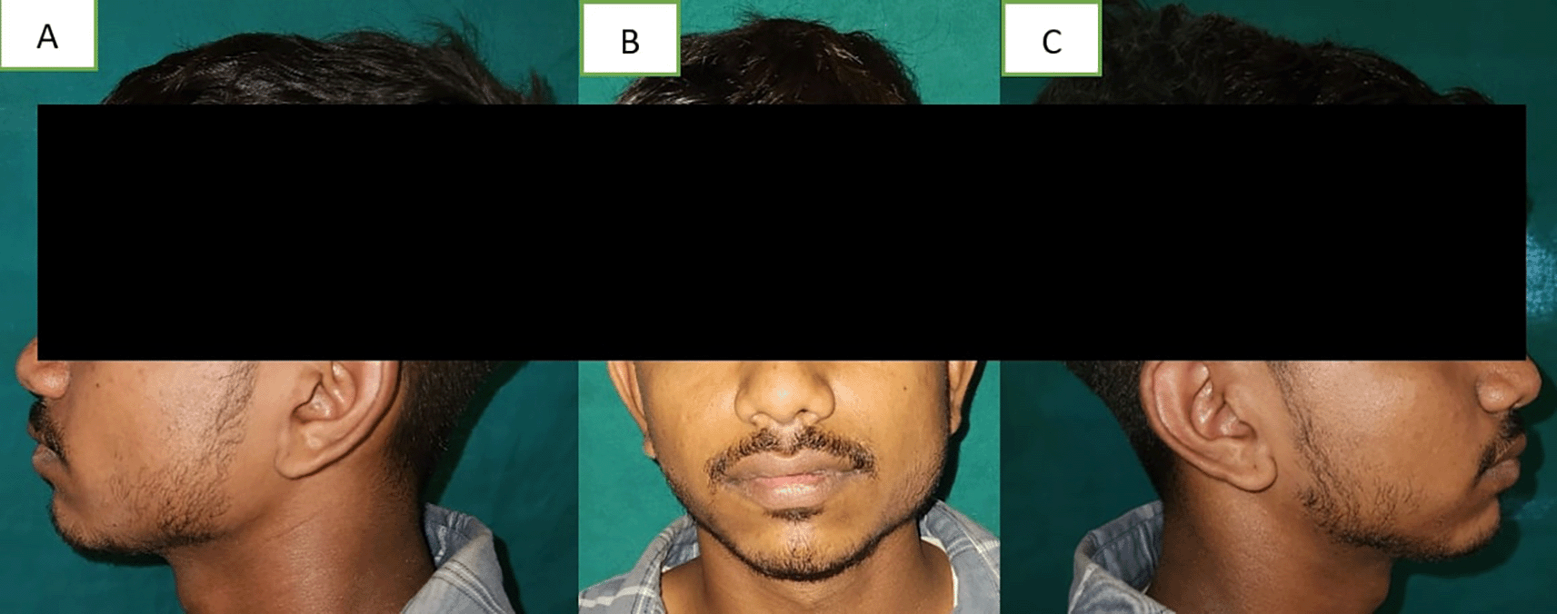
A 19-year-old male visited the dental hospital's oral medicine department. Due to unilateral facial swelling, the patient complained of an unattractive appearance (Figure 1). The patient explained that the enlargement, which had been asymptomatic until now, had gradually increased since birth. Additionally, he had never before experienced trouble opening his mouth or temporomandibular symptoms. There was no family history of any such swelling, no history of face injuries, dental anomalies, or clicking in the temporo-mandibular joint.
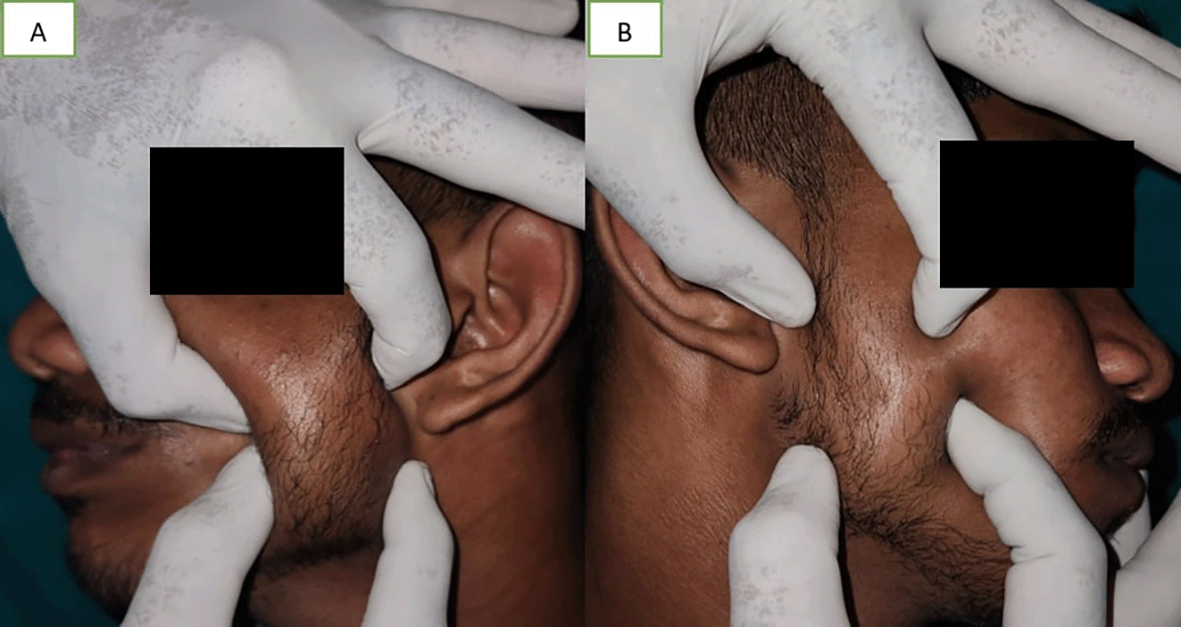
A soft to firm unilateral tissue mass was discovered during a clinical examination over the left body, close to the mandibular angle, which became prominent when the patient clenched their jaws (Figure 2). When the left masseter was palpated, the inferior portion of the muscle showed increased size. The masseter on the left side was broader than on the right side. Due to the increase in muscle size and stress, there was compensatory hypertrophy at the insertion of muscle. Bone spur growth and the prominence of the mandibular angle were felt (Figure 2).
The jaws moved normally as they opened and closed. During the occlusion, there was no midline deviation seen. Both an intra-oral and extra-oral examination revealed no abnormalities or possible etiological factors. There was no indication of a para-functional habit.
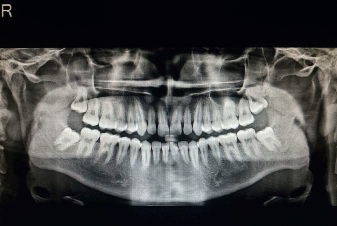
An orthopantomogram (OPG) was performed to look for any pathology (Figure 3). Compared to the right side, the antagonial notch on the OPG scans was slightly more pronounced. Other anomalies were not found in OPG.
When we informed the patient about the masseter hypertrophy in this case, the patient declined further therapy. As a result, there was no treatment given in this asymptomatic patient.
Asian Americans are more likely to be involved.7,8 Without regard to gender, the second and third decades of life have the highest incidence rates. In their analysis of 108 instances, Baek et al. showed that the average patient age was 30 years old, 60% of them had bilateral involvement, and 57% of them were men.8
According to some authors, the word “hypertrophy” may be deceptive because it refers to an increase in fiber count rather than cell size.9 Hypertrophy of the masseter muscle is thought to be an uncommon occurrence with no recognized etiology.
The cause of the majority of instances is unknown, despite the fact that numerous potential culprits, including malocclusion, bruxism, clenching, and abnormalities of the temporo-mandibular joint, have been proposed.4,9
In addition to being crucial for proper mastication, the masseter muscle is also crucial for facial aesthetics. Asymptomatic chronic expansion of one or both masseter muscles, known as masseteric hypertrophy, is typically the main complaint in cases of this condition.
The mandibular angle's bone spurs are frequently present observations. Bloem and Hoof asserted that this finding cannot be used as a diagnostic tool because 20% of healthy individuals have it. According to Guggenheim and Cohen,10 bone spurs are brought on by periostal irritation and new bone formation in response to the muscles bundles' elevated pressures.
It is important to correctly diagnose idiopathic masseter muscle hypertrophy because it can be mistaken for other conditions. Among these are masseter tumor, salivary gland disease, parotid tumor, parotid inflammatory disease, and intrinsic masseter muscle myopathy (caused by hypotrophy or hypoplasia on the contralateral side).11-13 There are two forms of Massetor Muscle Hypertrophy, according to Teixeira et al.: acquired due to functional hypertrophy and congenital or familial.12
A sialography is necessary to rule out this option since the correct diagnosis is more challenging in unilateral cases and necessitates a differential diagnosis with parotid gland abnormalities.14,15
Clinical examination, medical history, imaging modalities' findings, and muscle palpation can all be used to diagnose masseter hypertrophy. Visual inspection and palpation revealed a consistent muscle mass in masseteric hypertrophy, and the patient's forced bite could be felt to compress the muscle. Other benign and malignant neoplasms, on the other hand, are characterized by irregular and nodular development. The size and position of bucco-masseteric masses can be determined by sonography, computed tomography (CT), and magnetic resonance imaging (MRI).6,7 For the examination of hyperostosis at the point of muscle attachment in benign masseteric hypertrophy, CT scanning is essential.6 The panoramic radiograph is the most feasible diagnostic to support the clinical diagnosis when the physical examination points to masseteric hypertrophy.5 Ultrasonography of the region confirmed the uniform increased muscle mass with characteristics of normal musculature without any cystic, nodular or irregular foci.6
The use of CT is a well-established technique for delivering a wealth of details about nearby buildings and notable landmarks. Due to its high-quality imaging of bone structures and direct bone imaging, which is not achievable with MRI since cortical bone produces no detectable signal, CT scanning is essential in cases with masseter muscle hypertrophy with bone flaring. However, in this case report, the hypertrophied area and the border between the medial and lateral pterygoid could not be limited by CT. However, because muscle structural signals from the damaged side of the body are stronger than those from the unaffected side, MRI made it easier to diagnose the condition.10
There have been several reported treatments for masseter hypertrophy, ranging from non-invasive medical procedures to invasive surgical procedures. Some examples of conservative treatment include occlusal correction, relaxation therapy, botulinum therapy, spasmolytics, tranquilizers, and antidepressant therapy. Compared to surgical treatments, conservative techniques have both benefits and drawbacks.16,17 Despite being an intrusive treatment, surgical therapy is nevertheless favored for more dependable and long-lasting results. Masseteric enlargement typically doesn't require treatment. Reassurance, tranquilizers or muscle relaxants, psychiatric care, and injection of extremely modest dosages of botulinum toxin type A are examples of non-surgical therapeutic techniques.18 To fix malocclusions and premature contacts, dental restorations and occlusal modifications are crucial. Habits that are dysfunctional must be avoided. Patients may choose to have cosmetic surgery in this situation to lessen the prominence of the mandibular angle bone.19
A benign ailment called masseter muscle hypertrophy normally doesn't require surgery.
Due to improper diagnosis in these circumstances, needless biopsies, exploratory surgeries, and even radiotherapy for parotid tumors may be performed. To rule out other disorders, conventional radiography examinations, CT scans, and MRI scans are useful.20
Gurney made the initial surgical therapy suggestion in 1947. A submandibular incision is made, and 3/4 to 2/3 of the muscle mass is extracted. Adams proposed mandibular angle osteotomy in 1950. In 1950, Martensson removed the masseter muscle insertion using a triangle incision in a patient who had a history of unilateral masseter muscle hypertrophy and bruxism. In 1977, Beckers used the intraoral method to surgically treat 17 patients, excising the hypertrophied masseter’s internal muscular band. In 1982, Wood developed a surgical procedure in which he excised the mandibular angle’s bony protuberance without removing any masseter muscle tissue. According to Da Cruz et al. (1994), the intraoral technique provides superior cosmetic outcomes and a decreased risk of infection.5
Because it provided superior visibility, the extraoral approach was initially recommended frequently. Yet the intraoral approach has gained favor due to the advancement of new surgical tools and methods (such as particular retractors, rotating instruments, surgical saws, and, more recently, intraoral endoscopy). The scar created by the extraoral technique is unpleasant because the patients’ main complaint is about their face look. The intraoral technique has several benefits, including the avoidance of a visible scar in a procedure that is primarily cosmetic, the ability to safely make an incision in a surgical field away from the facial nerve’s marginal branch, and the ability to resect bone without severing the masseter-pterygoid sling.5
One of the alternative methods for treating maxillary muscle hypertrophy is injecting botulinum toxin type A into the hypertrophied muscle, as first suggested by Smyth12 and Moore and Wood.21 It is looked upon as a noninvasive method, which works upon the principle of inhibiting muscle contraction by hindering the release of acetylcholine.22 This results in paralysis of the muscle leading to “disuse atrophy” and reduction in the muscle volume. However, the disadvantage of this method is the temporary effect of the toxin proceeding to reversion to the original state in about 6 months.23 Also, botulinum is expensive and needs to be administered multiple times due to its reversible action.
When compared to surgical excision, masseter hypertrophy reduction using radiofrequency coagulator is a less invasive, safer, and far more durable technique than botulinum toxin therapy.24 This technique, which was initially presented by Powell et al.,25 employs radiofrequency energy to produce resistance in the tissue that eventually heats up. Heat between 50 and 90°C is produced as a result, which coagulates muscle tissue and creates scar tissue, which reduces the total volume of the affected muscle. This decrease in muscle mass starts three weeks following the procedure and lasts for up to eight weeks. Further research is need to determine the procedure’s long-term effectiveness, though.25 The main approach for the therapy, according to several studies, is surgical debulking of the hypertrophied muscle and, if necessary, excision of hyperostotic portions of the mandibular angle. Regarded as a necessary procedure to improve the patient’s appearance while undergoing treatment for masseteric hypertrophy, genioplasty.26
This case report provides an overview of the clinical presentation, diagnostic procedure, and therapy approaches for masseter hypertrophy, a rare illness that can have a major influence on a patient’s quality of life and look. The case study highlights the significance of taking masseter hypertrophy into account when making a differential diagnosis for individuals who exhibit facial asymmetry or jaw pain. Masseter hypertrophy can have a substantial impact on the aesthetics and functionality of the face, changing the jaw's appearance and function. To choose the best course of treatment, it is crucial to precisely evaluate the underlying problem.
| Views | Downloads | |
|---|---|---|
| F1000Research | - | - |
|
PubMed Central
Data from PMC are received and updated monthly.
|
- | - |
Provide sufficient details of any financial or non-financial competing interests to enable users to assess whether your comments might lead a reasonable person to question your impartiality. Consider the following examples, but note that this is not an exhaustive list:
Sign up for content alerts and receive a weekly or monthly email with all newly published articles
Already registered? Sign in
The email address should be the one you originally registered with F1000.
You registered with F1000 via Google, so we cannot reset your password.
To sign in, please click here.
If you still need help with your Google account password, please click here.
You registered with F1000 via Facebook, so we cannot reset your password.
To sign in, please click here.
If you still need help with your Facebook account password, please click here.
If your email address is registered with us, we will email you instructions to reset your password.
If you think you should have received this email but it has not arrived, please check your spam filters and/or contact for further assistance.
Comments on this article Comments (0)