Keywords
Optic disc swelling, obstructive hydrocephalus, tuberculoma
This article is included in the Datta Meghe Institute of Higher Education and Research collection.
Optic disc swelling, obstructive hydrocephalus, tuberculoma
A rare but unquestionably deadly form of tuberculosis is the central nervous system (CNS) disease brought on by Mycobacterium tuberculosis. Only 10% of tuberculosis patients are CNS cases, which have a significant mortality rate and distressingly severe neurological morbidity.1 Tuberculous meningitis (TBM) is a potentially fatal form of CNS infection causing death and severe disability up to 50%–60% of affected patients.1 Although there has been a recent come back of the disease in both developed and developing countries, the burden of CNS tuberculosis is primarily found in resource-poor regions of the world.2 The original Rich description2 suggested that CNS tuberculosis develops in two stages: first, tuberculous lesions (Rich’s focus) form in the brain during the bacteraemia stage or shortly thereafter, and later, CNS tuberculosis develops when one or more of these lesions burst or enlarge. This case demonstrates how disc edema and blurred vision might be tuberculoma’s early presenting symptoms. However, it can be difficult to detect and treat its morbidity. Early recognition and treatment improve the outcome.
A 19-year-old male Hindu by religion, student by occupation and no relevant family history presented with subacute onset of painless blurring of vision in both eyes for a two-week duration. This was associated with diplopia which later resolved spontaneously. Otherwise, there were no other complaints such as fever, headache, vomiting, cough, stiff neck, or any neurological symptoms. His best corrected visual acuity (BCVA) in both of his eyes was 6/12 upon evaluation. His dilated fundus examination revealed bilateral swelling of the optic disc, with few peripapillary shaped haemorrhages, tortuous vessels and multiple hard exudates confined to temporal to the disc suggestive of macular fan (Figure 1A and B). There were no other signs of uveitis such as vitritis, retinitis or choroiditis. Optic nerve function tests which included visual fields, pupillary light reflexes and colour vision were normal in both eyes. There was no relative afferent pupillary defect. Otherwise, his vital parameters were normal. There were no signs of meningeal irritation, such as neck rigidity or the Kernig’s sign. Higher mental functions were normal. The patient was oriented to time, place and person.
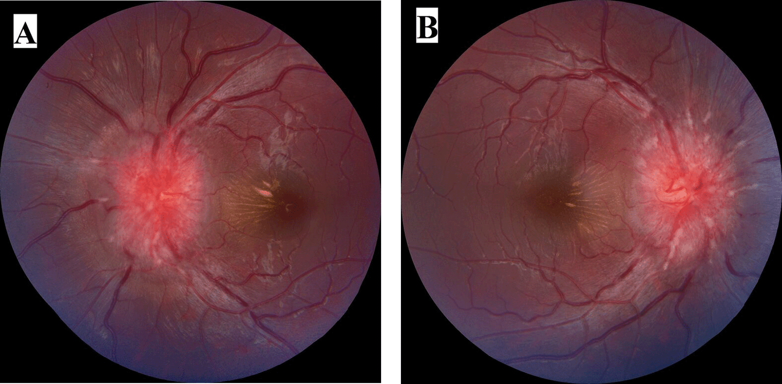
Fundus photograph was taken through Zeiss Visucam 524.
After obtaining written informed consent from the patient further intervention was started.
An evaluation of the cranial nerves indicated left sixth cranial nerve palsy that later spread to the right side. Treatment for tuberculosis was started. During the initial phase, he received isoniazid, rifampicin, pyrazinamide, and ethambutol for two months. Systemic steroids were also started. The continuation phase, which consisted of isoniazid and rifampicin, lasted for seven months after that.
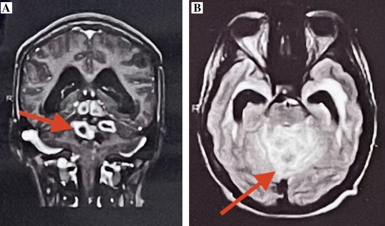
Laboratory investigations showed a raised erythrocyte sedimentation rate (ESR) of 109 mm with positive Mantoux reading of 20 mm at 72 hours. The Quantiferon-TB Gold in-tube test was positive. cerebrospinal fluid examination revealed an opening pressure of 12-20 cmH20, turbid in appearance; a lymphocytic predominant pleiocytosis. Total white blood cell counts ranged 60-500 cells/μL, with raised protein levels greater than 2.2 g/L and low glucose levels lower than 40 mg/dL. MRI imaging showed ring enhancing lesions noted in the cerebellar vermis, left cerebellar hemisphere and left middle cerebral peduncle with surrounding extensive perilesional edema appearing hyperintense on T2WI/FLAIR, hypointense on T1W1, showing central restriction on DWI with no blooming on SWI causing effacement of fourth ventricle leading to obstructive hydrocephalus suggestive of tuberculoma (Figure 2A and B).
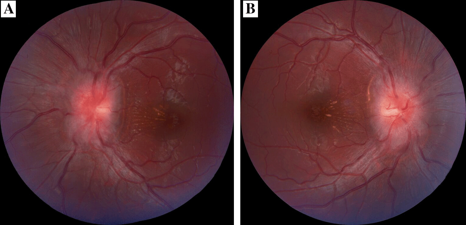
The clinical characteristics, laboratory results, CSF findings, and radiographic imaging are used to make the diagnosis of tuberculoma. However, if there are no signs of the typical clinical characteristics, the condition may prove difficult to diagnose. Tuberculomas are spherical, firm, avascular masses that range in size from 2 to 10 cm in diameter. They are well defined, and the gliosis and edema in the compressed brain tissue around them are visible.3 In our patient, the presentation of tuberculoma was associated with bilateral optic disc swelling. He presented with ocular signs of visual impairment, bilateral disc edema and sixth cranial nerve palsy. Our patient’s positive Mantoux test and positive TB-gold, elevated ESR indicated tuberculosis infection. After two months of anti-tubercular medications with steroids on tapering dose patient had marked reduced optic disc swelling and exudates with visual gain of 6/9 in both eyes (Figure 3A and B).
On follow-up visit liver function test was within normal limits (Table 1).
The majority of patients (80%) with TBM manifest in late stages (stages II and III), according to a review of 101 cases by Sinha et al.2 Late-stage TBM patients had papilloedema and cranial nerve palsy in 52% and 31% of cases, respectively,4 and 27% of patients had visual impairment. At the time of initial TBM presentation, visual impairment of less than 6/18, papilloedema, and cranial nerve palsy were found to be predictors of blindness and severe disability at 6 months. Kumar et al. have proposed that basal meningeal enhancement or tuberculoma or both were 89% sensitive and 100% specific for TBM.5 Commercial nucleic acid amplification assays for the diagnosis of TBM have shown a 98% specificity and 56% sensitivity, according to a recent meta-analysis.6 Our patient’s BCVA increased to 6/6, and the edema of his optic disc significantly decreased (Figure 3). Due to early initiation of anti-tubercular medication, adverse outcomes including blindness and impairment at 6 months were not observed in our patient. When left untreated, 30–50% of tuberculomas enter and continue to progress in a stationary course, according to reports published prior to the development of efficient anti-tuberculosis therapy.7
The majority of tuberculomas disappear after 12 to 24 months, while continued treatment lasting longer than two years may be necessary in patients with multiple or larger lesions. Anti-inflammatory drugs are administered in addition to the normal anti-tuberculosis regimen in some patients whose symptoms paradoxically deteriorate after therapy due to the release of inflammatory mediators.8 Surgery is necessary to remove particularly large tuberculomas, those that have a mass effect on the brain, and those that do not respond to medical treatment. In some cases, surgical excision is necessary for diagnosis as well as treatment.9
For individuals with clinically suspected TBM, the quantitative real-time PCR approach has been shown to have higher specificity (100%) and sensitivity (95.8%) than the traditional conventional PCR.10
| Views | Downloads | |
|---|---|---|
| F1000Research | - | - |
|
PubMed Central
Data from PMC are received and updated monthly.
|
- | - |
Provide sufficient details of any financial or non-financial competing interests to enable users to assess whether your comments might lead a reasonable person to question your impartiality. Consider the following examples, but note that this is not an exhaustive list:
Sign up for content alerts and receive a weekly or monthly email with all newly published articles
Already registered? Sign in
The email address should be the one you originally registered with F1000.
You registered with F1000 via Google, so we cannot reset your password.
To sign in, please click here.
If you still need help with your Google account password, please click here.
You registered with F1000 via Facebook, so we cannot reset your password.
To sign in, please click here.
If you still need help with your Facebook account password, please click here.
If your email address is registered with us, we will email you instructions to reset your password.
If you think you should have received this email but it has not arrived, please check your spam filters and/or contact for further assistance.
Comments on this article Comments (0)