Keywords
Emergency, Gastroenterology, Spondylodiscitis, Duodenal Perforation, Foreign Bodies, Psoas Abscess
Emergency, Gastroenterology, Spondylodiscitis, Duodenal Perforation, Foreign Bodies, Psoas Abscess
The ingestion of foreign bodies is a medical emergency; however, the majority of cases resolve spontaneously without causing damage to the gastrointestinal tract. Approximately 20% of cases require endoscopic extraction, and in 1% of cases, surgical intervention becomes necessary, especially when complications arise such as acute peritonitis.1,2 While fish bones are the most commonly implicated foreign bodies, occurrences involving toothpicks are much more rare.3 In this report, we present an unusual case of duodenal perforation caused by an ingested toothpick, which led to the formation of a collection in the psoas muscle and contiguous spondylodiscitis. Diagnosis and treatment were achieved through the utilization of endoscopy, radiology, and antibiotic therapy, without the need for surgical intervention.
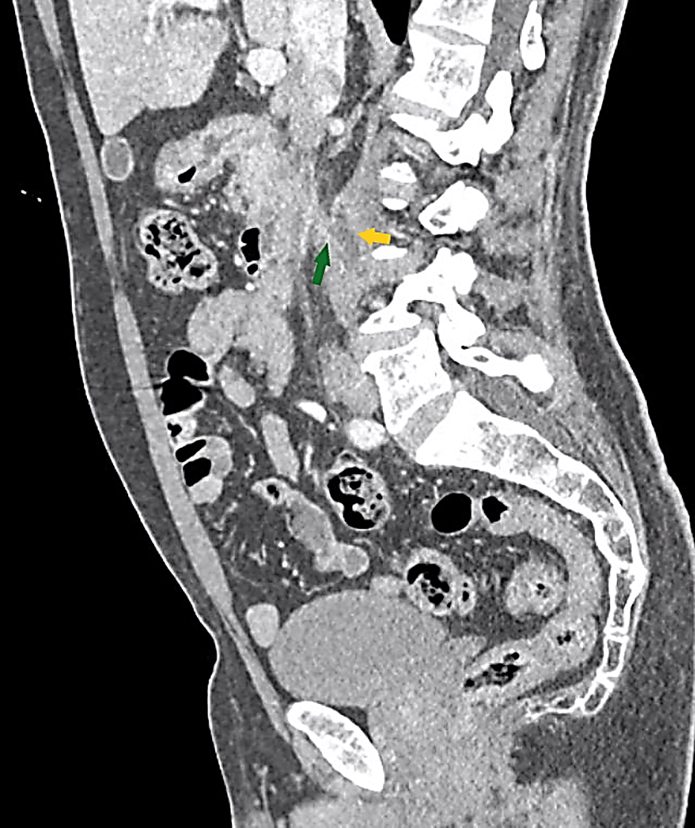
A 55-year-old north-African greengrocer man presented with a five-day history of right upper quadrant abdominal pain and functional impairment of the lower limbs. He had no significant medical history and was taking no medication. His symptoms were not accompanied by nausea or vomiting. Physical examination revealed an afebrile patient with tenderness in the right upper quadrant of the abdomen. Blood tests showed normal results. An abdominal computed tomography (CT) scan revealed the presence of a spontaneously dense linear foreign body measuring 6 cm in length. It was located retroperitoneally, with one end at the level of the superior duodenal flexure and the second portion of the duodenum, perforating the posterior wall of the second duodenum. The other end of the foreign body was situated at the level of the right psoas muscle, surrounded by a collection measuring 26 × 11 mm on the axial plane and extending over 47 mm in height. There was regular and circumferential thickening of the first and the second duodenum portions with an inflammatory appearance (Figure 1). No vessel injury was observed.

Upper gastrointestinal endoscopy revealed a wooden toothpick deeply embedded in the duodenal wall (Figure 2), which was successfully removed using biopsy forceps (Figures 2 and 3) without complications such as bleeding or purulent discharge.
No clinical improvement was noted after seven days of empirical antibiotic. A follow-up abdominal scan was conducted, which showed the collection size to be stable. Due to the persistent functional impairment, an MRI of the lumbar spine was performed, leading to a diagnosis of contiguous spondylodiscitis at the L4-L5 level (Figure 4).
A CT-guided fine-needle aspiration of the collection was carried out, and the puncture fluid was found to be purulent. A sample was sent for bacteriological examination, which revealed Escherichia coli as the causative agent. Subsequently, an eight-week antibiotic therapy regimen was initiated, adjusted based on the antibiogram results (Cefotaxime: 2 grams × 4/day, Ciprofloxacin 200 mg × 2/day. Immobilization of the lumbar spine was achieved using a corset.
The clinical and radiological progression showed improvement, with the functional impotence resolving and the psoas collection regressing on the follow-up CT scan. On the 13th day of hospitalization, the patient was discharged without further active treatment for spondylodiscitis. During the subsequent follow-up, one year after discharge, the patient remained asymptomatic.
The majority of ingested foreign bodies pass through the gastrointestinal (GI) tract without complications, symptoms, or requiring further intervention.1,2 GI perforation occurs in approximately 1% of cases, and the risk of perforation increases to 15–35% with thin and pointed objects such as toothpicks, needles, fish and chicken bones.2–4
Batteries can also cause chemical and electrical damage to the mucosal tissues.5,6 Other complications that may occur include obstruction, peritonitis, abscess, or perforation into adjacent organs.7
To the best of our knowledge, this is the second reported case of spondylodiscitis caused by toothpick ingestion in the literature. Toothpicks account for approximately 9% of ingested foreign bodies.7–9 Risk factors for toothpick ingestion include alcohol abuse, mental disorders, rapid eating, and consumption of foods containing toothpicks.7,10,11 Many patients are unaware of the ingestion, making the diagnosis of toothpick ingestion challenging.1 The clinical presentation varies widely depending on the site of perforation, with abdominal pain being the most prominent symptom.12 In the differential diagnosis, peptic ulcer perforation, acute appendicitis, and acute diverticulitis should be considered.13
Studies have shown that GI perforation most commonly occurs in the colon and ileocecal region, particularly in the appendix and Meckel’s diverticulum.2 Perforations in the gastric and duodenal regions are less frequent, and their presentations tend to be more chronic and less severe in nature.2,12,14 The occurrence of duodenal perforation is likely related to its anatomical morphology, characterized by an angulation and a C-loop shape.7
Plain radiographs are a simple and useful diagnostic tool for ingested foreign bodies; however, they may not detect radiolucent objects such as animal bones, glass, plastics, medications, and small metal objects.2,3,15,16 Radiographs can be sufficient to rule out free abdominal gas and determine the size, shape, location, and number of foreign bodies.11,17 However, identifying the localization of a toothpick using plain X-rays is challenging.11 In such cases, CT scanning and diagnostic endoscopy are generally preferred modalities.2
It is important to note that barium swallow studies are contraindicated in these cases due to the risk of GI perforation. Additionally, contrast agents used in these studies may interfere with endoscopic evaluation.2 The sensitivity of CT scans can be improved with 3D reconstruction.18 After performing a CT scan, endoscopic intervention can be carried out, allowing for both diagnosis and therapeutic removal of the foreign body to be done simultaneously.19
In our case, CT images were invaluable in detecting the foreign body in the duodenum, which appeared as a high-density linear object, but its exact nature could not be identified. The CT scan accurately determined the location of both ends of the toothpick and the depth of duodenal penetration. Furthermore, it confirmed the absence of vessel injury before endoscopic removal of the toothpick. Upper GI endoscopy is contraindicated when peritonitis or vascular penetration is suspected.7 Endoscopy confirmed the presence of a wooden toothpick, and our patient recalled possibly swallowing the object while he was asleep, intoxicated, with a toothpick in his mouth after consuming tapas containing toothpicks, one week prior.
Treatment modalities for foreign bodies in the GI tract are chosen based on the type and location of the object. Endoscopy is the recommended first-line management for duodenal or lower rectal perforation.11 Flexible endoscopic techniques are preferred over rigid endoscopes due to a lower risk of perforation.20,21 Commonly used tools include crocodile teeth forceps, polypectomy snares, magnetic probes, retrieval snare nets, Dormia panniers, and transparent cap-fitting devices.17,22,23 Endoscopists should be familiar with these tools and comfortable using them. In our case, a biopsy forceps easily grasped the toothpick. However, it is important to note that if there is suspected frank perforation or peritonitis, surgical treatment should not be delayed.
In our case, the duodenal perforation was associated with a collection in the psoas muscle, without signs of sepsis. Radiological drainage was preferred over surgery. Percutaneous drainage is the preferred method for treating retroperitoneal abscesses as it is better tolerated by patients, eliminates the need for general anesthesia, and is associated with shorter hospital stays.2,4,14,15 The mortality rate after surgical drainage of retroperitoneal abscesses is reported to be 39%–50%, whereas it is around 1.5%–10% for percutaneous drainage.2,14–16
Fortunately, the diagnosis of spondylodiscitis was quickly suspected and confirmed by an MRI of the lumbar spine. This allowed for the appropriate treatment of spondylodiscitis and halted its progression.
To the best of our knowledge, this is the first documented case of duodenal perforation complicated by a psoas muscle collection and spondylodiscitis. In summary, the overall disease course of this patient was unusual, primarily due to the delayed diagnosis and the patient’s lack of awareness regarding the ingestion of the toothpick. It is important to consider the possibility of foreign body ingestion and gastrointestinal perforation in patients presenting with abdominal pain. Thorough questioning and clinical suspicion can help avoid unnecessary surgical interventions.
Written informed consent to publish this case and associated images was obtained from the patient.
All data underlying the results are available as part of the article and no additional source data are required.
| Views | Downloads | |
|---|---|---|
| F1000Research | - | - |
|
PubMed Central
Data from PMC are received and updated monthly.
|
- | - |
Is the background of the case’s history and progression described in sufficient detail?
Yes
Are enough details provided of any physical examination and diagnostic tests, treatment given and outcomes?
Yes
Is sufficient discussion included of the importance of the findings and their relevance to future understanding of disease processes, diagnosis or treatment?
Yes
Is the case presented with sufficient detail to be useful for other practitioners?
Yes
Competing Interests: No competing interests were disclosed.
Reviewer Expertise: Innovative microbiome and gastrointestinal intervention therapy.
Alongside their report, reviewers assign a status to the article:
| Invited Reviewers | |
|---|---|
| 1 | |
|
Version 1 05 Oct 23 |
read |
Provide sufficient details of any financial or non-financial competing interests to enable users to assess whether your comments might lead a reasonable person to question your impartiality. Consider the following examples, but note that this is not an exhaustive list:
Sign up for content alerts and receive a weekly or monthly email with all newly published articles
Already registered? Sign in
The email address should be the one you originally registered with F1000.
You registered with F1000 via Google, so we cannot reset your password.
To sign in, please click here.
If you still need help with your Google account password, please click here.
You registered with F1000 via Facebook, so we cannot reset your password.
To sign in, please click here.
If you still need help with your Facebook account password, please click here.
If your email address is registered with us, we will email you instructions to reset your password.
If you think you should have received this email but it has not arrived, please check your spam filters and/or contact for further assistance.
Comments on this article Comments (0)