Keywords
COVID-19; Invasive fungal infections; Mucormycosis; Corticosteroids
COVID-19; Invasive fungal infections; Mucormycosis; Corticosteroids
We have removed parts of the results that looked repetitive from the tables, and the discussion has also been summarised to be less wordy.
See the authors' detailed response to the review by Prateek Nishant, Ranjeet Sinha and Sony Sinha
When the pandemic of coronavirus disease 2019 (COVID-19) started, therapeutic options were unavailable and many were recommended without robust evidence. With platform trials, several agents were tried, and corticosteroid therapy was found to be beneficial in a subset of COVID-19 patients who had severe illness-causing hypoxia and not across the entire spectrum of severity.1 It helped us to buttress the hypothesis that corticosteroids mitigate the detrimental effect of exuberant inflammatory response-related morbidity and mortality. However, the patient population included in these platform trials was predominantly from the Western world.
Having established the role of corticosteroids in severe COVID-19 infection, dexamethasone therapy became the standard of care for all hypoxic COVID-19 patients across the world. In India, while the use of corticosteroids was not routine in the first wave of COVID-19, corticosteroid therapy became a standard of care for the management of hospitalized COVID-19 patients during the second wave as evidence from RECOVERY trial was available by then.1 Being familiar, cheap, and the first drug to show evidence of survival benefit, corticosteroid therapy was welcomed with a lot of enthusiasm. Soon after, in India, several case reports of invasive fungal infections in COVID-19 patients emerged during the second wave. Though corticosteroid therapy was quite beneficial; in the Indian population, the real extent of benefit which is probably offset by the increased risk of fungal infections, is not known.2,3
There are very few studies from India showing the type of fungal infections and their risk factors in COVID-19 patients. Hence, we undertook the study to determine the frequency of invasive fungal infections in hospitalized COVID-19 patients from India. We also studied the spectrum of fungal infections, the risk factors of fungal infections, and their clinical outcomes in hospitalized COVID-19 patients.
Study design: We performed a single center, retrospective, cross-sectional analytical study, among patients admitted at a tertiary care hospital at Pondicherry (JIPMER) between April 1st, 2020, to August 31st, 2021.
Study participants: We defined a patient to have recent COVID-19 if they had tested positive for COVID-19 by RT-PCR/Antigen detection test either at admission or within 90 days of hospital admission. From our hospital electronic health records (EHR), all patients aged 13 and above hospitalized at JIPMER during the study period were identified using a structured query language (SQL) based query. These patients were screened for recent COVID-19 reports, and patients who had recent COVID-19 were included in the study. Patients with either unavailable clinical data or COVID-19 reports were excluded.
Ethical statement: The study protocol was reviewed and approved by the Institute Ethics Committee (Human Studies) of Jawaharlal Institute of Postgraduate Medical Education and Research, Puducherry (DHR REG.NO.EC/NEW/INST/2020/331). A waiver of Informed consent was approved by the Institute Ethics Committee (Human Studies).
Sample size estimation: Based on prior studies,4 the frequency of IFI among hospitalized COVID-19 patients was assumed to be 15%, with an error margin of 5%, with a level of significance at 0.05, the sample size estimated was 196 patients. Our study is a retrospective case record-based study and included all patients who met the selection criteria.
Study procedure: We had complete access to our hospital EHR, from which, all patients admitted during the study period were identified, and screened for COVID-19 reports. The data from EHR was exported in CSV format and cleaned using the software OpenRefine (version 3.6.2, for Microsoft Windows 11). For patients who had recent COVID-19, baseline characteristics including comorbidities and severity of COVID-19 illness, requirement for ICU admission, treatment details, duration of hospital stay, and clinical outcome at discharge were collected. Fungal culture, and histopathological biopsy reports were noted. In patients who underwent computed tomography based on clinical suspicion, image findings suggestive of fungal etiology were analyzed. Based on the European Organization for Research and Treatment of Cancer (EORTC) guidelines,5,6 which were modified accordingly for COVID-19 patients (see Extended data34), these patients were then categorized to have Possible, Probable or Proven Invasive fungal infection. For the study purpose, patients who had more than one group of IFI were classified as belonging to the IFI group with the highest category of likelihood. Clinical and laboratory data of patients with fungal infection were analyzed to identify the predictors of fungal infection among hospitalized COVID-19 patients.
Statistical analysis: The data were compiled using Microsoft Excel and analyzed using SPSS version 19.0 (SPSS for Windows, version 19.0, Chicago, SPSS Inc.) and R software (version 3.3.1, R Foundation for Statistical Computing). Categorical variables were expressed as percentages and frequencies, and continuous variables were reported as mean and standard deviation or median with the interquartile range. Categorical variables were compared by Chi-square test. The strength of association was expressed as odds ratios. Normality was assessed by the Kolmogorov-Smirnov test. To compare continuous normally distributed data, the two-tailed unpaired t-test was used. All tests were two-sided, and P<0.05 was considered statistically significant. Before performing multivariable analysis, we reduced the dimensions of the variables by multiple correspondence analysis. We found collinearity between the predictors 'corticosteroid and heparin' as well as 'diabetes mellitus and hypertension'. Considering biological plausibility, we chose corticosteroids and diabetes mellitus as predictors from each of the clusters for further analysis. We observed a similar clustering between 'end-stage renal disease (ESRD) and central venous catheter (CVC) usage'. We included both ESRD and CVC for the invasive fungal infection group and removed CVC for the subgroup analysis involving invasive mold infection. Multivariable logistic regression using Enter method was performed to identify the predictors.
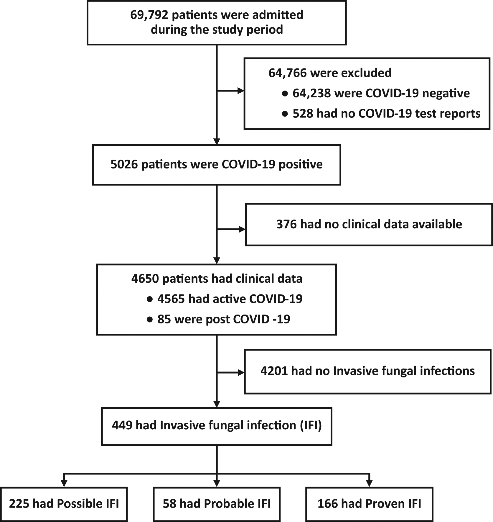
During the study period, 4650 patients were hospitalized with a history of recent COVID-19 (including patients admitted for COVID-19 and post-COVID-19 patients), among them, 4565 patients were hospitalized for active COVID-19 and 85 patients were admitted with post-COVID-19 complications.33 These post-COVID patients had received treatment for COVID-19 elsewhere and were hospitalized at JIPMER for post-COVID sequelae (Figure 1).
Among the hospitalized patients, who had recent COVID-19, the median age was 50 years (interquartile range [IQR], 35 to 62), with 27% of patients aged above 60 years; 59% were male. The most common comorbid illness in these patients was diabetes mellitus (31%) followed by systemic hypertension (25%) and chronic cardiac disease (8%). Ongoing immunosuppressive therapy (corticosteroid or other immunosuppressants) for other indications was present in 113 patients (2.4%) (Table 1).
| Patients with recent COVID-19 | All patients (n=4650) | With IFIa (n=449) | Unadjusted OR (95% CI) |
|---|---|---|---|
| Age, years | 50 (35 – 62) | 55 (44 – 64) | 1.02 (1.01 to 1.03) |
| Male sex | 2738 (58.9) | 294 (65.4) | 1.36 (1.11 to 1.67) |
| Coexisting conditions | |||
| Diabetes mellitesb | 1419 (30.5) | 269 (59.9) | 3.95 (3.23 to 4.82) |
| Diabetic keto-acidosis | 72 (1.5) | 26 (5.8) | 5.57 (3.41 to 9.10) |
| Hypertension | 1165 (25.1) | 151 (33.6) | 1.60 (1.30 to 1.97) |
| End-stage renal diseasec | 313 (6.7) | 62 (13.8) | 2.53 (1.88 to 3.40) |
| Chronic cardiac diseased | 372 (8.0) | 30 (6.7) | 0.81 (0.55 to 1.19) |
| Cerebrovascular accidente | 99 (2.1) | 21 (4.7) | 2.60 (1.59 to 4.25) |
| Asthma | 73 (1.6) | 3 (0.7) | 0.40 (0.13 to 1.27) |
| Other chronic lung diseases | 172 (3.7) | 12 (2.7) | 0.70 (0.38 to 1.26) |
| Chronic liver disease | 31 (0.7) | 1 (0.2) | 0.31 (0.04 to 2.29) |
| Autoimmune disease | 86 (1.8) | 12 (2.7) | 1.54 (0.83 to 2.85) |
| Immunosuppressive state | |||
| Prior systemic steroid usage | 95 (2.0) | 11 (2.5) | 1.23 (0.65 to 2.33) |
| Prior non-steroidal immunosuppressants usage | 95 (2.0) | 10 (2.2) | 1.11 (0.57 to 2.15) |
| HIV/AIDS | 13 (0.3) | 2 (0.4) | 1.71 (0.38 to 7.73) |
| Malignancyf | 203 (4.4) | 18 (4.0) | 0.91 (0.56 to 1.49) |
Among the patients admitted with active COVID-19 (n=4565), 1283 patients (28%) had severe illness at admission. Corticosteroids were used in 2014 patients (44%). Many of the patients with COVID-19, also received antibiotics (42%), and 10% of the patients received more than three antibiotics during their hospital stay (Table 2).
| Patients with Active COVID-19 | All patients (n=4565) | With IFI (n=366) | Unadjusted OR (95% CI) |
|---|---|---|---|
| Illness severity at admission | |||
| Hypoxia | 2184 (47.8) | 276 (75.4) | 3.69 (2.88 to 4.71) |
| Only supplemental oxygen | 1963 (43.0) | 234 (63.9) | - |
| Non-invasive ventilated | 111 (2.4) | 17 (4.6) | - |
| Mechanical ventilated | 110 (2.4) | 25 (6.8) | - |
| Severe COVID-19b | 1283 (28.1) | 172 (47.0) | 2.46 (1.99 to 3.06) |
| Maximal respiratory support | |||
| Only supplemental oxygen | 1295 (28.4) | 136 (37.2) | - |
| Non-invasive ventilation | 143 (3.1) | 18 (4.9) | - |
| Mechanical ventilation | 849 (18.6) | 137 (37.4) | - |
| Central venous catheter usagec | 638 (14.0) | 128 (35.0) | 3.89 (3.08 to 4.92) |
| ICU admission | 864 (18.9) | 146 (39.9) | 3.22 (2.57 to 4.03) |
| Pharmacotherapy | |||
| Corticosteroids | 2014 (44.1) | 233 (63.7) | 2.38 (1.91 to 2.97) |
| Dexamethasone | 1913 (41.9) | 217 (59.3) | 2.15 (1.73 to 2.67) |
| Methylprednisolone | 260 (5.7) | 69 (18.9) | 4.88 (3.61 to 6.58) |
| Prednisolone | 93 (2.0) | 12 (3.3) | 1.72 (0.93 to 3.19) |
| Hydrocortisone | 51 (1.1) | 13 (3.6) | 4.03 (2.13 to 7.64) |
| > 2 steroid formulations | 290 (6.4) | 75 (20.5) | 4.78 (3.58 to 6.37) |
| Parenteral anticoagulationd | 2064 (45.2) | 235 (64.2) | 2.33 (1.86 to 2.90) |
| Remdesvir | 448 (9.8) | 36 (9.8) | 1.00 (0.70 to 1.44) |
| Tocilizumab | 9 (0.2) | 3 (0.8) | 5.78 (1.44 to 23.19) |
| Baricitinib | 23 (0.5) | 3 (0.8) | 1.73 (0.51 to 5.84) |
| Antibiotic usage | 1935 (42.4) | 238 (65.0) | 2.74 (2.19 to 3.43) |
| Exposure to multiple antibioticse | 462 (10.1) | 95 (26.0) | 3.66 (2.83 to 4.73) |
Of the 4650 hospitalized patients with recent COVID-19 infection, 449 (9.7%) had an invasive fungal infection (Figure 1). Among those admitted for active COVID-19, 366 (8%) developed an invasive fungal infection during their hospital stay, whereas 83 out of 85 post-COVID-19 patients had an invasive fungal infection. The number of patients with proven or probable invasive fungal infections was 48.2 per 1000 COVID-19 patients.
The most common invasive fungal infections were due to mold infections (Mucorales & Aspergillus) occurring in 377 patients, followed by candidiasis (n=88). Out of 4650 patients, proven or probable mold infection occurred in 139 (3.0%) patients, among them 127 (91%) patients had mucormycosis and 36 (26%) had aspergillosis. Radiological evidence of pulmonary mold infection was present in 173 patients. In patients with mucormycosis, rhino-nasal or rhino-orbital-cerebral presentation (96.1%) was the most common presentation, followed by disseminated infection (3.9%), whereas among patients with isolated aspergillosis, pulmonary aspergillosis (67%) was common than rhino-nasal presentation (33%). Invasive candidiasis occurred in 88 patients, with positive fungal blood culture in 64 (73%) patients. Non-Albicans spp. (n=59) were more common than Candida albicans (n=7) (Table 3).
| Characteristics | Active COVID-19 (n=4565) | Post COVID-19 (n=85) | Total (%) (n=4650) |
|---|---|---|---|
| Total invasive fungal infection | 366 | 83 | 449 |
| Proven | 108 | 58 | 166 |
| Probable | 47 | 11 | 58 |
| Possible | 211 | 14 | 225 |
| Clinical syndrome of IFI | |||
| Mold infectionsa | |||
| Rhinosinusitis | 76 (1.7) | 73 (85.9) | 149 |
| Only Rhinosinusitis | 32 | 20 | 52 |
| Rhino-orbital | 21 | 41 | 62 |
| Rhino-orbito-cerebral/Rhino-cerebral | 23 | 12 | 35 |
| Pulmonary | 171 (3.7) | 5 (5.9) | 176 (3.8) |
| Disseminated | 3 | 4 | 7 |
| Candidiasis | |||
| Candidemia | 64 (1.4) | 0 | 64 |
| Others | 25 (0.6) | 1 | 26 |
| Fungal speciesb | |||
| Mucorales. spp | 60 | 67 | 127 |
| Aspergillus. spp | 19 | 17 | 36 |
| Candida. sppc | 64 | 0 | 64 |
| Candida albicans | 7 | 0 | 7 |
| Non albicans sppd | 59 | 0 | 59 |
| Candida auris | 20 | 0 | 20 |
After adjusting for confounding by multivariable logistic regression, diabetes, diabetic ketoacidosis, prior stroke, steroid usage, and central venous catheter usage were found to be independent predictors of IFI in patients with active COVID-19 (i.e., after excluding patients admitted for post-COVID sequelae). Diabetic ketoacidosis was the most important predictor (odds ratio, 3.31; 95%CI, 1.93 to 5.58). As compared to the risk of IFI among patients who received dexamethasone, the risk of IFI in patients who received methylprednisolone was 2.82 (95%CI, 2.07 to 3.85). In the subgroup of invasive mold infections (IMI), diabetes, diabetic ketoacidosis, steroid usage, antibiotic usage, and chronic kidney disease were identified as independent predictors and prior stroke did not show an association (Tables 4, 5).
| Characteristics | With IFI (n=366) | Without IFI (n=4119) | Univariate OR (95% CI) | Multivariate OR (95% CI) |
|---|---|---|---|---|
| Age | ||||
| <18 years | 1 (0.27) | 55 (1.31) | 0.21 (0.03 – 1.50) | - |
| 18 to 60 years | 236 (64.5) | 3043 (72.5) | 0.69 (0.55 – 0.86) | - |
| > 60 years | 129 (35.2) | 1101 (26.2) | 1.53 (1.22 – 1.92) | - |
| Male sex | 231 (63.1) | 2443 (58.2) | 1.23 (0.99 – 1.53) | - |
| Diabetes mellitus* | 195 (53.3) | 1149 (27.4) | 3.03 (2.44 – 3.76) | 2.06 (1.62 – 2.62) |
| Diabetic ketoacidosis* | 26 (7.10) | 46 (1.10) | 6.90 (4.22 – 11.31) | 3.31 (1.93 – 5.58) |
| End-stage renal disease† | 59 (16.1) | 251 (5.98) | 3.02 (2.23 – 4.11) | 1.44 (0.99 – 2.10) |
| Cerebrovascular accident‡ | 20 (5.5) | 78 (1.85) | 3.07 (1.81 – 4.98) | 2.23 (1.28 – 3.72) |
| Steroid usage for COVID§ | 233 (63.7) | 1781 (42.4) | 2.38 (1.91 – 2.97) | 1.39 (1.07 – 1.81) |
| Immunosuppression | 28 (7.65) | 282 (6.72) | 1.15 (0.77 – 1.72) | - |
| Antibiotic use* | 238 (65.0) | 1697 (40.4) | 2.74 (2.19 – 3.43) | 1.65 (1.27 – 2.14) |
| Severe COVID-19 | 172 (47.0) | 1111 (26.5) | 2.46 (1.99 – 3.06) | - |
| CVC* | 128 (35.0) | 510 (12.1) | 3.99 (3.08 – 4.92) | 1.96 (1.43 – 2.67) |
| Characteristics | With IMI (n=294) | Without IMI (n=4271) | Univariate OR (95% CI) | Multivariate OR (95% CI) |
|---|---|---|---|---|
| Age | ||||
| <18 years | 1 (0.34) | 55 (1.29) | 0.26 (0.04 – 1.90) | - |
| 18 to 60 years | 186 (63.27) | 3093 (72.42) | 0.66 (0.51 – 0.84) | - |
| > 60 years | 107 (36.39) | 1123 (26.29) | 1.60 (1.25 – 2.05) | - |
| Male sex | 188 (63.95) | 2486 (58.21) | 1.27 (1.00 – 1.63) | - |
| Diabetes mellitus* | 163 (55.44) | 1181 (27.65) | 3.26 (2.56 – 4.14) | 2.34 (1.80 – 3.05) |
| Diabetic ketoacidosis* | 21 (7.14) | 51 (1.19) | 6.37 (3.77 – 10.74) | 3.30 (1.87 – 5.64) |
| End-stage renal disease† | 42 (14.29) | 268 (6.27) | 2.49 (1.76 – 3.53) | 1.81 (1.24 – 2.59) |
| Cerebrovascular accident‡ | 14 (4.76) | 84 (1.97) | 2.51 (1.35 – 4.35) | 1.90 (1.00 – 3.36) |
| Steroid usage for COVID † | 192 (65.31) | 1822 (42.66) | 2.53 (1.98 – 3.24) | 1.65 (1.27 – 2.19) |
| Immunosuppression | 23 (7.82) | 287 (6.72) | 1.18 (0.76 – 1.83) | - |
| Antibiotic use§ | 180 (61.22) | 1755 (41.09) | 2.26 (1.78 – 2.89) | 1.46 (1.11 – 1.92) |
| Severe COVID-19 | 131 (44.56) | 1152 (26.97) | 2.17 (1.71 – 2.77) | - |
Compared to patients without IFI, patients with IFI had a longer duration of hospital stay (8 days vs 14 days, P <0.001), and the in-hospital mortality was more in the IFI group (21.2 vs 41.0%, P<0.001) (Table 6).
| Patients with Active COVID-19 | With IFI (n=366), n(%) | Without IFI (n=4199), n(%) | P value | Unadjusted OR (95% CI) |
|---|---|---|---|---|
| Escalation of respiratory support | 158 (43.2) | 887 (21.1) | <0.001 | 2.84 (2.28 to 3.53) |
| Duration of hospital stay, median (IQR) in days | 14 (10-23) | 8 (6-11) | <0.001* | 1.07 (1.06 to 1.08) |
| In-hospital mortality | 150 (41.0) | 892 (21.2) | <0.001 | 2.57 (2.06 to 3.21) |
At a global scale, both the absolute incidence of IFI as well as its geographical extent are increasing probably secondary to changing climatic conditions, ease of international travel, increased antifungal use, and frequent immunosuppression.7 Recently, the World Health Organization has enlisted several fungal infections as a major threat to public health.8 In the Indian context, even though IFI is associated with significant morbidity and mortality in hospitalized and immunocompromised patients, the real magnitude of IFI remains largely unknown. In India, during the second wave of COVID-19, there was a sudden increase in the number of IFI cases among COVID-19 patients. Numerous risk factors, such as corticosteroid therapy, diabetes mellitus, climatic conditions favouring fungal spread, genetic predisposition for developing IFI, and COVID-19 per se were implicated in such occurrences.9,10 A recent systematic review found that around 4.1% of Indians have some form of fungal infection and the occurrence of mucormycosis was 70 to 80-fold higher in India compared to Western countries.11–14 In our study, we found about 8% of the hospitalized COVID patients to have IFI and 60 out of 4565 patients (13 per 1000) hospitalized for COVID-19 to have mucormycosis, which are higher than that of previous estimates and the Western population.7,14
Invasive mold infections were present in around 8% of our patients hospitalized with recent COVID-19. While it has been established that pulmonary mold infections are associated with immunosuppression, the rhino-orbital form has previously shown an association with both corticosteroids and hyperglycemia.15 In our patients hospitalized for COVID-19, both rhino-orbital and pulmonary invasive mold infections were common, whereas, among patients hospitalized with post-COVID-19 complications, the rhino-orbital form was more common. The difference could be due to two possible explanations. Firstly, all of our post-COVID-19 patients were previously treated for active COVID-19 in outside hospitals, wherein the possible use of unwarranted and higher than the recommended dose of corticosteroids cannot be ruled out. Both high-dose corticosteroids and related hyperglycemia might have contributed to predominant rhino-orbital mucormycosis in this post-COVID-19 group. Secondly, in this subset of post-COVID-19 patients, early mortality compounded by the difficulty in the detection of pulmonary mold infections might have led to a lesser number of patients with pulmonary presentations among this group. Non-albicans spp. of candida were the predominant species causing candidemia and alarmingly, Candida auris constituted one-third of the entire isolates.
We found that both diabetes mellitus (adjusted OR, 2.06; 95%CI, 1.62 to 2.62) and diabetic ketoacidosis (adjusted OR, 3.31; 95%CI, 1.93 to 5.58) were associated with invasive fungal infections. Hyperglycaemia causes defective chemotaxis, phagocyte dysfunction, and impaired intracellular killing of fungi.16,17 In addition, the acidosis seen in ketoacidosis and CKD has been shown to predispose to mucormycosis.18–20 We also found corticosteroids to be associated with invasive fungal infections (adjusted OR, 1.39; 95%CI, 1.07 to 1.81). Steroids predispose to fungal infections by causing phagocyte dysfunction and hyperglycemia. Also, Indians are more susceptible than the Western population to diabetes as indicated by the Y-Y paradox.21,22 Hence, it is possible that corticosteroid therapy, by producing hyperglycemia in non-diabetics, may again increase the incidence of fungal infections.
We found no association between the occurrence of invasive fungal infections and other immunosuppressive states, however, the numbers were small. About 40% of patients admitted for COVID-19 received antibiotics during their hospital stay before the development of IFI and it was associated with IFI (adjusted OR, 1.65; 95%CI, 1.27 to 2.14). Exposure to antibiotics can disrupt the commensal flora and can predispose to IFI.23
Prior stroke, which has not been shown to be associated with fungal infections, emerged as an independent predictor of IFI (adjusted OR, 2.23; 95%CI, 1.28 to 3.72). Longer duration of hospital stay, a known risk factor for the development of Candida infections,24,25 is probably responsible for increased candidiasis in these patients with stroke. Both CVC usage (adjusted OR, 2.23; 95%CI, 1.28 to 3.72) and ESRD (adjusted OR, 2.23; 95%CI, 1.28 to 3.72) are known risk factors for invasive candidiasis and invasive mold infections, respectively. Unlike other studies, we did not find any association between sex, age, and severity of COVID-19.7,26,27 As expected, we found poor outcomes associated with IFI. The number of patients studied, and the proportion of patients with diabetes mellitus and end-stage renal disease in our study were similar to the RECOVERY trial. The dose of corticosteroids used were also similar.1 However, the incidence of IFI was higher among our patients. This may be interpreted as a unique susceptibility of Indians to invasive fungal infections and may be due to the high baseline risk of diabetes mellitus,28 and environmental and probable genetic factors.11,29–31
Our institute being one of the COVID care centres, catered to a large population during the pandemic. Thus, despite being a single-center study, we have included a large patient population. To make the study reliable, we have included only those patients with laboratory confirmation of COVID-19. Extensive workup (microbiological, histopathological, and radiological investigations) was done on every patient in whom IFI was suspected to classify based on EORTC guidelines for IFI. In our institute there was no routine screening of patients for fungal colonization was not done as it is not a recommended practice and hence exact prevalence of colonization/infection of IFI could not be estimated in this retrospective case record-based study. Possible confounding due to poor socioeconomic status, drug therapy before hospitalization, seasonal and environmental factors could not be adjusted for. Also, data pertaining to time interval of onset of COVID-19 is not available, its association with severity of illness could not be explored.
While there is no doubt that higher than the recommended dose or unwarranted use of corticosteroids in COVID needs to be avoided, the possibility that corticosteroid directly or indirectly by inducing hyperglycemia, poses an increased risk of IFI in the Indian population needs to be borne in mind before extrapolating the use of corticosteroid in severe COVID as per Western recommendations. Recently hydrocortisone has shown mortality benefits in severe community-acquired pneumonia.32 Given the high prevalence of IFI in the Indian population especially in the context of steroid usage, extreme caution has to be observed in extrapolating the above findings in the Indian population. This is one of the important implications of our study findings. In the Indian population, the benefits of corticosteroid therapy shown in clinical trials for various indications conducted in Western countries may be offset by fungal infections and may not be applicable. Due caution needs to be exercised before extending corticosteroid use in the Indian population.
The occurrence of IFI is higher in Indian COVID-19 patients as compared to the Western population. Corticosteroids appear to be a significant predictor of IFI, and methylprednisolone appears to have a slightly higher risk than dexamethasone (Table 2). We also found that diabetes mellitus, diabetic ketoacidosis, end-stage renal disease, and central venous catheterization as independent risk factors of IFI. Corticosteroid use, though shown to be beneficial, the increased incidence of IFI in the Indian population may negate the beneficial efforts of corticosteroids in the Indian population. To reduce the risk of IFI, we recommend adherence to the rational use of corticosteroids and antibiotics, along with monitoring of serum glucose levels.
| Views | Downloads | |
|---|---|---|
| F1000Research | - | - |
|
PubMed Central
Data from PMC are received and updated monthly.
|
- | - |
Provide sufficient details of any financial or non-financial competing interests to enable users to assess whether your comments might lead a reasonable person to question your impartiality. Consider the following examples, but note that this is not an exhaustive list:
Sign up for content alerts and receive a weekly or monthly email with all newly published articles
Already registered? Sign in
The email address should be the one you originally registered with F1000.
You registered with F1000 via Google, so we cannot reset your password.
To sign in, please click here.
If you still need help with your Google account password, please click here.
You registered with F1000 via Facebook, so we cannot reset your password.
To sign in, please click here.
If you still need help with your Facebook account password, please click here.
If your email address is registered with us, we will email you instructions to reset your password.
If you think you should have received this email but it has not arrived, please check your spam filters and/or contact for further assistance.
Comments on this article Comments (0)