Keywords
foreign bodies, ingestions, insertions, injuries, X-ray, CT scan, ultrasound, MRI.
This article is included in the Datta Meghe Institute of Higher Education and Research collection.
Foreign bodies are objects that do not typically belong in the human body but can be ingested, inserted, or entered due to injuries. This article presents various cases and examples of foreign bodies, including objects swallowed, objects inserted into the rectum, vagina, urethra, ear, and nose, or due to injuries caused by falls, puncture wounds, and gunshot wounds.
Foreign bodies can be difficult to detect, particularly if they are not inherently radio-opaque, and may be overlooked by patients who cannot provide an adequate history. These foreign bodies may cause harm to the patient. Interpretation is done on radiographs, computed tomography (CT), Ultrasonography (USG), and magnetic resonance imaging (MRI) studies.
Most foreign objects pass through the gastrointestinal tract without problem; sharp and elongated objects can cause significant injury, and even if they only partially perforate the bowel wall, they can produce chronic inflammatory processes that produce symptoms months or years later. Hence, searching for foreign bodies should be done throughout the gastrointestinal tract, particularly in children and people with mental illness who are more likely to swallow multiple items more than once.
Although rare, various materials can be left behind in the body of a patient after surgery, including large and small wire sutures, surgical drains, and retained sponges, which can cause potential complications and foreign body reactions.
This article highlights the importance of being aware of the presence of foreign bodies in clinical practice, and a thorough search should be carried out using different modalities, especially CT. Great suspicion and early diagnosis of foreign bodies can avoid potential complications and morbidity. In general, it provides information on the diagnosis and treatment of various types of foreign bodies.
foreign bodies, ingestions, insertions, injuries, X-ray, CT scan, ultrasound, MRI.
The human body is and forever will be an amazing mystery. But sometimes it is even more surprising to find things that do not normally belong in a human body, like a pen refill in the stomach or a sharp metallic object in the bladder. Although they are rare, foreign bodies are fascinating and significant. They can be overlooked and can cause harm to the patient. If one does not suspect the presence of a foreign body, interpreting radiograph, computed tomography (CT), ultrasonography (USG), and magnetic resonance imaging (MRI) investigations are especially prone to inaccuracy.1 Children, mentally challenged people, adults who exhibit atypical sexual behavior and even “normal” adults or children with risk factors are more likely to consume or introduce foreign bodies.2
This article discusses key concepts about foreign body ingestions, insertions, and injuries while illustrating a range of shocking foreign bodies.
Methods: This case series was carried out at tertiary care centre in central India. Radiography was done on digital and computerized radiography X-ray machine; multi-slice CT scanner and 1.5 Tesla MRI.
Ethical consideration: All ethical principles were followed during the study and all measures are taken to maintain anonymity. Institutional ethical committee of Shalinitai Meghe Hospital and research center, which is constituent unit of Datta Meghe Medical College have granted ethical clearance for study vide letter no. SMHRC/IEC/2023/02-59 dated 17/02/2023.
Consent: Written informed consent for publication of their clinical details and/or clinical images was obtained from the patient/parent/guardian/relative of the patient.
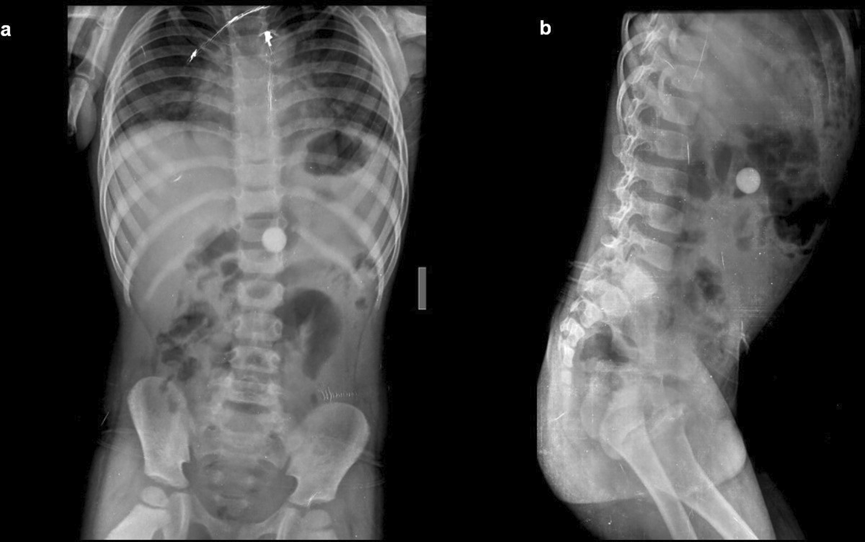
Case 1: A four-year-old child came with a history of abdominal pain. On a radiograph of the abdomen, frontal and lateral projections reveal circular radio opacity on the left side at the level of the L2-L3 disc suggestive of coin, which the patient had accidentally ingested. It passed through the gastro-intestinal tract without a problem.
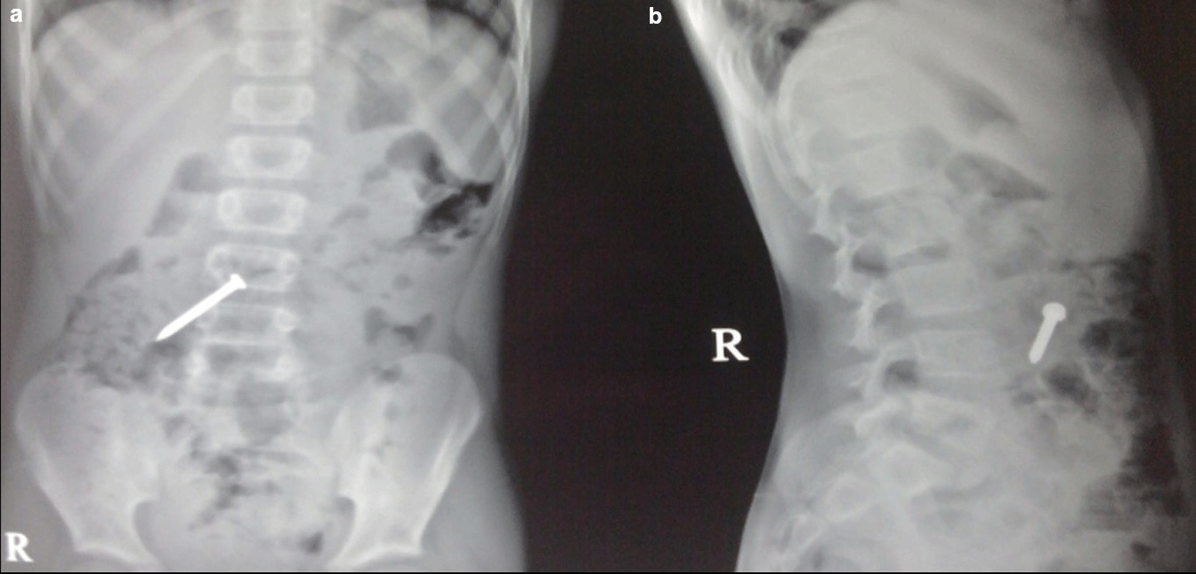
Case 2: A 34-year-old male carpenter by profession accidentally ingested a screw. A radiograph of the abdomen in frontal and lateral views reveals a nail at the level of the L4 and L5 vertebrae in the gastrointestinal tract, which passed without any problems.
Case 3: A 25-year-old male patient came for ultrasound examination with complaints of pain in the abdomen for two-three months. Radiograph of the chest and abdomen reveal multiple linear radio-opacities in the left hypochondriac and lumbar quadrants of the abdomen, and a plain CT scan shows multiple hyperdense linear metallic foreign bodies within the gastric lumen, many piercing the gastric wall partially without any evidence of perforation. On laparoscopy, multiple refills of the pen and wires were found in the stomach, which were removed.
Case 4: An 18-year-old woman patient came with a history of abdominal pain and vomiting on and off for 15 days. Contrast enhanced computed tomography (CECT) reveals a heterogeneous lamellated non-enhancing soft tissue density mass (with a wide attenuation range from -70 to 70 HU) intraluminally in the stomach, conforming to its shape and extending into the antrum, pylorus, and minimally into the duodenal cap suggestive of trichobezoar. Gastrotomy revealed the ball of hair in the stomach.
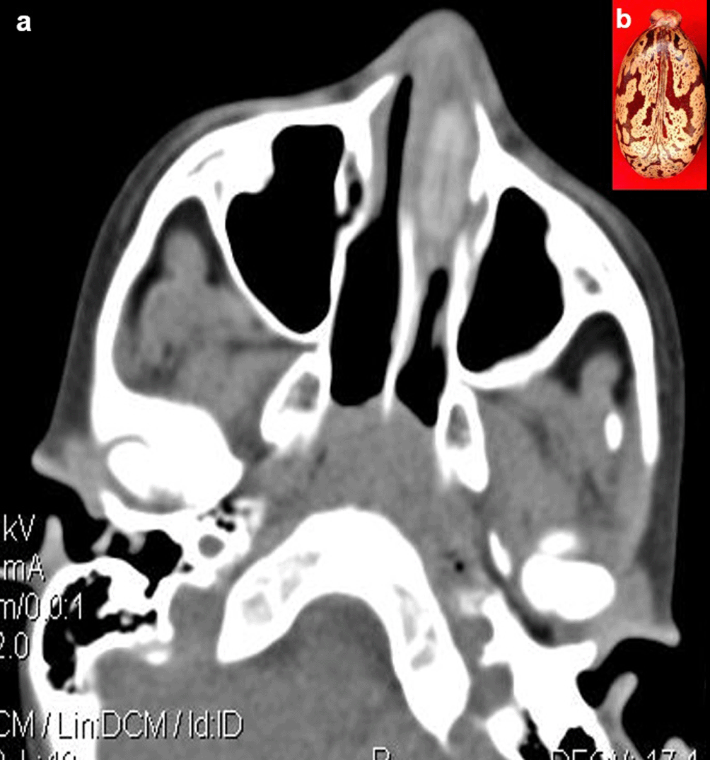
Case 5: A seven-year-old boy came with a history of epistaxis. CT paranasal sinuses (PNS) revealed a non-enhancing hyperdense lesion in the left nasal cavity, possibly a foreign body. It was removed under anaesthesia and found to be a castor seed.
Case 6: A 17-year-old woman came for an ultrasound examination in emergency hours with complaints of severe pain in her lower abdomen. Radiograph Pelvis anteroposterior (AP) view revealed long radio-opacity in the bladder with a radiolucent center that did not look like a calculus, but a foreign body. USG revealed a linear hyperechoic foreign body that penetrated the anterior wall of the bladder. The patient had a history that she had conceived three years ago and had tried abortion by a quack in her village. The patient was operated on and a shaggy piece of a long wooden stick with cotton wrapped around it was found.
Case 7: A 40-year-old male presented with a complaint of pain in the right heel region for two months. A radiograph of the lateral view of the right foot revealed there was evidence of calcific tendinitis of the Achilles tendon with thickening of the Kager fat pad and fat stranding. USG revealed that a well-defined thorn visualized in the Achilles tendon with associated surrounding tendinitis and increased fat echogenicity. USG-guided thorn removal was performed.
Case 8: A 50-year-old woman came for cervical spine. The patient was taken for an MRI scan when she complained of a severe headache. Radiograph skull AP & Lateral view was done, which showed a radiodense nail-like structure in scalp on right side. On asking, the patient gave a history of trauma ten years back and did not know that she had a nail in her scalp. It was removed under local anaesthesia.
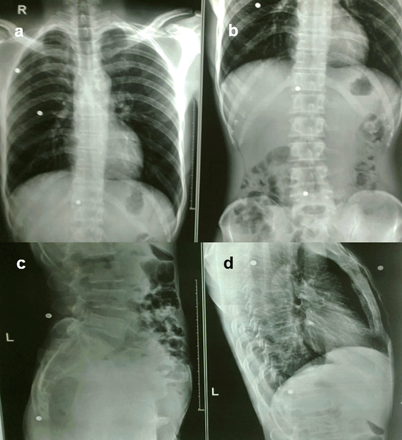
Case 9: A 37-year-old man had a history of bullet injuries. Radiographs of the chest and abdomen in frontal and lateral views revealed multiple pellets in the subcutaneous soft tissue of the thorax and abdomen.
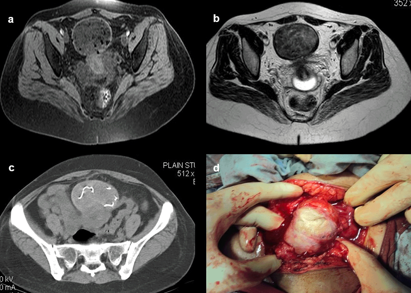
Case 10: A 33-year-old female came with a history of bleeding pervaginal for six months. MRI shows heterogeneous altered signal intensity soft tissue mass anterior to the uterus with multiple hypointense foci within. CT showed multiple linear metallic strings within a mass of soft tissue density anterior to the uterus, suggesting a foreign body (gossypiboma). The patient was operated on, and a large surgical sponge was removed.
A total of ten cases were studied, comprising of four females and six males of various age groups ranging from four years to 50 years (Tables 1 & 2). Four patients had ingested foreign bodies while two patients had history of insertion and four other had insertion due to injury (Table 3).
| Age distribution | Male | Female | Total No. of patients |
|---|---|---|---|
| <10 years | 02 | 00 | 02 |
| 11-20 years | 00 | 02 | 02 |
| 21 to 30 years | 01 | 00 | 01 |
| 31 to 40 years | 03 | 01 | 04 |
| 41 to 50 years | 00 | 01 | 01 |
| Total | 06 | 04 | 10 |
Foreign bodies are objects that do not typically belong in the human body but can be ingested, inserted, or entered due to injuries. This article presents various cases and examples of foreign bodies, including objects swallowed, objects inserted into the rectum, vagina, urethra, ear, and nose, or due to injuries caused by falls, puncture wounds, and gunshot wounds.
The swallowing of foreign bodies is a common condition in children and mentally challenged individuals.3–5 Fortunately, most ingested objects move through the digestive system without causing any problems (Figure 1a,b). Sharp and elongated objects can pass uneventfully (Figure 2a,b); however, they can pierce the mucosal lining and seriously damage or completely perforate the intestinal wall (Figure 3a-e). The object may just partially puncture the gut wall, resulting in a chronic inflammatory condition with few symptoms that is diagnosed months or years later.5–7
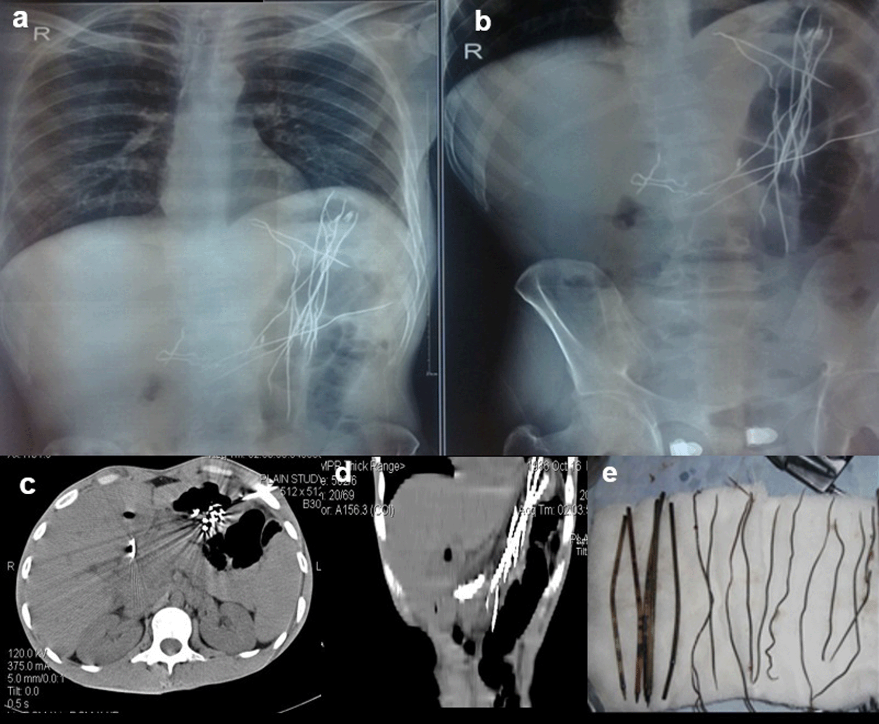
On laparoscopy, multiple refills of the pen and wires were found in the stomach, which were removed (e).
When a patient cannot provide a sufficient history or has swallowed things that are not naturally radio-opaque, the diagnosis of an ingested foreign body is frequently missed. If a foreign body is suspected and is not visible on a Radiograph because of its radiolucent nature, a CT scan of the abdomen or chest may be beneficial8 (Figure 4a,b).
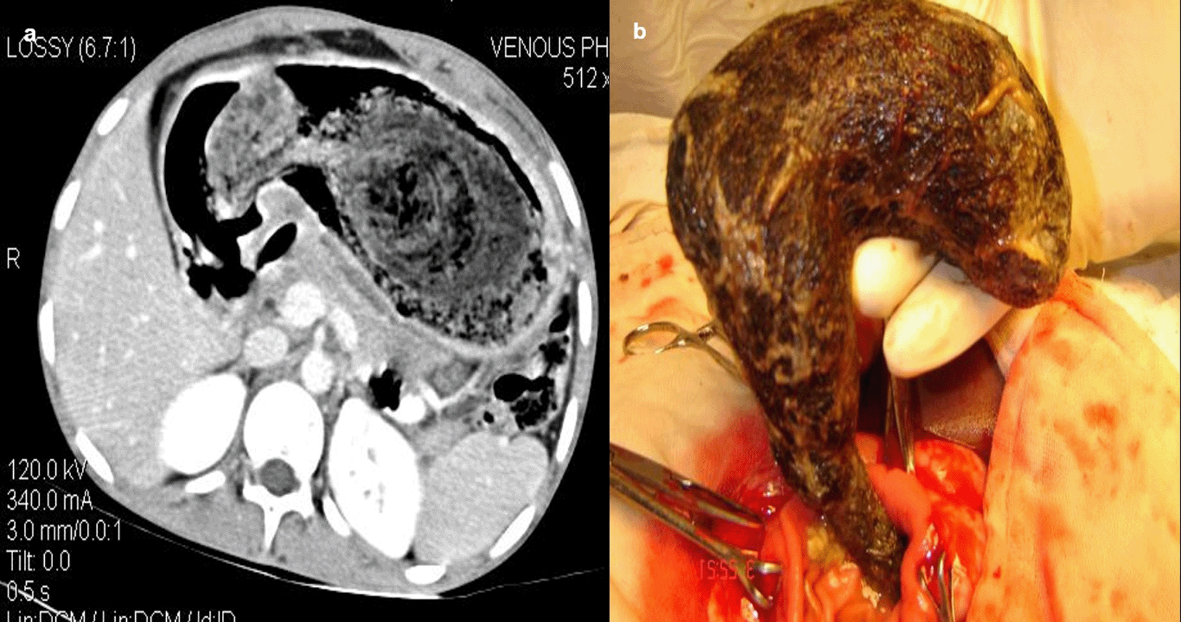
Gastrotomy revealed the ball of hair in the stomach (b).
Sometimes you may not have a proper history of the ingestion of sharp objects. When a patient has a history of ingesting a foreign body, whether it is an adult or a kid, they should be checked for the entire body, from the base of the skull to the anus, from the nasopharynx to the rectum. The hunt for other foreign bodies should not stop just because one has been discovered because youngsters are particularly prone to eating items in multiples.9
The rectum, vagina, urethra, ear, and nose are common places for foreign items to be inserted. These are especially common in children (Figure 5a,b) but can also be seen in adults. The deposition of mineral salts is especially likely to occur in foreign bladder substances, resulting in the formation of bladder calculi (Figure 6a,c). In fact, when a child or young adult develops a bladder calculus, the presence of an embedded foreign body should be suspected.10

USG revealed a linear hyperechoic foreign body that penetrated the anterior wall of the bladder (b). The removed foreign body was a long wooden stick with cotton wrapped around it (c).
Most people may have experienced at least one or two minor injury incidents, such as falls, abrasions, cuts, scrapes, and burns. Few of them may have experienced injuries from firearms and may have experienced puncture wounds from splinters, thorns, needles, or glass.2
On ultrasound, all foreign bodies in soft tissue are initially hyperechoic. Sonography is important for the correct localization of all kinds of soft tissue foreign bodies and the detection of non-radiopaque foreign bodies. Accurate localization can help minimize surgical exploration and can also direct the percutaneous removal of a foreign body11 (Figure 7a,b)

USG revealed that a well-defined thorn visualized in the Achilles tendon with associated changes of tendinitis (b). Thorn removed under ultrasound guidance (c).
Some metallic foreign bodies can be accidentally diagnosed during an MRI or CT study due to artefacts or sometimes due to pain as they enter the magnetic field12 (Figure 8a,b).
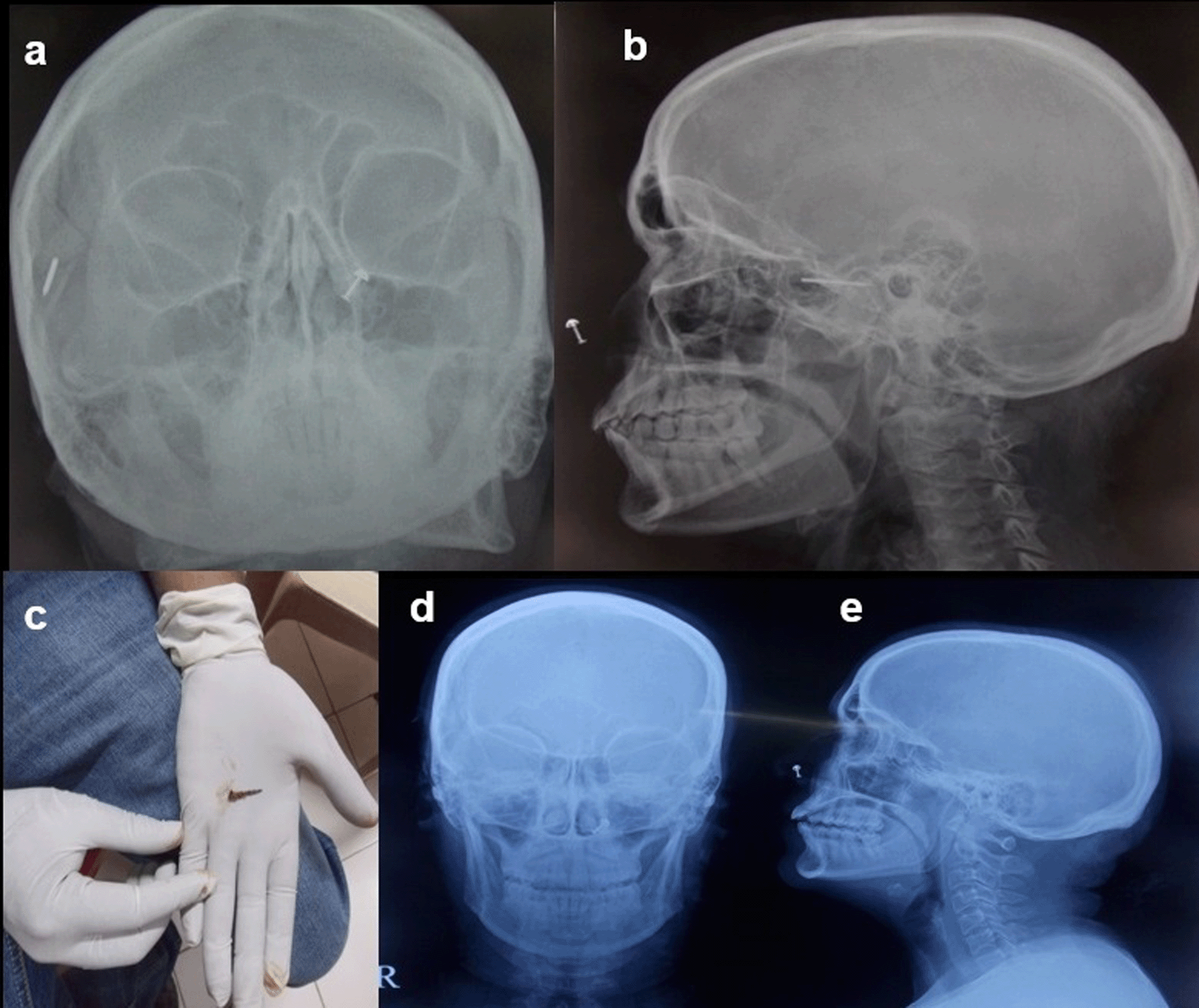
The nail removed under ultrasound guidance (c). Normal radiograph skull A-P & Lateral view post removal of the foreign body (d,e).
The gauge of a shotgun pellet determines its size, the higher the number, the smaller the pellet. Serious soft tissue and bone damage can result from the combined mass striking a target close to the gun barrel (Figure 9a-d). Because steel pellets are ferromagnetic, they could move dangerously if such a patient with embedded steel pellets was exposed to a magnetic field, making magnetic resonance imaging potentially dangerous.2
After surgery, not infrequently, patients have surgical items inside their bodies. Surgical drains, wound gauze packs, bandages, skin staples, small surgical staples, intra-arterial, intravenous, intra-spinal, and intraabdominal catheters are among the postoperative supplies that are most frequently seen. Other uncommon materials, such as retained abdominal sponges (Figure 10a-d) and needles, that were unintentionally left behind after surgery, are challenging to find clinically and radiographically because patients have vague symptoms, these objects are difficult to see on radiographs, and the radiologist and referring physician have a low level of suspicion for such objects. The nursing staff may perform a comprehensive sponge count at the conclusion of a surgical procedure and identify any remaining surgical sponges right away. A misplaced sponge may not be identified for months or even years after surgery if it is not found at that time. The foreign body reaction to a surgical sponge left inside the body for a long time is frequently called a gossypiboma. The sponge’s cotton matrix is what creates the foreign body reaction’s nidus. There is the development of a foreign body granuloma with surrounding fibrosis and retraction around the cotton nidus. Many people have no symptoms, and the retained sponge is often only unintentionally found when the patient has a radiological examination for another reason.13–17
Foreign bodies are interesting, and most of them are diagnosed incidentally in various parts of the human body and can cause significant harm if not properly managed. The diagnosis and management of foreign bodies can be challenging and require a high index of suspicion. Imaging studies such as Radiographs, CT scans, USG, and magnetic resonance imaging can be helpful in detecting and localizing foreign bodies. The management of foreign bodies can involve a variety of interventions, including endoscopy, surgical exploration, and percutaneous removal. Prevention is also key, particularly in children and mentally handicapped adults who are at increased risk of foreign body ingestion or insertion. It is important for healthcare providers to be aware of the potential for foreign bodies and to maintain a high level of vigilance when evaluating patients. Ultimately, early detection and appropriate management can prevent serious complications and improve patient outcomes.
All data underlying the results are available as part of the article and no additional source data are required.
CARE guidelines for case reports: 13-item checklist
| Views | Downloads | |
|---|---|---|
| F1000Research | - | - |
|
PubMed Central
Data from PMC are received and updated monthly.
|
- | - |
Is the background of the cases’ history and progression described in sufficient detail?
Yes
Are enough details provided of any physical examination and diagnostic tests, treatment given and outcomes?
Yes
Is sufficient discussion included of the importance of the findings and their relevance to future understanding of disease processes, diagnosis or treatment?
Partly
Is the conclusion balanced and justified on the basis of the findings?
Partly
References
1. Birk M, Bauerfeind P, Deprez PH, Häfner M, et al.: Removal of foreign bodies in the upper gastrointestinal tract in adults: European Society of Gastrointestinal Endoscopy (ESGE) Clinical Guideline.Endoscopy. 2016; 48 (5): 489-96 PubMed Abstract | Publisher Full TextCompeting Interests: No competing interests were disclosed.
Reviewer Expertise: surgery, endoscopy,
Is the background of the cases’ history and progression described in sufficient detail?
Partly
Are enough details provided of any physical examination and diagnostic tests, treatment given and outcomes?
Partly
Is sufficient discussion included of the importance of the findings and their relevance to future understanding of disease processes, diagnosis or treatment?
Partly
Is the conclusion balanced and justified on the basis of the findings?
Partly
Competing Interests: No competing interests were disclosed.
Reviewer Expertise: Pediatrics, Pediatric Cardiology, Pediatric Emergency-Critical Care, Pediatric Pulmonology, Neonatology, Perinatology, Ultrasound/Echocardiography
Alongside their report, reviewers assign a status to the article:
| Invited Reviewers | ||||
|---|---|---|---|---|
| 1 | 2 | 3 | 4 | |
|
Version 3 (revision) 18 Jul 24 |
read | read | read | |
|
Version 2 (revision) 06 Jun 24 |
read | read | ||
|
Version 1 11 Oct 23 |
read | read | ||
Provide sufficient details of any financial or non-financial competing interests to enable users to assess whether your comments might lead a reasonable person to question your impartiality. Consider the following examples, but note that this is not an exhaustive list:
Sign up for content alerts and receive a weekly or monthly email with all newly published articles
Already registered? Sign in
The email address should be the one you originally registered with F1000.
You registered with F1000 via Google, so we cannot reset your password.
To sign in, please click here.
If you still need help with your Google account password, please click here.
You registered with F1000 via Facebook, so we cannot reset your password.
To sign in, please click here.
If you still need help with your Facebook account password, please click here.
If your email address is registered with us, we will email you instructions to reset your password.
If you think you should have received this email but it has not arrived, please check your spam filters and/or contact for further assistance.
Comments on this article Comments (0)