Keywords
spontaneous coronary artery dissection, concomitant, carotid artery dissection, Clinical: Cardiology, Medical Direction: International EMS, Medical Direction: Interfacility / critical care transport
spontaneous coronary artery dissection, concomitant, carotid artery dissection, Clinical: Cardiology, Medical Direction: International EMS, Medical Direction: Interfacility / critical care transport
Spontaneous coronary artery dissection (SCAD) has been under-diagnosed and unknown for decades.1 It is an uncommon cause of acute coronary syndrome, generally affecting young or middle-aged women2 and individuals with few atherosclerotic risk factors.3 In recent years, the more frequent and earlier use of coronary angiography and advanced intracoronary imaging in acute coronary syndrome (ACS) has led to increased detection of SCAD. In the general population, SCAD can account for up to 4% of ACS cases.4 SCAD typically arises from an underlying predisposing arterial disease that weakens the wall, with or without previous stress factors,5 and its association with dissection of the internal carotid artery remains exceptional.
Here we are reporting a case of a young man who suffered from spontaneous dissection of both coronary and internal carotid arteries in the absence of an aortic dissection.
Through our observation of concomitant spontaneous coronary and carotid dissection, we discuss its clinical presentation, therapeutic management as well as its pathogenesis and the factors favoring its occurrence.
A 34-year-old Tunisian man who worked as a primary school teacher, with no personal or family history of cardiovascular disease was admitted to emergency with sudden altered consciousness. He was a smoker and suffered from type I diabetes. On examination, the patient appeared tired. His vitals and temperature were normal. It was oriented towards place, time and person. There were no speech abnormalities. He had no chest pains and the family reported no alcohol or drug use.
An electrocardiogram was performed within 30 minutes of arrival at the emergency department and showed ST-segment elevation (Figure 1). Troponin I levels increased to 1343 ng/l. The diagnosis of STEMI was initially considered and the patient received fibrinolytic therapy (Tenecteplase administered as a single 5-second intravenous bolus in weight-based doses of 0.50 mg/kg, with a maximum dose of 50 mg.) because reperfusion therapy by primary per-cutaneous coronary intervention (PCI) was not available.
Treatment for the acute coronary syndrome (ACS) was initially integrated including: Aspirin, 300 mg, Clopidogrel, 300 mg and Low-Molecular-Weight Heparins (Enoxaparin: Lovenox®) 1 mg/kg subcutaneously.
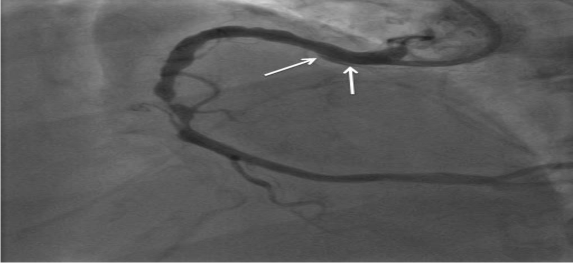
After two hours, the patient developed right hemiplegia and aphasia. A cerebral CT scan revealed a left ischaemic stroke in the anterior junctional territory. CT angiography of the supra-aortic arteries showed a dissection of the left internal carotid artery. However, CT angiography of the aorta revealed no abnormalities (Figure 2). Coronary angiography performed after 48 hours showed dissection of the anterior inter-ventricular artery (Figure 3).

no further interventions, as distal coronary flow was reasonable. After a multidisciplinary staff meeting, a consensus was reached in favor of conservative management. A statin (atorvastatin at 80 mg/day) was started and anti-coagulant therapy was discontinued. The course was marked by persistent ST-segment elevation despite an increase in Troponin I levels (2000 ng/l).
Echocardiography showed a preserved ejection fraction at 60%. However, control head CT revealed signs of a second cerebral ischemia. The patient was admitted to the cardiology department and subsequently treated with only statin and aspegic (160 mg per day).
The outcome was favourable and the patient had no sequelae. Physical rehabilitation sessions were scheduled on discharge.
The patient was informed about diagnoses and the risk of recurrence. Multidisciplinary follow-up was arranged, including referral to genetic services. He was discharged on day 10. A coronary CT angiogram to determine the resolution of coronary dissection is expected. The patient has not yet been followed up on a long-term basis, but is thought to be well.
SCAD is rare. Long misidentified and therefore underestimated, it is now recognized as a possible etiology of ACS with elevated myocardial markers and non-obstructive coronary arteries.6 The angiographic appearance of a SCAD is variable and the diagnosis can be made by OCT (Optical Coherence Tomography) in complex cases.7,8 Management should be as conservative as possible.9
Spontaneous dissection of the internal carotid artery is a cause of ischemic stroke, especially in young patients.10 The test of choice for a positive diagnosis is MRI.11 Treatment includes anticoagulants or antiplatelet drugs.12
The etiology of spontaneous coronary and carotid dissections is multifactorial. It is believed that there is an underlying “arterial disease” which may be associated with a precipitating factor. Some hypothesize that spontaneous dissections are due to an abnormality in the connective tissue of the vascular wall,13 as it has been found in most skin biopsies from patients with spontaneous cervical artery dissection concluding to a molecular deficit in the biosynthesis of the extracellular matrix.14 Predisposing connective tissue diseases include Marfan disease, Ehlers-Danlos syndrome, autosomal dominant poly cystic kidney disease, alpha1-antitrypsin deficiency and hereditary hemochromatosis.15 Other predisposing conditions have been identified, including fibromuscular dysplasia,16,17 pregnancy and post-partum,18 systemic inflammatory diseases and connectivitis (infection, lupus, Horton’s disease, etc.).19
We have described the case of a patient with concurrent dissection of the anterior inter-ventricular artery and the internal carotid artery. This patient had no history of drug use, trauma or infection and all markers of inflammation were normal. There was no history or clinical signs of connective tissue abnormality.
This clinical case shows the difficulty encountered in the etiological diagnosis of ACS and ischemic strokes. In our case, the associated ischemic stroke led to a more complicated management.
Due to the notable prevalence of extra-coronary arterial anomalies associated with coronary dissection, it is advisable to supplement with further imaging of the vascular system.
The choice of the most appropriate treatment cannot be standardized, as several factors must be taken into consideration. Thrombolysis in the acute phase remains controversial.20
Conservative treatment may not be appropriate in high-risk patients with ongoing ischaemia, left main artery dissection or haemodynamic instability as our case.21 In such cases, the medical satff must consider urgent PCI or coronary artery bypass grafting (CABG), but these decisions must be individualised and made on a case-by-case basis because we hadn’t expertise in such operations. Literature on CABG after SCAD is poor limited to case reports and small cases serie.
The concomitant dissection of the coronary and the internal carotid arteries with an uninjured aorta is a rare if not exceptional entity. Outside of the postpartum, toxic or post-traumatic context causing an isolated dissection of the coronary artery, this association suggests a congenital pathology of the vessels such as Marfan’s disease or Ehlers-Danlos syndrome. Treatment is complex and needs collegial decision making.
Written informed consent for publication of their clinical details and/or clinical images was obtained from the patient
All data underlying the results are available within the scope of the article and no additional source data is required.
| Views | Downloads | |
|---|---|---|
| F1000Research | - | - |
|
PubMed Central
Data from PMC are received and updated monthly.
|
- | - |
Is the background of the case’s history and progression described in sufficient detail?
Yes
Are enough details provided of any physical examination and diagnostic tests, treatment given and outcomes?
No
Is sufficient discussion included of the importance of the findings and their relevance to future understanding of disease processes, diagnosis or treatment?
No
Is the case presented with sufficient detail to be useful for other practitioners?
Partly
Competing Interests: No competing interests were disclosed.
Reviewer Expertise: Coronary artery disease
Alongside their report, reviewers assign a status to the article:
| Invited Reviewers | |
|---|---|
| 1 | |
|
Version 2 (revision) 13 Nov 23 |
read |
|
Version 1 12 Oct 23 |
read |
Provide sufficient details of any financial or non-financial competing interests to enable users to assess whether your comments might lead a reasonable person to question your impartiality. Consider the following examples, but note that this is not an exhaustive list:
Sign up for content alerts and receive a weekly or monthly email with all newly published articles
Already registered? Sign in
The email address should be the one you originally registered with F1000.
You registered with F1000 via Google, so we cannot reset your password.
To sign in, please click here.
If you still need help with your Google account password, please click here.
You registered with F1000 via Facebook, so we cannot reset your password.
To sign in, please click here.
If you still need help with your Facebook account password, please click here.
If your email address is registered with us, we will email you instructions to reset your password.
If you think you should have received this email but it has not arrived, please check your spam filters and/or contact for further assistance.
Comments on this article Comments (0)