Keywords
Pregnancy, femoral head, avascular necrosis, hip pain, hip replacement
This article is included in the Datta Meghe Institute of Higher Education and Research collection.
Pregnancy, femoral head, avascular necrosis, hip pain, hip replacement
Bone marrow cells and osteocytes die as a consequence of impaired blood supply to the bone affected. This pathological process is osteonecrosis, acknowledged as aseptic necrosis, ischemic necrosis, or avascular necrosis (AVN).1 This impaired blood supply induces demineralization and trabecular thinning, leading to consequent joint surface collapse and subchondral bone fracture. It was first reported in 1957 by Pfeiffer.2 AVN most commonly affects men between the ages of 30 and 50 years old.
Hip osteonecrosis has a number of known causes, including trauma, a genetic predisposition, sickle cell disorder, coagulation abnormalities, alcoholism, and high-dose corticosteroid therapy. Pregnancy is not a well-known risk factor for femoral head AVN, and it should be distinguished from one of the more prevalent hip pathologies in pregnancy, the so-called “Pelvic pain syndrome” and transient osteoporosis of the hip (TOH). Other than pregnancy, all other risk factors were ruled out for our patient. Not much is known regarding the development or management of AVN related to pregnancy since there have only been a few cases reported until now. Here, we have a case of neglected hip pain in a pregnant patient that resulted in AVN of the femoral head due to the uncommon association between pregnancy and the condition, which, if properly investigated early on, could have been treated more conservatively. According to the symptoms, examination, and imaging findings, a clinical diagnosis compatible with the disease is made.
In this study, we present the case of a 25-year-old healthy Asian woman, a homemaker, who had complaints of mechanical pain in the left hip that started shortly postpartum, after caesarean section giving birth to a 4.1 kg female baby (primigravida, G1). This caused discomfort in the left hip when ambulating, which aggravated on movement and was relieved on rest. She consulted her gynecologist about the symptoms and her complaints were neglected as hip pain of a benign cause. Her symptoms eventually worsened, which caused a painful limp and hindered her daily activities. The patient had no previous medical history. There was no history of trauma, alcohol, smoking, coagulation abnormalities, sickle cell disease, or steroid usage.
On physical examination, the patient was 150 cm in height and her weight was 64 kg. She had a painful limp and a range of painful hip movements, with restrictions of mainly the abduction (30°), internal rotation (20°), and flexion (90°) of the hip.
The patient, a 25-year-old female came to our hospital in February 2023 for assessment of pain in her left hip. Her pain began a year and a half ago at the end of her first pregnancy following a caesarean section through which she delivered a healthy 4.1 kg female baby. The pain was aggravated on movement and initially relieved on rest, which gradually worsened and radiated to the knee. The patient consulted her gynecologist and was treated conservatively considering it a benign cause of pain. Her symptoms were not relieved and hence the patient came for further management, where she was advised to undergo blood tests, X-ray, and magnetic resonance imaging (MRI) study.
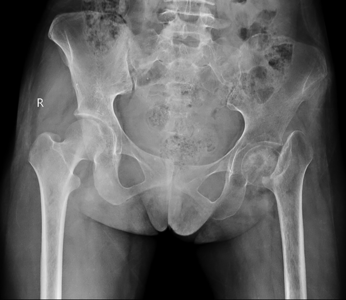
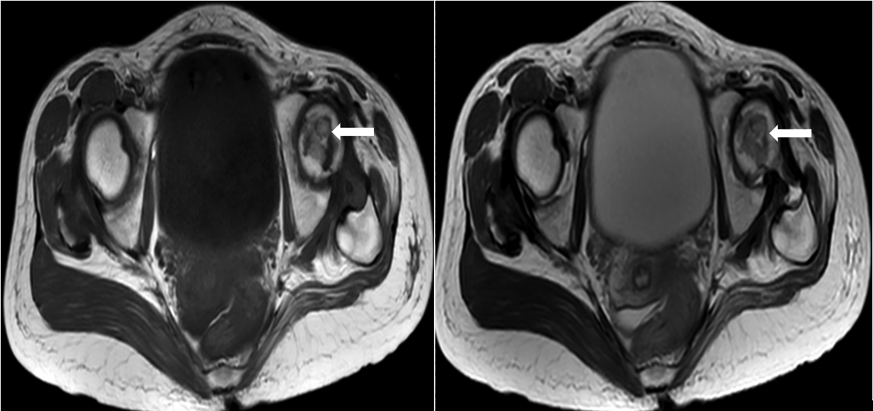
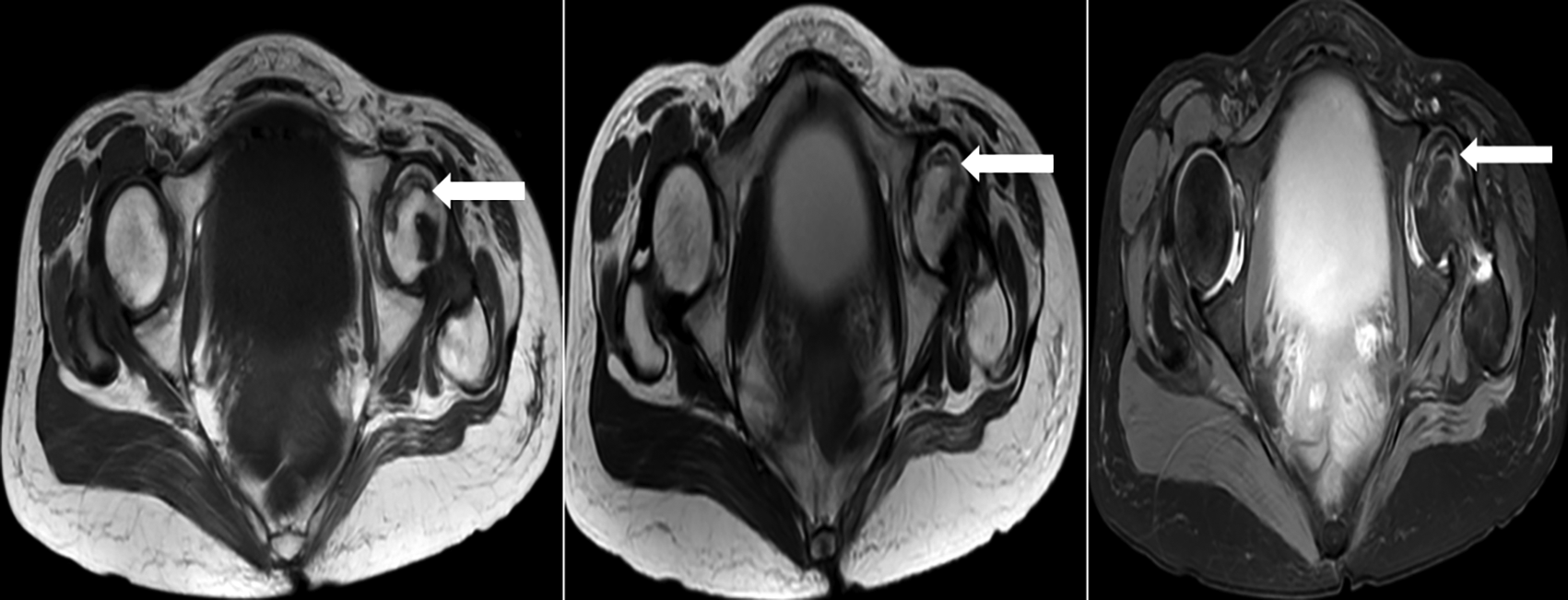
Radiograph showed left femoral neck fracture with superolateral displacement of the greater trochanter and the rest of the shaft of the left femur (Figure 1). T1 and T2 weighted imaging sequences MRI of the femoral heads showed an irregular geographical area in the anterosuperior part of the left femoral head (Figure 2). T2, T1, and PD FatSat MRI images show the double line sign (Figure 3). Radiograph images showed a hip replacement implant prosthesis on the left side post-hip replacement surgery (Figure 4). The laboratory tests—complete blood count (CBC), erythrocyte sedimentation rate, liver function tests, c-reactive protein (CRP), creatinine, lipid panel, thyroid-stimulating hormone (TSH), rheumatoid factor and antinuclear antibody were all within normal limits. There weren’t any abnormities in the abdominal ultrasound. The patient was diagnosed with AVN of the femoral head on the left side and associated secondary osteoarthritis. One other pathology that could be confused with AVN is the TOH, which is self-resolving.


T1WI, T1 weighted image; T2WI, T2 weighted image.

T1WI, T1 weighted image; T2WI, T2 weighted image.
The blood test results of the patient were normal. The patient had been prescribed pain killers that had not resolved the patient’s symptoms. The patient had her X-ray done, which showed fracture of neck of the left femur with displacement of the shaft. The orthopedic advised for MRI of the hip, which showed the features of osteonecrosis of left hip. The patient then underwent a total hip arthroplasty. The surgery was uneventful.
The most common causes of atraumatic hip osteonecrosis include alcohol intake and corticosteroids.3,4 Other risk factors include genetics, vasculitis, hyper coagulopathy, cocaine use, and micro emboli.5 While idiopathic AVN of the hip accounts for around 20% of cases,3,4 femoral head osteonecrosis is rarely associated with pregnancy.
AVN of the hip associated with pregnancy has been linked to a number of pathophysiological mechanisms, but none of them have been proven to be the sole contributing factor. Pregnancy commonly results in increased coagulability and venous congestion.6–8 Also, some endocrine changes that increase the levels of unbound cortisol during pregnancy have been linked to an increased risk of hip AVN.6,7 Hyperplasia of parathyroid can also occur during pregnancy, which increases the parathyroid hormone levels thus increasing the risk for developing osteonecrosis.9 The endogenous lipoproteins can be destabilized by the placenta, which can eventually lead to fat embolism.10
The right common iliac artery lies right above left common iliac vein, making the vein more susceptible to compression with excessive weight gain and during pregnancy, especially on the left side. This mechanical factor has also been suggested as a contributing factor to the increased risk of hip osteonecrosis in pregnancy.6,7,11,12 The literature indicates that the left hip is most frequently affected and this involvement mostly occurs in the third trimester or soon after delivery,6,7,12,13 which supports the mechanical theory. In addition, hip AVN more commonly develops in primigravida,7,14 and in older women6,7,13,14 in contrast to our case, where the patient is young. From current studies, another cause may be ovulation induction, which activates both fibrinolytic and coagulation systems.15,16 It has been reported that a difficult delivery or the compression of a growing uterus can lead to injury of the artery in the round ligament or the femoral joint.17 Biochemical, hematological and coagulation parameters were all within the normal limits in our patient.
The initial complaint is generally unilateral and can either be a gradually worsening or a sudden groin pain that radiates to the back, thigh or knee. Physical examination shows a restriction of active and passive joint movements associated with pain, more pronounced with rotation and less with flexion and abduction.18
TOH is another common hip pathology in pregnancy and is sometimes mistaken for hip AVN.6,14,19–21 It is a rare cause of acute hip pain in pregnancy and most commonly occurs in the third trimester,19 characterized by sudden onset of pain in the hip that can be severe without movement restriction. TOH is a condition that is self-limiting and resolves gradually within seven to eight months. The management comprises of weight bearing restrictions.6,21,22 By contrast, AVN onset is insidious, with pain increasing progressively, leading to movement limitation, and will eventually need early surgical intervention.19,20,21 Hence it is very important to differentiate between these entities since the treatment and prognosis differ greatly.6,12,19,21
Since the changes of osteopenia in TOH are evident only four to eight weeks from the onset of symptoms, the standard X-ray lacks sensitivity in distinguishing AVN and TOH.20,21 With specificity and sensitivity of more than 99%, MRI is the gold standard imaging modality for diagnosing AVN in its early stages.23 The MRI features of TOH are diffuse edema of the head and neck of the femur and, by contrast, MRI features of AVN include diffuse edema with focal defects and subchondral changes.7,19–21 Double line sign is a pathognomonic MRI finding in AVN and refers to two adjacent serpentine lines demarcating the boundary between viable and devitalized bone marrow. The inner line corresponds to reparative granulation tissue, while the outer line corresponds to reactive sclerosis.24
The best way to treat AVN associated with pregnancy is not very clear. Early diagnosis and advice are significant. Conservative methods such as physical therapy, restricted weight bearing, medication, electrical stimulation, extracorporeal shock-wave treatment, and hyperbaric oxygen treatment are usually started if there is no evidence of collapse.25 The prognosis appears to be good following this conservative therapy, however surgical treatment would eventually be required for the secondary osteoarthritis or degenerative changes at a later age.18
Although AVN developing post-pregnancy is a rare occurrence, AVN of the femur should be one of the top differentials for post-partum hip pain. This could aid medical professionals in identifying the disease earlier before the collapse of the femur head occurs, which may aid in the preservation of the joint as opposed to replacement.
Written informed consent for publication of their clinical details and clinical images was obtained from the patient.
All data underlying the results are available as part of the article and no additional source data are required.
| Views | Downloads | |
|---|---|---|
| F1000Research | - | - |
|
PubMed Central
Data from PMC are received and updated monthly.
|
- | - |
Is the background of the case’s history and progression described in sufficient detail?
Partly
Are enough details provided of any physical examination and diagnostic tests, treatment given and outcomes?
Partly
Is sufficient discussion included of the importance of the findings and their relevance to future understanding of disease processes, diagnosis or treatment?
No
Is the case presented with sufficient detail to be useful for other practitioners?
Partly
References
1. Turgay T: Avascular necrosis of the bilateral femoral head associated with pregnancy: a case report. Ağrı - The Journal of The Turkish Society of Algology. 2018. Publisher Full TextCompeting Interests: No competing interests were disclosed.
Reviewer Expertise: Radiology, ultrasound, Nonvascular interventional Radiology
Is the background of the case’s history and progression described in sufficient detail?
No
Are enough details provided of any physical examination and diagnostic tests, treatment given and outcomes?
No
Is sufficient discussion included of the importance of the findings and their relevance to future understanding of disease processes, diagnosis or treatment?
No
Is the case presented with sufficient detail to be useful for other practitioners?
No
Competing Interests: No competing interests were disclosed.
Reviewer Expertise: Trauma & Orthopaedic Hip Surgeon
Alongside their report, reviewers assign a status to the article:
| Invited Reviewers | ||
|---|---|---|
| 1 | 2 | |
|
Version 1 18 Oct 23 |
read | read |
Provide sufficient details of any financial or non-financial competing interests to enable users to assess whether your comments might lead a reasonable person to question your impartiality. Consider the following examples, but note that this is not an exhaustive list:
Sign up for content alerts and receive a weekly or monthly email with all newly published articles
Already registered? Sign in
The email address should be the one you originally registered with F1000.
You registered with F1000 via Google, so we cannot reset your password.
To sign in, please click here.
If you still need help with your Google account password, please click here.
You registered with F1000 via Facebook, so we cannot reset your password.
To sign in, please click here.
If you still need help with your Facebook account password, please click here.
If your email address is registered with us, we will email you instructions to reset your password.
If you think you should have received this email but it has not arrived, please check your spam filters and/or contact for further assistance.
Comments on this article Comments (0)