Keywords
aesthetic, gingival contour, ovate pontics
This article is included in the Datta Meghe Institute of Higher Education and Research collection.
The period of time between extraction and recovery following tooth loss in the esthetic zone can be awful to the patient's appearance and mental health. A suitable alternative to extraction and replacement of a maxillary anterior tooth is quick tooth replacement with an ovate pontic on a temporary fixed partial denture. The interdental papilla, which is preserved by ovate pontic, in turn protects the contour of gingiva that after extraction would have been lost. Utilizing an ovate pontic, an instantaneous tooth replacement removes the emotionally distressing partial edentulous stage, while also producing a considerably more aesthetically acceptable, hygienic, and natural-looking replacement tooth. Ovate pontics have the additional benefit of avoiding the dissatisfaction associated with an unattractive ridge lap pontic. The case study demonstrated an ovate pontic replacement for an upper anterior tooth that helped a patient whose mobile anterior tooth was advised for extraction and finally resulting to both improved esthetics and health.
aesthetic, gingival contour, ovate pontics
When an individual loses their anterior teeth, it has a profoundly negative impact on their ability to integrate into society as usual. Any patient who extracts or loses a single anterior tooth may find it difficult in social integration, but unless extensive bone and soft tissue loss is present along with the tooth, replacing the tooth with a fixed partial denture (FPD) or an implant yields an expected esthetic result.1 Even so, the outcome is typically acceptable when handled by a qualified practitioner. To succeed, many things must be taken into account. These characteristics include the pontic's size, form, color, and location, as well as its profile as it emerges from the soft tissues.2
Pontics that look natural and are hygienic have been the subject of many different designs over the years. We have the chance to fulfill both of the above objectives because of the ovate pontic's design. The ovate pontic, also known as the emergence profile, is a method used to give the appearance that the tooth is emerging from the gums. It can also aid to form and preserve the interdental papilla. The contour is a cleanability-enhancing design in ovate pontic.3
The ovate pontic is a method for giving the appearance that the tooth is poking through the gum.4 However, clinicians don't typically use an ovate pontic design on a regular basis. Preserving inter-proximal tissue following tooth removal is one of the difficult aspects of a dental treatment plan. In restorative dentistry, it is highly desirable to prevent alveolar bone collapse.
An Asian female patient, 33 years old, reported to the dental hospital with complaint of a loosening of tooth in anterior region. Patient also wished to replace the mobile tooth with a firm tooth. There were not any previous medical interventions or treatments or any systemic history. On examination, right central incisor (11) was grade III mobile with supraeruption of the same tooth (Figure 1). On radiographic examination, orthopantomogram (OPG) revealed that there was severe bone loss in 11 region (Figure 1). A diagnostic impression recorded and treatment planning was done.

The appearance of the patient's maxillary anterior teeth bothered her. Patient was advised and recommended to remove the mobile tooth and replace it with an ovate pontic that, in terms of form, functionality, and appearance, exactly mimics the missing tooth.
With maxillary right lateral incisor (12) and maxillary left central incisor (21) teeth, final tooth preparation was completed [Figure 2A]. Then, the right central incisor (11) was extracted under local anesthesia with no complication [Figure 2B]. During extraction, considerable care was taken to avoid damaging the lingual and buccal plates. This process is essential to maintaining both the interdental papillae and the bone. The cast was scored around the tooth to be removed, creating a 3 mm depression to represent the extraction socket. Fabrication of a temporary restoration using luxatemp material was done [Figure 3]. On the prepared tooth, a temporary fixed partial denture (FPD) was placed with the pontic portion immersing into the extraction socket by 2-3 millimeters, resulting in a depression of 1-1.5 millimeters once the tissues receded throughout the healing phase. The required changes, including the placement of the ovate pontic, were made to enable the appropriate placing of the FPD. The tissue surface of the pontic was cleaned and polished to avoid irritating the socket's healing tissues and to stop the buildup of bacterial plaque. A temporary restoration substance was used to solidify the work [Figure 3].
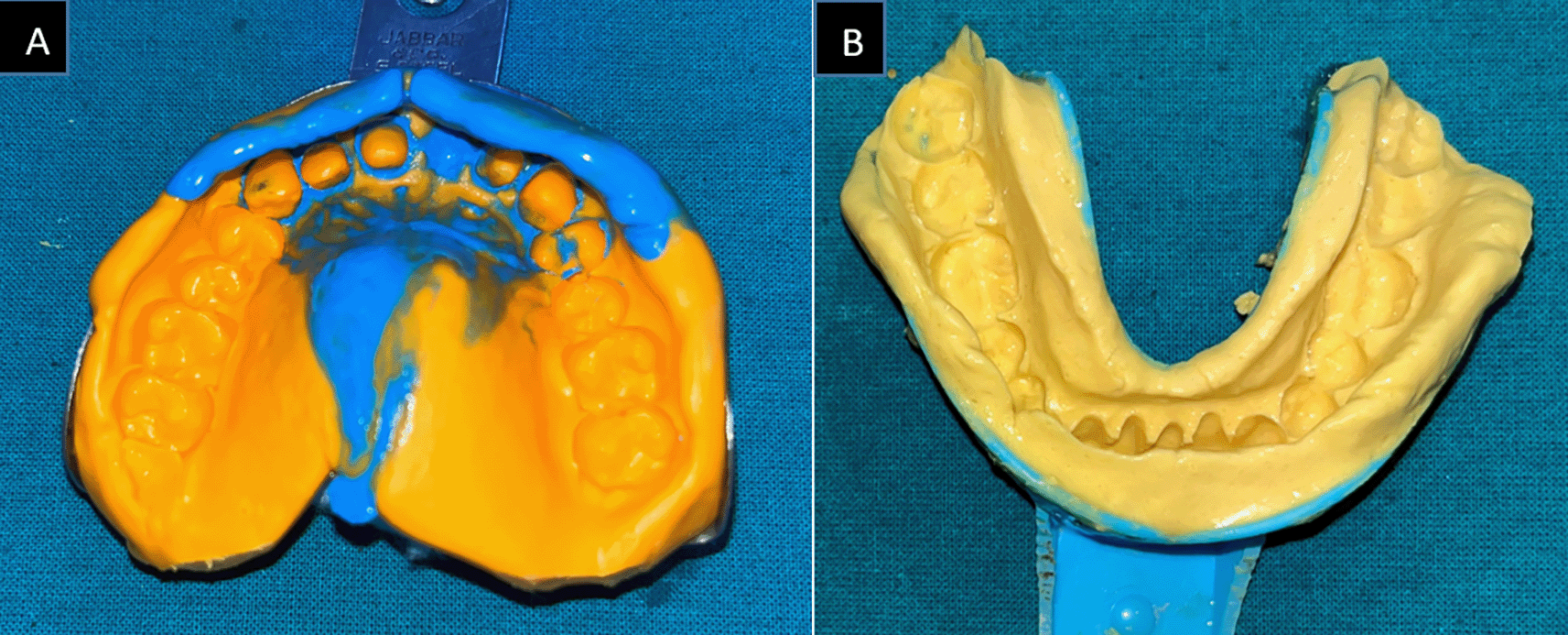
After extraction and temporary prosthesis, the patient was told to come back in 48 hours to have the temporary restoration removed and have the healing socket checked [Figure 4]. Following evaluation, the temporary restoration was restored using temporary luting material. To manipulate the soft tissues and reproduce the ovate pontic receptor location, a succession of temporary fixed partial denture was made. An elastomeric material was used to create an impression of the abutment tooth and the prepared soft tissue site after three to four months, until the soft tissues had developed and the extraction site had healed [Figure 5]. GIC was used to build and cement the ceramic-metal repair. After giving post-cementation instructions, the patient was asked to come back for routine dental follow-up. The patient was happy and satisfied with the prosthesis [Figure 6].

Abrams invented the ovate pontic in 1980.5 Both implant prostheses and fixed and removable partial dentures have been utilized effectively using the concept itself. Super floss can be used to keep the pontic's tissue surface clean in order to avoid any tissue inflammation. Although some have questioned its viability, the tissue surface of the pontic's convex form always allows for appropriate cleansing of the area beneath it.6 Counselling may take time, and some patients may not accept having teeth created close to missing teeth.
For a satisfactory marginal fit, careful attention to the preliminary repair is required. It is crucial to take final impressions for the FPD as soon as the temporary reconstruction is removed. If not, tissue may revert to its original state and result in an ovate pontic space on the model that is considerably smaller than the temporary pontic.7
For a good esthetic result in this case, the ovate pontic shape was intended to draw attention to the mucosa's position on the alveolar ridge by recreating a concave soft tissue outline. For directed papilla growth and stabilization, the tissues were molded.2
Ovate pontics have the following advantages when used in the anterior aesthetic zone:
1. Maintenance of the normal gingival contour and the interdental papilla
2. Gets rid of the emotionally distressing partial edentulous phase
3. A substitute that is hygienic and aesthetically appealing and seems natural
4. Discards the dissatisfaction brought on by an unappealing ridge lap pontic
5. Gets rid of unsightly “black triangles”.2
The height of the apical pontic was chosen and selected by the tissue/bone complex already present, aesthetics, support, cleaning convenience, and prevention of food impaction. For the majority of pontic forms, passive ridge contact was recommended; however, the ovate pontic requires close contact in order to support, shape, and preserve tissue.8,9
Ovate pontic can meet the aesthetic, functional, and hygienic specifications of an artificial tooth in FPD. For individuals with a high smile line or for the replacement of missing teeth in the cosmetic zone, ovate pontics are advised. The pontic minimizes the black triangles while creating the impression of a free interdental papilla and gingival border. The patient's oral hygiene routine will determine if the prosthesis is ultimately successful.
The patient was provided with an informed consent form and provided a thumb print signature to consent for the publication of their clinical details and images.
All data underlying the results are available as part of the article and no additional source data are required.
| Views | Downloads | |
|---|---|---|
| F1000Research | - | - |
|
PubMed Central
Data from PMC are received and updated monthly.
|
- | - |
Is the background of the case’s history and progression described in sufficient detail?
Partly
Are enough details provided of any physical examination and diagnostic tests, treatment given and outcomes?
Partly
Is sufficient discussion included of the importance of the findings and their relevance to future understanding of disease processes, diagnosis or treatment?
No
Is the case presented with sufficient detail to be useful for other practitioners?
No
Competing Interests: No competing interests were disclosed.
Reviewer Expertise: Prosthodontics, Dental materials, Maxillofacial prosthodontics, tooth wear and oral rehabilitation
Alongside their report, reviewers assign a status to the article:
| Invited Reviewers | |
|---|---|
| 1 | |
|
Version 1 30 Oct 23 |
read |
Provide sufficient details of any financial or non-financial competing interests to enable users to assess whether your comments might lead a reasonable person to question your impartiality. Consider the following examples, but note that this is not an exhaustive list:
Sign up for content alerts and receive a weekly or monthly email with all newly published articles
Already registered? Sign in
The email address should be the one you originally registered with F1000.
You registered with F1000 via Google, so we cannot reset your password.
To sign in, please click here.
If you still need help with your Google account password, please click here.
You registered with F1000 via Facebook, so we cannot reset your password.
To sign in, please click here.
If you still need help with your Facebook account password, please click here.
If your email address is registered with us, we will email you instructions to reset your password.
If you think you should have received this email but it has not arrived, please check your spam filters and/or contact for further assistance.
Comments on this article Comments (0)