Keywords
Accessory Spleen, Acute Abdominal Pain, Radiologic Diagnosis, Laparoscopy Surgery, Case Report
Accessory Spleen, Acute Abdominal Pain, Radiologic Diagnosis, Laparoscopy Surgery, Case Report
The spleen is a single mass of lymphatic tissue between the 9th and 11th ribs in the abdominal cavity. 1 , 2 It regulates the homeostasis of the immune and hematologic systems. 3
The accessory spleen is an ectopic splenic tissue that develops in the fifth week of fetal development as a result of the incomplete fusion of splenic masses. 3 , 4 It carries out similar functions as the normal spleen because of the similar subset of structures. Similar to the normal spleen, the accessory spleen is vascularized by the splenic artery. 5
About 95 % of ASs are located in the splenic hilum proximal to the tail of the pancreas. However, only 5% of accessory splenic tissue can be found in the gastrosplenic ligaments, the lienorenal ligaments, the wall of the stomach, the wall of the intestines, the greater omentum, the mesentery, or even the pelvis or scrotum. 6 , 7
Although earlier research found that about 10-15% of people in the general population have an accessory spleen, they typically don't cause any symptoms. However, if they experience accessory spleen torsion, surgery may be necessary. 7 Less invasive laparoscopy surgery is preferred to treat accessory spleen torsion with no abdominal complication. Accessory spleen torsion uncommonly happens that only accounts for 0.2-0.3% of splenectomy cases. 8
The torsion of the accessory spleen that may lead to infraction is caused by the long pedicle anatomical structure. 9 , 10 This abnormality might be recognized by radiological examination that could be a consideration for early surgery for the purpose of torsion prevention. 10
In March 2023, an Austronesian 22-year-old college student who was administered to the emergency department admitted to have crampy abdominal pain in the left upper quadrant (LUQ) for three days of onset. The symptom was periodically worsened and relieved for the past year. He could not find a comfortable position to relieve his pain. The recurrent symptom occurred five times a year. The patient experienced a decreasing appetite and vomiting of liquid and food contents every time the symptoms worsened. The previously given prescription of spasminal, hyosin, braxidin medicine had no relieving effect on the symptoms.
Physical examinations revealed tenderness in LUQ with stable hemodynamic condition (Blood Pressure: 138/85 mmHg; Heart rate: 100 beats per minute; Respiratory rate: 20 breaths per minute; Temperature: 36,5oC; Saturation of O2: 100%)
All laboratory findings were in the normal range. The radiological findings from both ultrasound and an abdominal multislice CT scan reveal that the accessory spleen may be found in the lienorenal area, where it makes up around 5% of the total accessory splenic tissue. The size was about 1.6 x 1.8 x 1.4 cm and the vascular accessory spleen extends from the splenic pedicle to the left splenorenal region.
The patient was transferred to the surgery department, the oral diet was started the day after the laparoscopic surgery was performed, and he was able to outpatient two days later. The follow-up two weeks after the surgery showed no problem was found. The pathology report showed the clinical findings listed in Figure 4.
Figure 1 shows the registration for abdominal laparoscopy conducted with a 5 mm incision in three locations including sub-umbilical, left lower quadrant in the midclavicular line, and right upper quadrant in the midclavicular line. This procedure aimed to access the posterior-medial aspect of the spleen as the accessory spleen took place. The image also shows the appearance of a long pedicle that causes several recurrent torsion and detorsion of the accessory spleen.
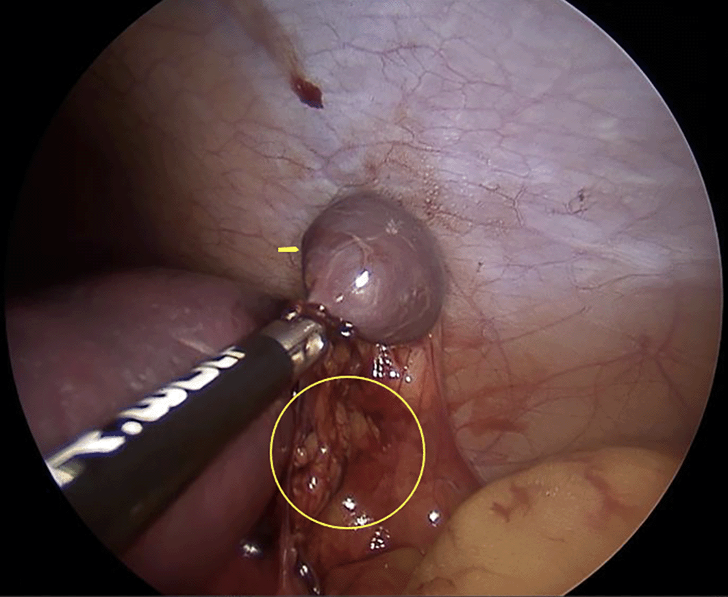
Surgery of the laparoscopy. Acessory spleen ( ), vascular pedicle (
 ).
).
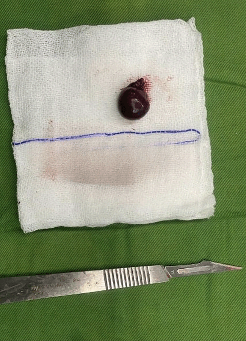
Figure 2 shows the excised accessory spleen in size of 1.8x1.5x1.5cm and 0.5cm length of the pedicle. The macroscopic features show the blackish-brown color with a spongy consistency.
CT Scan MX200CT III series and USG was used to access this patient. Accessory spleen location, size, and vascularization were captured from the process as shown in Figure 3.
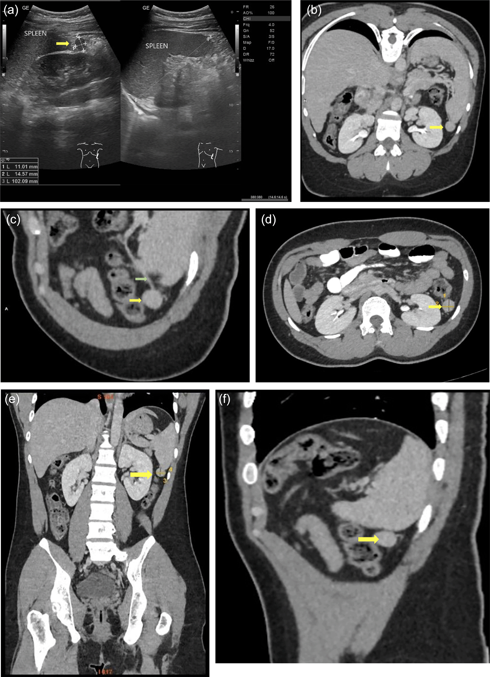
Radiologic Findings (a: USG findings; b-f: CT scan findings). Acessory spleen ( ), vascular pedicle (
 ).
).
From both ultrasound and abdominal multislice CT scan, the location of the accessory spleen in the region of lienorenal is about 5% location of accessory splenic tissue. The size was about 1.6 x 1.8 x 1.4 cm and the vascular accessory spleen extends from the splenic pedicle to the left splenorenal region.
Figure 4 shows the microscopic features of the excised accessory spleen. The accessory spleen is in the form of an encapsulated tissue, containing white pulp and red pulp, with areas of bleeding, accompanied by necrotic foci, covered with neutrophil inflammatory cells, lymphocytes, and histiocytes. No signs of malignancy was seen. Those macroscopic and microscopic features are commonly found in accessory spleen torsion that confirmed the diagnosis.
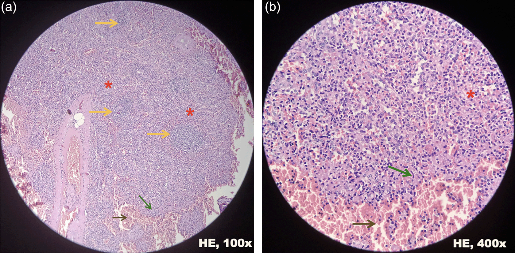
Histopathology findings showing the white pulp ( ), red pulp (
 ), necrotic foci (
 ), and areas of bleeding (
 ) in the patient’s excised accessory spleen.
Figure 5 shows the minimally invasive laparoscopic procedure. This allows the patient to do the outpatient care one day after the surgery.
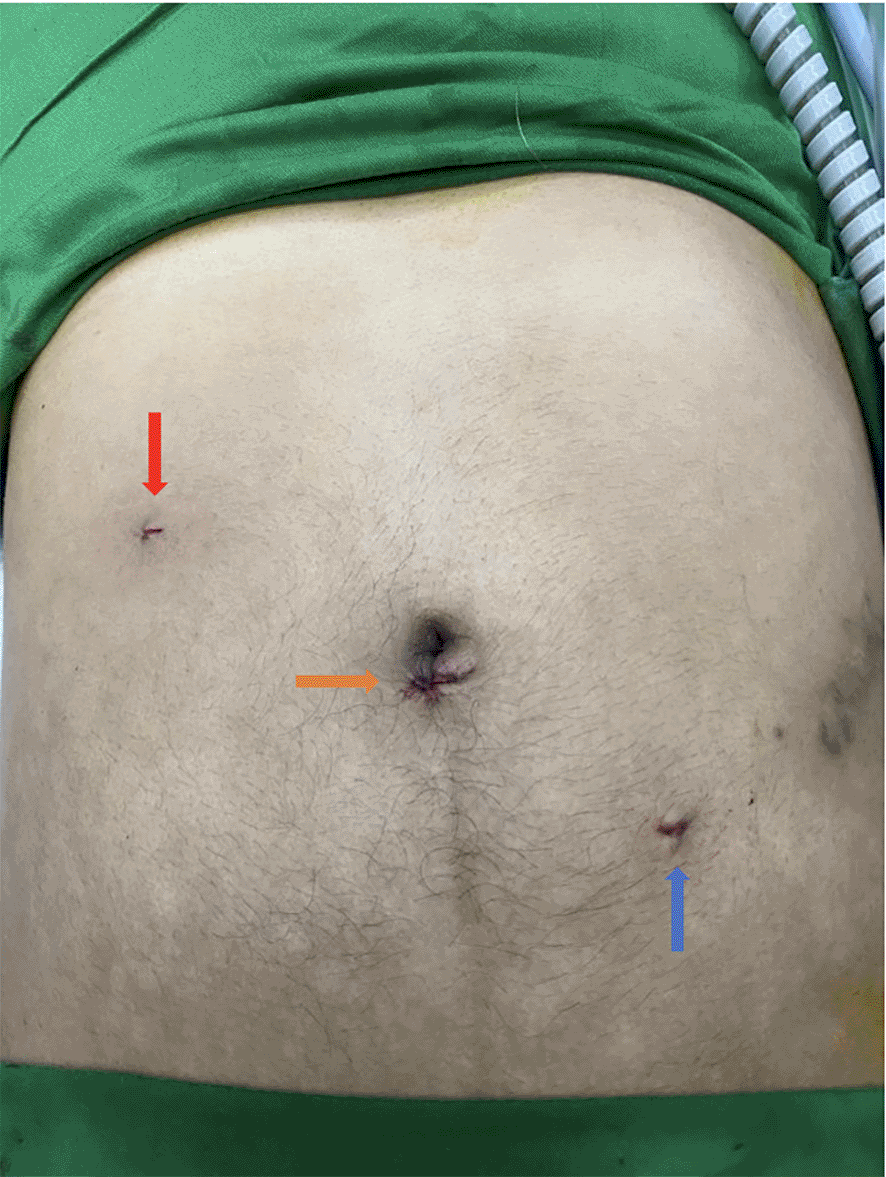
Post Surgery appearance showing the 3 incision locations; right upper quadrant in the midclavicular line ( ); sub-umbilical incision (
 ); left lower quadrant in the midclavicular line (
 ).
).
This case serves an Austronesian 22-year-old college student who was diagnosed having recurrent serial torsion and detorsion of the accessory spleen. This condition made the patient feel crampy abdominal pain. The worst clinical manifestation happened 3 days before being administered to a hospital.
Compared to other cases of acute abdominal pain, this condition has no pathognomic signs that directly refer to accessory spleen torsion diagnosis. 11 Furthermore, late diagnosis and treatment may cause several complications, such as spontaneous rupture, embolization, and abdominal bleeding that may lead to acute peritonitis. That case has a more complicated treatment and poorer prognosis. 3 Thus early diagnosis and treatment strategy is needed to solve the case before the complication occurred.
The early modalities to detect accessory spleen could be started from the USG test. Further CT scan can be conducted after the accessory spleen finding from the USG test. The venous phase of the CT scan with multiplanar and 3D reconstruction is highly recommended to determine the surgery indication since it can show a clear picture of the accessory spleen (isodense nodule besides of spleen) as well as the Jokari sign, the typical sign of accessory spleen, which shows the obvious link between the nodule and the splenic vascular pedicle. 3 This long pedicle makes the accessory spleen easily twisted and untwisted, so it is highly recommended to be treated as soon as possible using laparoscopy surgery before the complication appears. Accessory spleen anatomical location is highly correlated to its pedicle length. Atypical anatomical position, such as gastrosplenic ligaments, the lienorenal ligaments, the wall of the stomach, the wall of the intestines, the greater omentum, the mesentery, or even the pelvis or scrotum, is far departed from the main splenic pedicle that causes the AS’s pedicle being long. 6 , 7
Those recurrent torsion and detorsion phenomenon causes micronecrosis and micro bleeding within the organ, leading to periodically recurrent atypical abdominal pain the patient felt. However, this condition may worsen into massive necrosis and hemorrhage if not diagnosed and treated as soon as possible. 12
A good early diagnosis strategy will also affect the effective minimally invasive treatment strategy with a good prognosis. Accessory spleen torsion without complication is preferred to be treated using laparoscopy rather than laparotomy which is a more invasive procedure. Laparoscopy surgery is proven to be able to effectively excise the accessory spleen, and even the patient can do outpatient care one day after surgery. 13
Besides of good diagnosis strategy, an appropriate treatment strategy is needed. modified anti-Trendelenburg 3-port laparoscopic excision of the accessory spleen (LEAS) could be the best option to evacuate the accessory spleen. It is because this technique is less invasive than laparotomy surgery and 4-port laparoscopic surgery. It costs lower and is more accessible to most of the healthcare providers. This procedure required an anti-Trandelenburg position of the patient during the surgery to expose the accessory spleen clearly. Three ports were placed in the sub-umbilical, left lower quadrant in the midclavicular line, and right upper quadrant in the midclavicular line. The open Hasan technique was used to access the abdomen through the supraumbilical port and exploration was focused on the preoperative imaging-identified area. The removal of accessory spleens laparoscopically identified that were confirmed to be splenic tissue by both microscopic examination and permanent section pathologic diagnosis was considered a technical success. 2 , 14
This case shows that laparoscopic accessory splenectomy is a safe and effective treatment for accessory spleen torsion and radiologic findings can be used for early diagnosis.
Recurrent torsion and detorsion of the accessory spleen is an extremely rare case that may cause acute abdominal pain. Lack of diagnosis and treatment procedure may lead to further complications, such as spontaneous rupture, embolization, and abdominal bleeding, leading to acute peritonitis. A combination of USG and CT scan with venous phase is preferred to early diagnose the presence of accessory spleen as well as its vascularization to determine the risk of torsion. While the findings of the accessory spleen with long pedicle are highly suggested to be treated using modified anti-Trendelenburg 3-port laparoscopic excision of the accessory spleen (LEAS)
Written informed consent was obtained from the patient for publication of this case report and any accompanying images. A copy of the written consent is available for review by the Editor-in-Chief of this journal.
Data sharing is not applicable to this article as no datasets were generated or analyzed during the current study.
Mendeley Data: CARE checklist for “Case report of severe intermittent acute abdominal pain caused by extremely rare case of lienorenal accessory spleen torsion and detorsion: an accurate diagnostic and treatment strategy”. DOI: 10.17632/bh8bg4h78k.1. 15
KCT and DA: examining the medical literature, drafting and revising the case report, and assessing the patient's clinical data. AAA and LK: direct supervision throughout the development and modification of the case report, as well as a review of the medical literature. Reviewing the case report and medical literature, reading the final manuscript, and approving the manuscript was conducted by all writers.
| Views | Downloads | |
|---|---|---|
| F1000Research | - | - |
|
PubMed Central
Data from PMC are received and updated monthly.
|
- | - |
Provide sufficient details of any financial or non-financial competing interests to enable users to assess whether your comments might lead a reasonable person to question your impartiality. Consider the following examples, but note that this is not an exhaustive list:
Sign up for content alerts and receive a weekly or monthly email with all newly published articles
Already registered? Sign in
The email address should be the one you originally registered with F1000.
You registered with F1000 via Google, so we cannot reset your password.
To sign in, please click here.
If you still need help with your Google account password, please click here.
You registered with F1000 via Facebook, so we cannot reset your password.
To sign in, please click here.
If you still need help with your Facebook account password, please click here.
If your email address is registered with us, we will email you instructions to reset your password.
If you think you should have received this email but it has not arrived, please check your spam filters and/or contact for further assistance.
Comments on this article Comments (0)