Keywords
Paget's Disease, Nipple and Areolar, Metastasis, Pleural Effusion
This article is included in the Datta Meghe Institute of Higher Education and Research collection.
A rare type of breast cancer is called Paget’s disease of the breast. It is a nipple-areolar complex condition that frequently coexists with in situ or invasive cancer. The most frequent presenting symptom is itching. The nipple-areolar complex frequently experiences eczematous alterations, which in more advanced stages result in ulcer formation and the eventual loss of the nipple and areola. The epidermotropic theory states that Paget’s cells are ductal cancer cells that have moved from the breast parenchyma to the nipple epidermis. The in situ transformation idea says that without any other pathologic process in the breast parenchyma. This case is unique because the right side of Paget’s disease of the breast was advanced and led to malignant right pleural effusion.
A 71- year-old female came with complaints of itching, wound, and destruction of the right nipple and areola of the breast for three years. She also had complaints of breathlessness, weakness, and decreased appetite. This was clinically diagnosed as advanced metastatic Paget’s disease of the breast. Ultrasonography and mammography did not reveal any lump.
A detailed clinical examination was done, which revealed left-sided decreased breath sounds on chest auscultation. The rest of the vitals of the patient were within normal limits. A chest X-ray posteroanterior (PA) view was done, which revealed the presence of a right-sided moderate pleural effusion. Chemotherapy was accepted well, and outcome was good.
Early detection is necessary for Paget’s illness of the breast. About 1%–3% of primary breast cancer cases are caused by Paget’s breast disease. Breast cancer is sporadic in nature. Biopsy is a must for diagnosis of disease in patients with normal ultrasound and mammogram. Disease management depends on the underlying tumor, the spread of the disease, the presence or absence of an underlying lump, and age of the patient.
Paget's Disease, Nipple and Areolar, Metastasis, Pleural Effusion
Nipple Paget’s illness was originally identified in 1874. It is a malignant condition that causes the disappearance of the nipple and eczematous changes over the nipple and areolar and is frequently linked to underlying cancer in situ.1 In the epidermal layer, typical large clear cells (Paget’s cells) can be seen under a microscope. These cells have pleomorphic and hyperchromatic nuclei, prominent nucleoli, and an abundance of pale cytoplasm. Paget’s breast disease is most common in postmenopausal women with age group of >50 years.2,3 The most frequent association with Paget’s disease of the breast is ulceration and destruction of the nipple and areola. Ultrasonography and mammography of the breast help identify the underlying cause. Biopsy is done for a confirmatory diagnosis. Our case is Paget’s disease without any lump underneath.
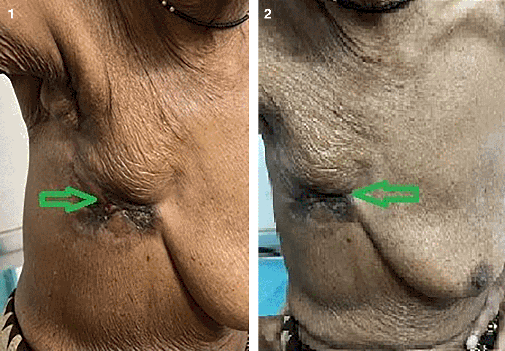
We report the case of a 71-year-old woman. She is a house-wife and is Caucasian with no past medical or family history who reported to the outpatient clinic with chief complaints of itching, wound, and destruction of the right nipple and areola of the breast for three years. She also had complaints of breathlessness, weakness, and decreased appetite. The examination revealed ulceration, destruction of the nipple-areolar complex, irregular margins, uneven surface, blackening of surrounding skin, and right axillary lymphadenopathy present (Figures 1 and 2). The results of routine blood investigations suggestive of raised WBC (white blood cells) counts, 21,900 per microliter (normal range: 4,500 to 11,000 WBCs per microliter). There was no palpable breast mass. The left breast and axilla were normal, with no evidence of any lump. Ultrasonography and mammography did not reveal any lump. A respiratory examination revealed decreased right-sided breath sounds. The patient had not undergone any intervention in the past.

The patient was conscious, oriented to time, place, and person, with no lymphadenopathy, pallor, icterus, cyanosis, or oedema. They were clinically diagnosed with advanced metastatic Paget’s disease of the breast. Ultrasonography and mammography did not reveal any lump.
She was afebrile. Her pulse rate was 110 bpm. Her blood pressure was 130/70 mmHg. Her respiratory rate was 42 breaths per minute. A detailed clinical examination was done which revealed left-sided decreased breath sounds on chest auscultation. The rest of the vitals of the patient were within normal limits. A chest X-ray posteroanterior (PA) view was done, which revealed the presence of a right-sided moderate pleural effusion (Figure 3). Due to the radiological findings of a right-sided moderate pleural effusion and clinical findings of worsening dyspnea of the patient, an urgent intercostal chest drain (ICD) insertion was done, and approximately 1 L of yellowish fluid and pleural fluid samples were sent for all routine pleural fluid investigations.
Per abdomen - soft, non-tender, no guarding/rigidity/distension, bowel sounds were present in all four quadrants.
Routine pleural fluid investigations of the patient were done, which were as follows:
• Pleural fluid total white blood cell count – 440 cells/cmm (normal range – 440 cells/cmm)
• Pleural fluid lactate dehydrogenase – 210 IU/L (normal range – 105 to 333 IU/L)
• Pleural fluid adenosine deaminase (ADA) – 2.103 IU/L (normal range – less than 40 IU/L)
• Pleural fluid, differential cell count: Lymphocyte-predominant pleural effusion (65% lymphocyte)
• Pleural fluid Xpert (resistance to rifampicin) RIF assay (cartridge-based nucleic acid amplification testing – CBNAAT) – Mycobacterium tuberculosis (Mtb) – not detected
Pleural fluid on wet mount – (red blood cells) RBC – 2–3/HPF, WBC – 2–3/HPF, (differential leukocyte count) DLC –lymphocytes = 65%, polymorphs – 35%.
Nuclei showed mild Pleomorphism and infrequent nucleoli. The cytoplasm was modest to scant. It showed cytoplasmic lumina in a few reactive mesothelial cells, cells with smudged nuclei, and the background showed few macrophages, lymphocytes and thin proteinaceous material, and few red blood cells. This was suggestive of infiltrates of invasive ductal carcinoma.
Incisional biopsy concluded Paget’s disease of the breast with infiltration of invasive ductal carcinoma of the breast (Figure 4). This was clinically diagnosed as advanced metastatic Paget’s disease of the breast with malignant pleural effusion. Patient had received adjuvant chemotherapy. Patient was unexpectedly discharged on request of relatives with ICD (intercostal chest drain) in situ. Patient was haemodynamically and vitally stable on discharge.
Nipple Paget’s illness was originally identified in 1874. It is most common in the age group between 50 to 70 years. Paget’s disease is a malignant disease. The nipple-areolar complex initially develops eczematous alterations due to the disease, and in later stages, the nipple-areolar complex will be destroyed by ulceration.4 Paget’s nipple illness may be connected to primary invasive or in situ breast cancer.5 As the condition progresses, it first manifests as eczematous alterations of the nipple-areolar complex, changes in skin tone, ulceration, burning sensations, and bleeding of the nipple. It might or might not be connected to a bulge underneath. Only 2% to 3% of all cases of breast cancer are caused by the condition. The migration of ductal carcinoma from which Paget’s cells originated is the most widely accepted explanation.1
What is extremely unusual about this case is the malignancy had spread to the adjacent structures and led to malignant pleural effusion.
The generally recognized migratory idea states that ductal carcinoma in situ cells migrate from the primary tumor to the nipple and surrounding skin via milk ducts. The initial presentation of Paget’s disease of the breast is an eczematous lesion, similar to contact dermatitis, in the skin of the breast at the areola and/or nipple and is refractory to usual topical treatments.6
The underlying duct carcinoma cells share similarities with Paget cells in terms of HER2 oncogene positive and epithelial cell characteristics. The precise mechanisms are less clear, although interactions between the HER2 on the tumor cells and the heregulin-alpha protein produced by nipple epidermal keratinocytes have been linked to the chemotaxis.7
If conservative breast surgery is intended, it is imperative to confirm preoperatively through clinical, radiological, and histological investigation that the patient is tumor-free.8–10
The biopsy is crucial for making a Paget’s disease diagnosis. Clinical breast examination is more significant than a biopsy. A clinical breast exam can detect an associated breast lump in about 50% of people with Paget’s disease. When a breast lump can be felt, a mammography, magnetic resonance imaging (MRI) scan, or ultrasound examination is required. In Paget’s disease, CD138 and p53 are regarded as positive and detrimental in the typically present Toker cells.2
Chemotherapy – for people with invasive ductal carcinoma, chemotherapy may be given before surgery to shrink the tumor or after surgery to reduce the chance of cancer returning. Chemotherapy may also be recommended as the main treatment for people with metastatic breast cancer.11
Radiation – in most breast cancer cases, radiation therapy is used after surgery to kill any remaining cancer cells. Occasionally, it may be used to shrink a tumor before surgery. Radiation therapy may also be recommended when a tumor can’t be surgically removed due to the size or location.12
Mastectomy with or without the removal of axillary lymph nodes on the same side was considered the standard surgery for Paget’s breast disease. Most of the patients suffer along with the underlying tumor. So removing only the nipple and areola doesn’t cure the disease. Recent studies have demonstrated that radiation therapy followed by conservative breast surgery is an effective choice for individuals with limited disease. Long-term breast-conserving surgery would be comparable to mastectomy in terms of overall survival without disease.13
Patients with Paget’s breast disease and a breast tumor should undergo a mastectomy along with a sentinel lymph node biopsy to check for axillary lymph node dissemination. If the sentinel node is positive, axillary clearing is performed.14
Early detection is necessary for Paget’s illness of the breast. About 1%–3% of primary breast cancer cases are caused by Paget’s breast disease. Breast cancer is sporadic in nature. The biopsy is a must for diagnosis of disease in patients with normal ultrasound and mammogram. Disease management depends on the underlying tumor, the spread of the disease, and the presence or absence of an underlying lump. The presence of a palpable tumor, the extent of primary cancer, and the presence of distant metastases all affect the prognosis.
Written informed consent for publication of their clinical details and clinical images was obtained from the patient.
All data underlying the results are available as part of the article, and no additional source data are required.
| Views | Downloads | |
|---|---|---|
| F1000Research | - | - |
|
PubMed Central
Data from PMC are received and updated monthly.
|
- | - |
Provide sufficient details of any financial or non-financial competing interests to enable users to assess whether your comments might lead a reasonable person to question your impartiality. Consider the following examples, but note that this is not an exhaustive list:
Sign up for content alerts and receive a weekly or monthly email with all newly published articles
Already registered? Sign in
The email address should be the one you originally registered with F1000.
You registered with F1000 via Google, so we cannot reset your password.
To sign in, please click here.
If you still need help with your Google account password, please click here.
You registered with F1000 via Facebook, so we cannot reset your password.
To sign in, please click here.
If you still need help with your Facebook account password, please click here.
If your email address is registered with us, we will email you instructions to reset your password.
If you think you should have received this email but it has not arrived, please check your spam filters and/or contact for further assistance.
Comments on this article Comments (0)