Keywords
Lithium disilicate, marginal fit, internal fit, cement space, fabrication method.
This article is included in the Datta Meghe Institute of Higher Education and Research collection.
Lithium disilicate, marginal fit, internal fit, cement space, fabrication method.
There was a change in the inclusion criteria.
In the previous manuscript the inclusion criteria selected was 1st molar, in the revised manuscript the inclusion criteria that is taken in consideration is maxillary incisors in need of prosthetic restoration. Also there are changes in the finish lines configuration. In the previous manuscript shoulder finish line with supragingival margin was used. In the revised manuscript shoulder finish line with subgingival margin is used.
See the authors' detailed response to the review by Tariq Abuhaimed
The conventional crown fabrication method is a time-consuming and technique sensitive method, that requires meticulous fixation of the negative replica of the tooth by a stable means of registration, to minimize errors in manufacturing process of dental crowns. The transportation of impressions to the dental laboratory is governed by factors such as ambient temperature, transit time, distance from the laboratory, etc. These factors can affect impression accuracy.1 The time required to pour the cast, the physical properties of gypsum product also affect the impression quality causing minor distortions in the impression. The use of die hardener, die spacer and the entire casting process may be accompanied with some amount of error creating marginal discrepancy or any kind of discrepancy in dental prosthesis that may lead to periodontal problems and endodontic problems.
Computer aided designing (CAD) & computer aided manufacturing (CAM) are currently growing and widely establishing technology. In the current scenario increased interest of dental surgeon and engineers in technology and artificial intelligence is taking over and developing a techno-societal approach towards dental problems. Both CAD and CAM are increasingly used in dental laboratory for fabrication of crown and bridge prosthesis. The use of this technology has reduced the efforts and time in fabrication of the prosthesis, making the treatment affordable both in terms of time & money compared to conventional methods.
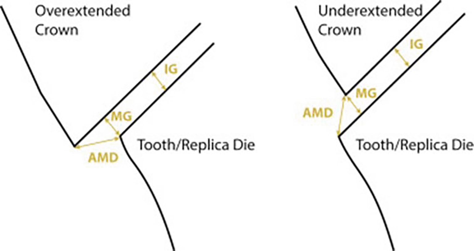
The indirect restorations have long term success rate depending on “internal” and “marginal fit”. The poor fit will result in marginal dissolution, leading to secondary caries and failure of the prosthesis.2 Holmes et al. in his study, quoted marginal gap as perpendicular measurement from cervical margin of casting to preparation margin. He further defined absolute marginal discrepancy as angular combination of marginal gap & extension error (over and under extension).2 If the cement is exposed to saliva, it can be dissolved leading to weakened prosthesis, improper marginal gap on the other side.2 Marginal discrepancy will indicate marginal crown extension in relation to abutment margin and is of importance in overextension or under extension cases.
The “lithium disilicate crowns” are also widely used in dental practice in the current scenario. It is a glass ceramic material that is gaining popularity. This material is commonly used in dental laboratories according to pressing technology or manufacturing crowns. It has high fracture toughness, flexural strength and is very aesthetic. It is commonly known as “IPS e.max. Lithium disilicate (IPS e.max)” requires minimal invasive preparation that justifies the statement of Sir DeVan that “Perpetual preservation of what remains is more important than the meticulous replacement of what is missing.”
In this study we will be focusing on the properties of marginal fit, internal fit and cement space of lithium disilicate (IPS e.max) crowns fabricated by conventional and digital method respectively. The null hypothesis will state there is no difference in the internal fit, marginal fit and cement space of lithium disilicate crowns in the methods of fabrication.1
The replica technique is reliable, non-invasive and is a valid technique for calculating marginal fit and internal fit of crowns by an in vivo method. But, the disadvantage with this is that it is technique sensitive and there are chances of distortion of the replica impression.3
Rationale: The purpose of this study is to determine the “marginal fit”, “internal fit” and “cement space” of lithium disilicate crowns as these are important parameters for determining fit by two different methods of fabrication.
Aim: To evaluate and compare the “marginal fit”, “internal fit” and “cement space” of single unit lithium disilicate crown with shoulder finish line with two different methods of fabrication.
Objective: To evaluate and compare,
The experimental study is being conducted in the department of prosthodontics and crown & bridge, Sharad Pawar Dental College and Hospital Sawangi(M).
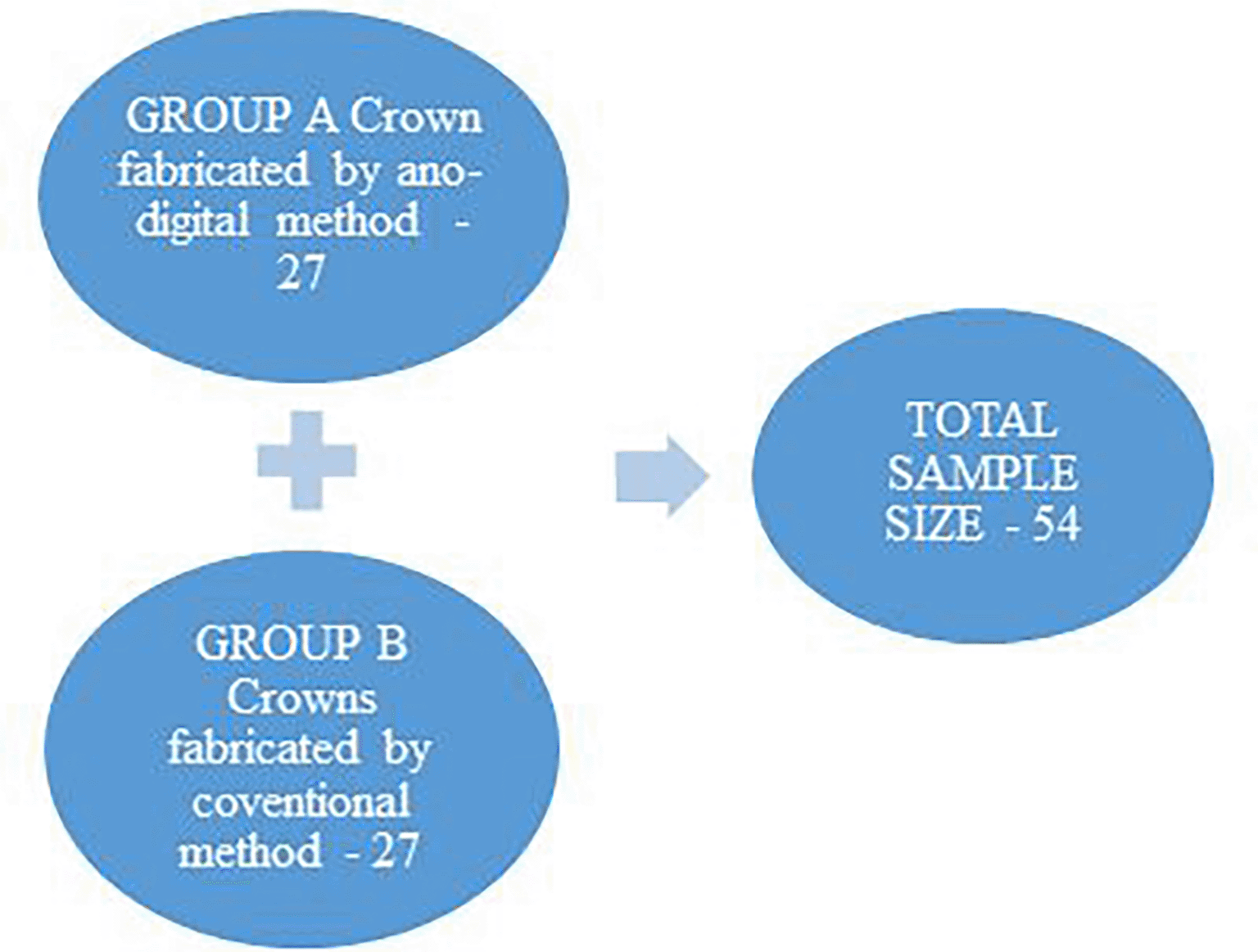
A total of 54 patients within the age group of 18-54 years, will be selected from patient coming to out-patient’s department (OPD) of prosthodontics and crown & bridge. The patients will be the residents of vidarbha region Maharashtra The members will be recruited from the outpatient department of prosthodontics and crown and bridge of Sharad Pawar Dental College. The sample population considered will be the residents of Vidarbha region Maharashtra. Further these 54 patients will be divided into two groups of 27 patients in each group randomly group A will consist of the subjects where lithium disilicate crowns are fabricated using CAD and CAM. Group B consisted of subject with whom lithium disilicate crowns will be fabricated using a pressed technique. Figure 1 represents the study design.
To compare 2 means, using mean and standard deviation.
where α and β are constant,The value of α = 0.05
β = 0.1
The mean in group 1, μ1 = 100
group 2, μ2 = 63
Standard deviation in group 1, σ1 = 54
group 2, σ2 = 24
Ratio (group 2/group 1) = 1
Z1-α/2 = 1.96
Z1-β = 1.28
At 90% power
Substituting the values.
Minimum sample size needed is 27 per group.
Total number of sample size needed 54.
The “marginal fit”, “internal fit” and “cement space” of “lithium disilicate crowns” will be calculated with the help of stereomicroscope. There will be 2 groups and 27 patients will be randomly distributed in each group.
The test used to perform statistical analysis will be unpaired t test.
Armamentarium
Burs – TF11, SF-11, SF-41, EX11, EX12, WR13, end cutting bur.
Elastomeric impression material (light body & heavy body).
Alginate impression material.
Equipped CAD – CAM lab.
Equipped Ceramic lab.
Lithium disilicate ingot.
Burs for finishing & polishing ceramic
Die stone.
Dental stone.
Dental plaster.
Stereomicroscope.
Two groups will be made group A & B. Group A will consist of the lithium disilicate crown fabricated by ano-digital method. Group B will consist of lithium disilicate crowns fabricated by a conventional method. All patients to be examined will be selected from patients coming to out-patient department (OPD) of prosthodontics and crown and bridge.
Every new patient coming to OPD will be checked for the inclusion and exclusion criteria.
According to this criteria, selected patients will be allocated in group A and group B randomly. Informed consent will be taken from patients then only he/she will be included in the sample. Tooth preparation will be planned on the tooth of interest. Before starting preparation, a silicon putty will be adapted to the entire tooth and adjacent tooth on each side & an index of the silicon putty will be taken. A diagnostic wax up will be done if necessary. Incisal reduction will be done by marking depth orientation grooves. A no 171 bur of fine girt round end tapered diamond will be used to smooth the planes of occlusal reduction. Proximal reduction will be done with a diamond bur.
The lingual axial wall will be reduced with coarse grit tapered torpedo diamond & a shoulder finish line with subgingival margin will be prepared. After the completion of tooth preparation an impression will be taken with elastomeric impression material and a study cast and working cast with a die will be made. There are 2 methods of crown fabrication digital and conventional. The material used for the fabrication of the crown prosthesis is lithium disilicate ingot. Lithium disilicate will be used for crown fabrication with CAD-CAM and lithium disilicate ingot will be used to fabricate the crown by pressed method. The digital method is as follows. The cast will be scanned by the extra oral scanner in the CAD-CAM lab and the scans will be recorded and saved in stl/g-code format. The cast would be then articulated digitally with the help of virtual articulator using software and the designing of the prosthesis would be done. The material used for crown prosthesis is the lithium disilicate ingot, the crown will be fabricated by a subtractive method with 5 axis milling from an industrially fabricated block of lithium disilicate.1 With a few more subsequent steps, of sintering and finishing and polishing crown prosthesis is made using digital method.3
After obtaining the cast a pattern will be fabricated. Investing and burn out procedures will be carried out. Ceramic ingots are then heat pressed in the mold cavity in a special ceramic press furnace, cooled and retrieved. Sub structure is obtained by pressing. As our inclusion criteria includes maxillary incisors staining and glazing procedures are made to get aesthetic result. Then the internal fit of the crown fabricated with digital and conventional method will be measured & the 2 groups will be compared.2
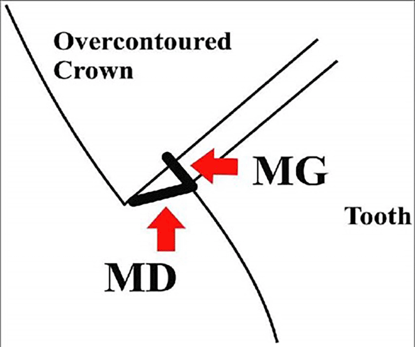
Figures 2 & 3 gives a diagramatic representation of over and under extended crowns, marginal gap and internal gap.
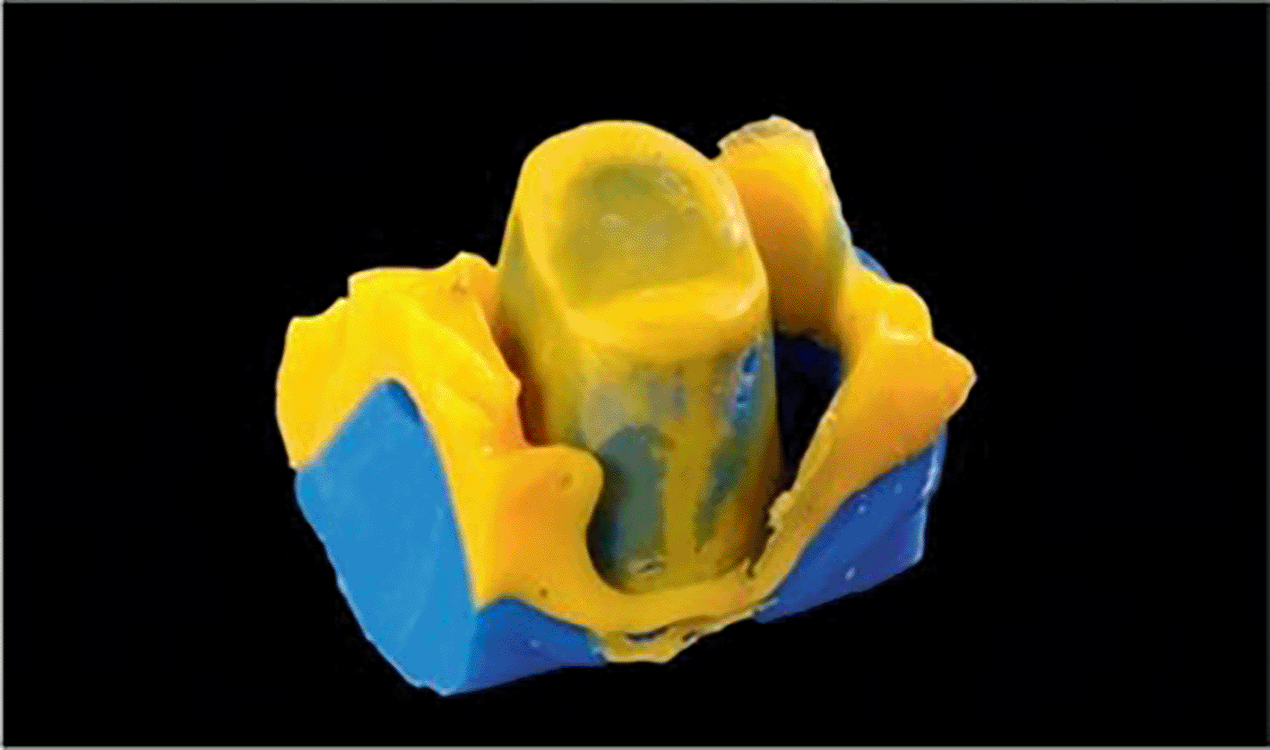
The measurement of the internal fit, marginal fit and cement space will be done with the help of elastomeric impression material (light body and heavy body).4 The prepared crown prosthesis will be loaded with a light body elastomeric impression material and the impression of the prepared tooth will be taken with the processed crown.4 Then silicon putty will be used as a tray to retrieve the impression. Figure 4 is demonstrating the replica technique.
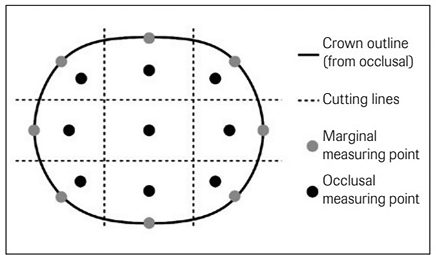
The impression is divided into 4 different parts mesial, distal, buccal, lingual, occlusal and the thickness of each segment is measured under a stereomicroscope to calculate the marginal fit, internal fit and cement space. Figure 5 determines the marginal and occlusal measuring points.
An in vivo study was conducted by KW Boening et al. (2000) to assess the clinical fit and accuracy of ceramic crowns of anterior & posterior teeth. On evaluating the clinical fit of 80 anterior & posterior teeth, the author calculated the median marginal gap width of anterior and posterior teeth using an identical technique. Based on the computations made with the light microscope for each part, he concluded that anterior teeth had width of median marginal gap between 80 & 95 microns, while posterior teeth had median marginal gap widths between 90 and 145 microns. A median marginal gap width of 80–180 microns and 115–245 microns was observed respectively in anterior & posterior region.4
In the year 2014 Jonathan et al. studied comparison of marginal fit, internal fit, and cement space of crowns made by digital and conventional methods. The upper second premolar was prepared on a typodont for full ceramic crown. He scanned the typodont with a lab scanner & used a digital file to replicate maxillary arch from a monolithic block of yttria-stabilized zirconia that served as the master cast. He obtained a digital impression of the prepared upper right second premolar in his scanning unit. The scan file was exported & emailed to the dental lab. Digital articulation, digital waxing & final crown design. Fifteen crowns were fabricated by CAD technology designed with lithium disilicate glass-ceramic blocks & fifteen lithium disilicate glass-ceramic crowns were manufactured using laboratory and conventional techniques. The original zirconia die was removed from the zirconia master cast to assess the margins of crown. Circumferential edge gap measurements were taken at eight locations. Vertical component determined the measurements. An entire measure of 240 images (2 groups, 15 crowns per group, 8 sites per group) were done.1
Necla et al. in 2014 assessed marginal gaps & two finish line design shoulder and chamfer of all ceramic crowns with absolute marginal discrepancy, using micro-CT pre and post cementation, they divided 60 extracted maxillary premolar teeth into two groups, and extended grouping was done based on the crowns they received. The results of this study concluded that the all-ceramic system exhibited clinically acceptable margins. The Feldspar-Cerec-Inlab ceramic system showed the least variance, except for MG values in the coronal mesial region. The MG and MD values of all ceramics increased significantly after cementation, except for the MG shoulder-prepared design and the MD chamfer-prepared design. The conclusion of Necla et al. In the studies conducted, all-ceramic crowns had a clinically acceptable marginal fit. The fieldpathic cerac inlab system showed minimal variance compared to other ceramics.5
Stefanie Anke Rau et al in 2018 analysed the clinical fit of monolithic zirconia single crowns in a prospective cohort study. A total of 30 molars were restored with monolithic zirconia single crowns. Silicone impressions were made to measure clinical fit using a stereomicroscope. Measurements were taken at 17 points per crown on the marginal and occlusal surfaces. The average clinical fit was 0.104 mm at the coronal margin and 0.101 mm at the occlusal surface. The conclusion of the study was that zirconia monolithic single crowns have shown acceptable clinical fit.6
Ji-Eun Ryu et al. in the year 2020 evaluated marginal and internal fit of 3D printed provisional crowns according to assembled directions using silicon replica technique. In this technique they scanned the prepared resin tooth and single crown was designed using computer aided design (CAD) software. Provisional 10 crowns were printed using a DLP based 3D printer at 6 directions (120 degree, 135 degree, 150 degree, 180 degree, 210 degree, 225 degree) respectively with. In total 60 crowns were printed to measure marginal and internal fit. A duplicate silicon model was fabricated and thickness was measured using a digital microscope. Sixteen reference points were set and divided into 4 groups marginal gap, cervical gap, axial gap and occlusal gap. Measurements were analyzed using one-way ANOVA and Dunnet T3. The conclusion of the study was that the 3D printed provisional crowns varied in the marginal and internal fit depending upon the assembled angle and best fit was achieved with assembled angles of 150 and 180 degrees.7
Recently in year 2021 Maria Rizonaki et al. evaluated marginal and internal fit of CAD-CAM ceramic crowns made from lithium disilicate based on 3 different finish lines (rounded shoulder, chamfer, feather-edge). Thirty anterior lithum disilicate crown (n = 10 per finish line group) were fabricated with the help of TRIOS intraoral scanner. An attempt was made by the author to cement the crowns on the respective prepared typhodont teeth with fabricated duplicate dies the absolute marginal discrepancy, marginal gap & internal gap was evaluated by using microcomputed tomography. Values obtained from each specimen from sagittal and trans axial section were 66 and a rendering software program was used to calculate the volume of cement gap for each specimen by means of 3D region growing. Marginal discrepancy & gap values were statistically significant between group. The differences in cement space between groups were not statistically significant.2
| Views | Downloads | |
|---|---|---|
| F1000Research | - | - |
|
PubMed Central
Data from PMC are received and updated monthly.
|
- | - |
Provide sufficient details of any financial or non-financial competing interests to enable users to assess whether your comments might lead a reasonable person to question your impartiality. Consider the following examples, but note that this is not an exhaustive list:
Sign up for content alerts and receive a weekly or monthly email with all newly published articles
Already registered? Sign in
The email address should be the one you originally registered with F1000.
You registered with F1000 via Google, so we cannot reset your password.
To sign in, please click here.
If you still need help with your Google account password, please click here.
You registered with F1000 via Facebook, so we cannot reset your password.
To sign in, please click here.
If you still need help with your Facebook account password, please click here.
If your email address is registered with us, we will email you instructions to reset your password.
If you think you should have received this email but it has not arrived, please check your spam filters and/or contact for further assistance.
Comments on this article Comments (0)