Keywords
hamartoma, nevus sebaceous, scalp, case report
This article is included in the Datta Meghe Institute of Higher Education and Research collection.
Epidermal nevus sebaceous, commonly known as the nevus sebaceous of Jadassohn, is a congenital sebaceous hamartoma. It typically manifests as a single yellowish plaque across the head and neck and is composed of sebaceous glands. It commonly occurs during infancy and grows during puberty. Usually, it follows a benign course; however, in a few cases, it can be malignant. This is the case of a 13-year-old child with verrucous plaques on the temple and scalp.
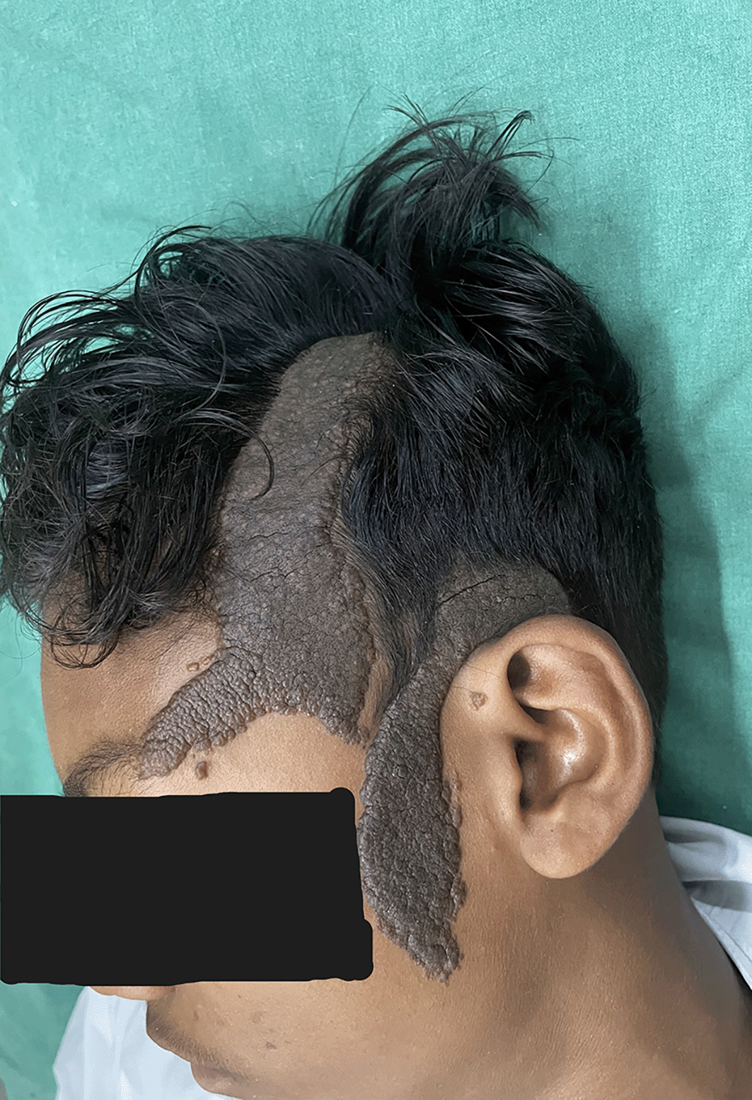
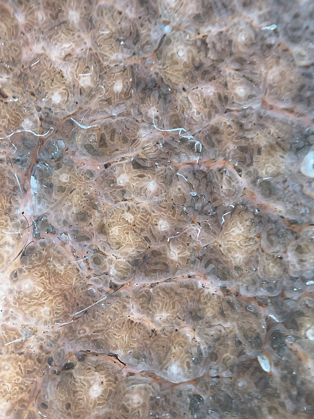
We report the case of a 13-year-old boy with a steadily developing hyperpigmented verrucous plaque on the scalp and ipsilateral side of his face. A dermoscopic examination revealed ridges and fissures in a cerebriform pattern with yellowish-gray globules and a papillary appearance. Physical examination and laboratory tests revealed no abnormalities. Biopsies were taken from the scalp and temple area, and the findings were consistent with the diagnosis of nevus sebaceous. The patient was referred to a plastic surgeon for a staged excision.
We describe a unique example of a sebaceous nevus that affected the scalp and ipsilateral side of the face. As this hamartomatous growth carries the risk of cancer development, a dermatologist must identify the condition and begin treatment before malignant transformation occurs. This example of multiple verrucous plaques is an exception.
hamartoma, nevus sebaceous, scalp, case report
We have added the differentiating points between the close differential diagnosis of Nevus Sebaceous, how to clinically identify the malignant transformation in the case of Nevus Sebaceous, and various non-invasive investigations for the same with genetics.
See the authors' detailed response to the review by Aswath Rajan
See the authors' detailed response to the review by Dipanjan Basu
Nevus sebaceous (NS), initially described by Jadassohn, is a complicated hamartoma that typically develops on the face or scalp and has an epithelial or adnexal origin.1
It can appear at birth or develop in infancy and increases during puberty, suggesting a hormonal influence. It can occasionally be found in other locations, such as the trunk or the oral or vaginal mucosa, although it mostly affects the scalp. Less frequently, it affects the preauricular area and neck.2
Nevus sebaceous of Jadassohn (NSJ) develops in three stages. It manifests as isolated, well-circumscribed, smooth, yellowish plaques without hair during the infantile period. It becomes more noticeable with a verrucous or mamillated appearance during puberty. The last stage is characterised by peripheral telangiectasias and a nodular or tumoral appearance.3
Many neoplasms develop alongside NS as proliferative growth begins. Both benign and malignant tumors have been reported to grow in NS. NS can be a site of basal cell cancer, syringocystadenoma papilliferum, trichoblastoma, and hidradenoma.4
A 13-year-old boy visited the dermatology outpatient department on 8th September 2023 with a raised lesion on his scalp since birth and a lesion that had spread to the left side of the face over ten years. The ophthalmological, neurological, or cutaneous systems did not exhibit any abnormalities during physical examination. These skin lesions had not previously occurred in the family. The results of all laboratory tests, including the kidney function test, liver function test, urine examination, and complete blood count were within normal ranges. The patient had no other complaints.
On cutaneous examination, a well-demarcated hyperpigmented verrucous plaque with a size of 8 × 4 cm was present on the frontal area of the scalp extending down to involve the forehead and a 7 × 3 cm plaque was present on the temporoparietal area and left preauricular area [Figure 1]. Based on the patient’s medical history and physical examination, the possible differential diagnoses were identified as congenital melanocytic nevus, giant seborrhoeic keratoses, and verrucous epidermal nevus. However, a thorough examination through dermoscopy and histology conclusively ruled out these possibilities. In seborrheic keratosis, other than ridges and fissures multiple milia and comedone-like openings will be there. In acanthosis nigricans, there will be papillary projections with hyperpigmented dots and perifollicular pigmentation. We confirmed on clinical grounds that there were no clinical signs in the lesion to undergo a malignant transformation like ulceration, bleeding from the lesion or sudden increase in the size of the lesion. On dermoscopic examination, ridges and fissures were present in a cerebriform pattern with yellowish-grey globules and a papillary appearance [Figure 2]. Histopathological examination revealed acanthosis, papillomatosis, and mild hyperkeratosis. There were immature and mature sebaceous glands with sebaceous hyperplasia and primitive hair follicles [Figure 3]. The diagnosis of nevus sebaceous was established based on clinical presentation, dermoscopic findings, and histological analysis. The patient was referred to a plastic surgeon on 8th September 2023 for a staged surgical excision of the nevus sebaceous. Our dermatology department does not offer plastic surgery services, hence the referral. Unfortunately, the patient was lost to follow-up after the referral, and we do not have any further information available.

(Written informed consent for publication of their clinical details and clinical images was obtained from the relatives of the patient).

Nevus sebaceous is a condition that appears at birth and increases in size with age. The exact cause of this condition is still uncertain, but recent studies have shown that it may be linked to women who have tested positive for the human papillomavirus or carry mutations in the PTCH gene.5,6
Nevus sebaceous can present as one of the manifestations of Epidermal Nevus Syndrome.7 There are some hereditary syndromes, including didymosis aplasticosebacea and SCALP (sebaceous nevus, central nervous system malformations, aplasia cutis congenita, limbal dermoid, and pigmented nevus) syndrome, that may present nevus sebaceous as a symptom. This condition typically appears as a smooth, yellowish-orange, round, oval, or linear plaque, mostly on the scalp, leading to alopecia.5
A previous study found that nevus sebaceous can occur in multiple locations, similar to verrucous epidermal nevi.8 Nevus sebaceous is rarely reported in the literature to affect the scalp and ipsilateral side of the face.9 In our case, the scalp and the ipsilateral side of the face were affected.
Several discussions have taken place regarding the emergence of secondary benign and malignant tumors inside the nevus sebaceous. While basal cell carcinoma development has been documented by multiple authors in adults, recent reports have also identified atypical malignant neoplasms such as eccrine porocarcinoma, sebaceous carcinoma, apocrine carcinoma, and squamous cell carcinoma developing inside the NS.10,11
There is a risk of developing malignant tumors in the Nevus Sebaceous. To detect these tumors accurately, non-invasive techniques like High-frequency Ultrasound and Reflectance Confocal Microscopy are used. These techniques help in visualizing the skin and skin appendages for accurate depth and lateral border detection. Reflectance Confocal Microscopy is particularly useful as it allows for in vivo evaluation of lesions and shows both anatomical features and individual cells.12,13 The presence of PTCH deletion, HRAS, and KRAS mutation can lead to malignant transformation in the nevus sebaceous.14
Although the timing of resection for nevus sebaceous therapy is debatable, most researchers feel that surgical excision is the preferred course of action. However, surgical excision to remove nevus sebaceous creates a linear scar. There are various therapeutic options, such as CO2 laser therapy, to reduce scarring. However, CO2 laser vaporization completely eradicates the sebaceous section of the nevus, which is located in the epidermis or papillary dermis.15
The primary take-away lesson from our case is as follows: We describe a unique example of a sebaceous nevus that affected the scalp and ipsilateral side of the face. As this hamartomatous growth carries the risk of cancer development, a dermatologist must identify the condition and begin treatment before malignant transformation occurs. This example of multiple verrucous plaques is an exception.
Written informed consent for publication of their clinical details and clinical images was obtained from the relatives of the patient.
All data underlying the results are available as part of the article and no additional source data are required.
| Views | Downloads | |
|---|---|---|
| F1000Research | - | - |
|
PubMed Central
Data from PMC are received and updated monthly.
|
- | - |
Competing Interests: No competing interests were disclosed.
Reviewer Expertise: dermatology
Is the background of the case’s history and progression described in sufficient detail?
Yes
Are enough details provided of any physical examination and diagnostic tests, treatment given and outcomes?
Partly
Is sufficient discussion included of the importance of the findings and their relevance to future understanding of disease processes, diagnosis or treatment?
Partly
Is the case presented with sufficient detail to be useful for other practitioners?
Partly
Competing Interests: No competing interests were disclosed.
Reviewer Expertise: dermatology
Is the background of the case’s history and progression described in sufficient detail?
Yes
Are enough details provided of any physical examination and diagnostic tests, treatment given and outcomes?
Yes
Is sufficient discussion included of the importance of the findings and their relevance to future understanding of disease processes, diagnosis or treatment?
Partly
Is the case presented with sufficient detail to be useful for other practitioners?
Yes
References
1. Lena C, Kondo R, Nicolacópulos T: Do you know this syndrome? Schimmelpenning-Feuerstein-Mims syndrome. Anais Brasileiros de Dermatologia. 2019; 94 (2): 227-229 Publisher Full TextCompeting Interests: No competing interests were disclosed.
Reviewer Expertise: Cell biology and therapeutic strategies of Large /giant congenital nevi and neurocutaneous melanocytosis.
Alongside their report, reviewers assign a status to the article:
| Invited Reviewers | ||
|---|---|---|
| 1 | 2 | |
|
Version 2 (revision) 08 Apr 24 |
read | |
|
Version 1 27 Nov 23 |
read | read |
Provide sufficient details of any financial or non-financial competing interests to enable users to assess whether your comments might lead a reasonable person to question your impartiality. Consider the following examples, but note that this is not an exhaustive list:
Sign up for content alerts and receive a weekly or monthly email with all newly published articles
Already registered? Sign in
The email address should be the one you originally registered with F1000.
You registered with F1000 via Google, so we cannot reset your password.
To sign in, please click here.
If you still need help with your Google account password, please click here.
You registered with F1000 via Facebook, so we cannot reset your password.
To sign in, please click here.
If you still need help with your Facebook account password, please click here.
If your email address is registered with us, we will email you instructions to reset your password.
If you think you should have received this email but it has not arrived, please check your spam filters and/or contact for further assistance.
Comments on this article Comments (0)