Keywords
systemic lupus erythematosus, interleukin – 2, natural killer cells, CD56bright, CD56dim, interferon – γ
This article is included in the Cell & Molecular Biology gateway.
systemic lupus erythematosus, interleukin – 2, natural killer cells, CD56bright, CD56dim, interferon – γ
Systemic lupus erythematosus (SLE) remains a serious health issue. The global morbidity of SLE was predicted to be between 9.0 and 366.6 cases per 100,000 individuals, and of them, the mortality was reported ranging from 6.7 to 37.8%.1 The management of SLE presents considerable difficulties, both in terms of diagnosing the condition and providing effective treatment. It has been widely known that SLE is a disease with 1000 faces, suggesting that SLE has a wide variety of clinical manifestation, and therefore, the diagnosis of SLE requires the carefully holistic investigation.2 On the other hand, the treatment of SLE remains a challenging global issue. The current treatment for SLE patients only targets remission, no single effective treatment has been proven in curing SLE. This challenging treatment issue is the implication of the complicated pathogenesis of SLE.3 Theoretically, the pathogenesis of SLE is complex and may involve interferon regulatory factor, toll-like receptor, protein tyrosine phosphatase, natural killer (NK) cells, and interferon-γ (IFN-γ).4 Studies suggested that, in SLE patients, NK cells dysfunction was observed, and the activity of NK cells was governed by the balance expression between CD56bright (NK cells regulator) and CD56dim (NK cells cytotoxic).5–7 Moreover, CD56bright had been proven to have the ability to initiate the production of IFN-γ.8 Therefore, a therapeutic modality by targeting the activity of NK cells might provide a beneficial impact.
Interleukin-2 (IL-2) is one of the primary cytokines having pleiotropic impact on the immune system.9 IL-2 has been reported as the potential treatment for the management of several diseases with immunological dysfunction such as: chronic graft-versus-host disease (GVHD)10 and acute myeloid leukemia.11 The possible mechanism of action of IL-2 in NK cells is by binding to NK cell activating receptors to affect the expression of NK cell subsets such as CD56bright and CD56dim, and as a result, the proliferation and the production IFN-γ may occur.5 In the case of GVHD and acute myeloid leukemia; IL-2 had been reported to affect the expression of CD56bright and CD56dim NK cells as well as the levels of IFN-γ.10,11 On the other hand, in the case of SLE, since a previous report revealed that the levels of IL-2 was decreased,12 the potential treatment of IL-2 in SLE patients by assessing NK cells subsets has never been investigated. Therefore, our present study sought to assess the implication of IL-2 administration to the expression of CD56bright and CD56dim as well as the levels of IFN-γ.
The study was approved by the local Ethical Committee of Saiful Anwar Malang Hospital (No. 400/231/K.3/302/2019; approved on 22 October 2019)13 and conducted in concordance with the Declaration of Helsinki.14 Before being involved in the study, patients provided written informed consent. A total of 18 mL of peripheral venous blood was collected and placed in a vacutainer tube containing EDTA.
During January–July 2020, lymphocytes were collected from six female SLE patients in Saiful Anwar Hospital, Indonesia. SLE patients in our present study conformed with the classification criteria of the 2019 European League Against Rheumatism and the American College of Rheumatology (2019 EULAR/ACR).15 The baseline characteristics of SLE patients in our study were as follows: age 18–49 years old (mean = 36,17±6,494 years) and in active disease with Mexico-SLE Disease Activity Index (MEX-SLEDAI) score > 5 (mean = 9,5±2,345 points). The subjects were divided into four different groups of IL-2 stimulation: D0 (control/0 U/ml), D1 (50 U/ml), D2 (150 U/ml), and D3 (250 U/ml).
Peripheral blood mononuclear cells (PBMCs) were collected by Lymphoprep™ Density Gradient Medium 1.07 (Axis – Shield, Oslo, Norway) within 6 hours of blood drawing. PBMCs were then incubated in the medium of Roswell Park Memorial Institute (RPMI) 1640 (Sigma-Aldrich, Missouri, US, RRID:SCR_013728) containing L-Glutamine and 10% Gibco™ Fetal Bovine Serum (Sigma-Aldrich, Missouri, US, RRID:SCR_013728) and supplemented with penicillin (100 U/ml)/streptomycin (100 mg/ml; Sigma-Aldrich, Missouri, US, RRID:SCR_013728) and Hepes (Sigma-Aldrich, Missouri, US, RRID:SCR_013728) overnight. Subsequently, PBMCs were stimulated with rh-IL-2 (BioLegend, California, US, RRID:SCR_001134), with four different doses (0, 50, 150, 250 U/ml) and incubated for 72 hours at 37°C with 5% CO2.
Following incubation with four different doses of IL-2, cells were collected, washed, and rehydrated for coloration. PBMCs obtained from the incubation were collected and distributed into four separate Eppendorf tubes. Subsequently, they were subjected to centrifugation at 4000 rpm for a duration of 3 minutes at a temperature of 22°C. The pellets were then washed using cell staining buffer (CSB) and centrifuged once again at 4000 rpm for 3 minutes at 22°C. Cells were colored at 4°C for 30 minutes with anti-CD3 antibody (Biolegend, SanDiego, CA, RRID:SCR_001134) conjugated with Peridinin-Chlorophyll-Protein (PerCP) (Biolegend, SanDiego, CA, RRID:SCR_001134) and anti-CD56 antibody (Biolegend, SanDiego, CA, RRID:SCR_001134) conjugated with Fluorescein Isothiocyanate (FITC) (BioLegend, San Diego, CA, RRID:SCR_001134). Cells were washed twice and analyzed using Fluorescence-activated Cell Sorting (FACS) Melody (BD Bioscience, Haryana, India, RRID:SCR_018091). The cell number was analyzed by the BD Cell Quest software (BD Biosciences, San Jose, CA, US, RRID:SCR_014489). The analysis yielded a percentage (%) of the cells. The lymphocyte population was targeted to identify the CD3+ and CD3- lymphocyte populations. The CD3-negative lymphocyte populations were then further analyzed to determine the CD56 expression patterns. By using the FACSMelody (BD Bioscience, Haryana, India, RRID:SCR_018091), the lymphocyte population was analyzed and separated into CD3-positive and CD3-negative subgroups. Following that, only the CD3-negative lymphocyte population was selected to evaluate the expression levels of CD56. The results were expressed as percentages of isolated NK cells (CD3-CD56+). The isolated NK cells were further sorted as CD56dim and CD56bright (see the Underlying data).13
Supernatants from IL-2 stimulated by PBMC incubation were collected and stored at -80°C to determine the levels of cytokines. The secretion of IFN-γ (BioLegend, San Diego, CA, RRID:SCR_001134) was measured with the ELISA kits according to the instructions (Legend Max™ Human IFN-γ ELISA Kit, Catalog no: 430107, BioLegend, San Diego, CA, RRID:SCR_001134). Measurement of IFN-γ levels was conducted on all culture plates, taken from the culture supernatant. The cell culture results were centrifuged first, and the supernatant present at the top was isolated, followed by measurement using ELISA. A 500 μL volume of the 1,000 pg/mL top standard was prepared by diluting 25 μL of the standard stock solution in 475 μL of Assay Buffer A. Six two-fold serial dilutions of the 1,000 pg/mL top standard were conducted in separate tubes, using Assay Buffer A as the diluent. This resulted in the following concentrations of IFN-γ in the tubes: 1,000 pg/mL, 500 pg/mL, 250 pg/mL, 125 pg/mL, 62.5 pg/mL, 31.3 pg/mL, and 15.6 pg/mL. Assay Buffer A was utilized as the zero standard (0 pg/mL). To prepare the plate, it was washed four times with a minimum of 300 μL of 1X Wash Buffer per well. Any residual buffer was removed by tapping the plate firmly upside down on absorbent paper. Subsequent washes were carried out in the same manner. A 50 μL volume of Assay Buffer A was added to each well designated for standard dilutions and samples. Similarly, a 50 μL volume of the appropriate standard dilutions or samples was added to their respective wells. The plate was sealed using the provided Plate Sealer and incubated at room temperature for 2 hours while shaking at 200 rpm. After the incubation, the contents of the plate were discarded into a sink, and the plate was washed four times with 1× Wash Buffer. A 100 μL volume of Human IFN-γ Detection Antibody solution was added to each well, the plate was sealed, and incubated at room temperature for 1 hour with shaking. The contents were then discarded into a sink, and the plate was washed four times with 1× Wash Buffer. Subsequently, a 100 μL volume of Avidin - Horseradish Peroxidase (HRP) A solution was added to each well, the plate was sealed, and incubated at room temperature for 30 minutes with shaking. The contents were discarded into a sink, and the plate was washed five times with 1× Wash Buffer. During the final wash, the wells were soaked in 1× Wash Buffer for 30 seconds per wash to minimize background. Furthermore, a 100 μL volume of Substrate Solution F was added to each well and incubated for 15 minutes in the dark. Wells containing human IFN-γ exhibited a blue color, with intensity correlating to their concentration. To stop the reaction, a 100 μL volume of Stop Solution was added to each well. The color of the solution changed from blue to yellow. Within 30 minutes, the absorbance was measured at 450 nm.
The difference of CD56bright NK cells, CD56dim NK cells, and IFN-γ levels between groups was analyzed using ANOVA, and the post hoc Tukey was used to analyze the comparison between groups and to determine effect estimate. A p-value less than 0.05 was considered significant impact. The effect estimate was presented using mean difference (MD) and 95% confidence interval (95%CI). We used Statistical Product and Service Solution 18 (SPSS 18, SPSS inc. Chicago, IL, USA, RRID:SCR_002865) to analyze the data. A scatter plot was used to describe the comparison of CD56bright NK cells, CD56dim NK cells, and IFN-γ between groups.
The levels of CD56bright NK cells after the administration of different dose of IL-2 such as D0, D1, D2, and D3 were as follows: 57.27 ± 37.27, 241.16 ± 64.41, 256.94 ± 50.95, and 259.37 ± 36.44 ×1000 cells/mm3 respectively (Table 1). We found that the levels of CD56bright NK cells were higher in D1, D2, and D3 compared to D0 and the MDs were 183.89, 199.67, and 202.10 x1000 cells/mm3; respectively (Figure 1).
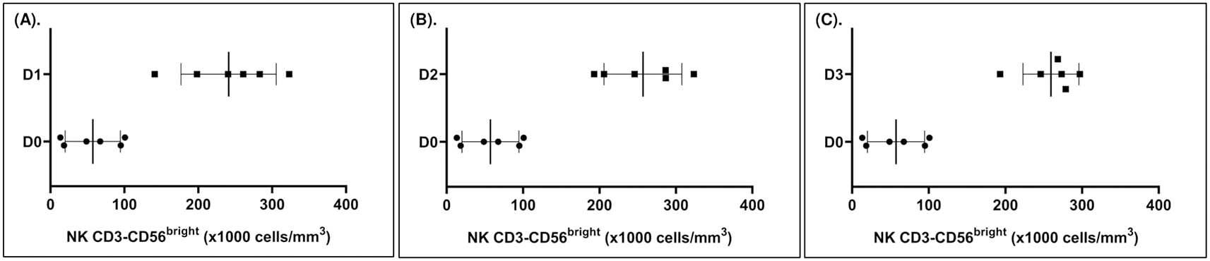
(A) D1 vs. D0 [MD: 183.89; CI: 105.29, 262.48; p<0.0001]. (B) D2 vs. D0 [MD: 199.67; CI: 121.08, 278.26; p<0.0001]. (C) D3 vs. D0 [MD: 202.10; CI: 123.51, 280.70; p<0.0001].
After the administration of IL-2, the levels of CD56dim NK cells in D0, D1, D2, and D3 groups were 812.85 ± 167.37, 631.98 ± 129.90, 616.42 ± 157.97, and 615.90 ± 155.57 ×1000 cells/mm3 respectively. We were unable to determine the impact of IL-2 administrations on the levels of CD56dim NK cells when comparing D1, D2, or D3 to D0 (Table 1).
The levels of IFN-γ in groups D0, D1, D2, and D3 were 24.01 ± 2.56, 26.09 ± 4.79, 30.11 ± 5.34, and 32.43 ± 7.14 pg/ml respectively. In our analysis, we found that the administration of IL-2 had no significant impact on the levels of IFN-γ (Table 1).
Our current study found that the administration of IL-2 had potential impact to the levels of CD56bright NK cells, but not to the levels of CD56dim NK cells and IFN-γ. Compared to the control group, we elucidated that the higher the dose of IL-2 administration the higher the impact to the levels of CD56bright. Our present study was the first report on the role of IL-2 on the regulation of CD56bright in the case of SLE. However, our current findings were consistent to what has been reported by previous studies in other disease settings and healthy individuals. In the case of chronic GVHD, Hirakawa et al. conducted a study to assess the administration of IL-2 and its impact on the expression of CD56bright NK cells. They found that a low dose of IL-2 was sufficiently effective to affect the expression of CD56bright NK cells.10 Additionally, similar findings on the impact of IL-2 administration to the regulation of CD56bright NK cells in the case of chronic GVHD were also reported by Kubo et al.16 On the other hand, in the case of healthy individuals, the induction of CD56bright NK cells regulation after the administration of IL-2 has also been reported by McQuaid et al.17 Similar findings in the case of healthy individuals has also been reported by Saito et al.18 Moreover, in the case of acute myeloid leukemia, the treatment by using the combination of histamine dihydrochloride and IL-2 provided the reconstitution of a deficient NK cell fraction through the specific amplification of CD56bright NK cells.11 Thus, the results of our study might add insight into the role of IL-2 in the regulation of CD56bright in various diseases.
The theoretical basis for our findings regarding the impact of IL-2 on the regulation of CD56bright in SLE cases remains inadequately explained. However, several possible mechanisms might help to elucidate the potential role of IL-2 on CD56bright. IL-2 is a cytokine required for the activation and proliferation of immune cells, including NK cells, and NK cells have the receptors to interact with IL-2 subunits, i.e: IL-2Rα, IL-2Rβ, and IL-2Rγ.19 To establish the interaction, the high affinity of T cells and activated NK cells was needed, and the affinity of T cells is expressed by a small T cell subunit and CD56bright NK cells. IL-2 received by the receptor complex in the NK cells may activate Janus tyrosine-kinase (JAK)-1 and JAK-3 which is required for the signal transduction. Furthermore, the activation of JAK-1 and JAK-3 may initiate the phosphorylation of signal transducer and activator of transcription (STAT)-5, the control center and regulation of specific gene transcription.20 On the other hand, IL-2 signal may also induce phosphorylation from the adapter protein Shc which will activate ras-raf map kinase and PI3K signals,21 and subsequently, this circumstance may cause the transmission of IL-2 signals from the membrane to the core.22,23 In addition, the complex interaction between IL-2 and IL-2Rα/β/γ may induce the phosphorylation of STAT-5 and increase CD25 and Foxp3 expression in Treg, and as a result, this condition may activate its suppression activity.24 In SLE patients, it has been known that dysfunction of NK cells may develop,25 and has been associated with an impaired release of a soluble cytotoxic factor.26 Therefore, the administration of IL-2 may help to alleviate the number and function of Tregs cells, and as expected, this condition may control autoreactive cells by modulating the activation of NK cells.27 IL-2 may increase NK cell reactivity, while Tregs cells can limit the intrinsic production of IL-2 from T cells, and thereafter, they may regulate the cytotoxicity of NK cells. This mechanism was likely to underlie our findings showing that the administration of IL-2 was associated with upregulation of CD56bright.
To the best of our knowledge, our current investigation is the first study reporting the role of IL-2 on CD56bright NK cells, CD56dim NK cells, and IFN-γ in the case of SLE; and we revealed that only CD56bright NK cells were affected by the administration of IL-2. Our current finding might add new insight on the impact of IL-2 administration to CD56bright NK cells. Moreover, the findings of our present study might serve as the initial reference for further studies in the context of the implication of IL-2 administration to the expression of NK cells in the case of SLE. Based on several previous studies suggesting that a therapeutic option by targeting NK cells might provide benefits for SLE patients,28,29 we expected that our present findings could serve as a foundation for future studies, potentially leading to the establishment of SLE treatment strategies that target NK cells.
Our study had several important limitations. First, in our present study, some potential factors involved in the pathogenesis of SLE that might affect the final findings were not analyzed, for example: human leukocyte antigen, STAT-5, JAK-1, and JAK-3.20 Second, the design of our study was a post-test only control group design. A study by applying a pre-test post-test control group design might be required to obtain better evidence. Third, we only assessed IL-2 as the potential cytokine in our study. Further studies involving other cytokines might be required to obtain more comprehensive findings.
Our study reveals that the administration of IL-2 provides the beneficial impact in the expression of CD56bright NK cells in the case of SLE. Our current study may establish the additional insight on the role of IL-2 in the treatment of SLE by targeting NK cells.
Conceptualization: DS; Data Curation: DS; Formal Analysis: DS, JKF; Investigation: DS; Project Administration: DS; Resources: DS; Methodology: DS, JKF; Software: JKF; Visualization: JKF; Supervision: HS, HK, KH; Validation: HS, HK, KH; Writing – Original Draft Preparation: DS, JKF; Writing – Review & Editing: JKF. All authors have critically reviewed and approved the final draft and are responsible for the content and similarity index of the manuscript.
| Views | Downloads | |
|---|---|---|
| F1000Research | - | - |
|
PubMed Central
Data from PMC are received and updated monthly.
|
- | - |
Provide sufficient details of any financial or non-financial competing interests to enable users to assess whether your comments might lead a reasonable person to question your impartiality. Consider the following examples, but note that this is not an exhaustive list:
Sign up for content alerts and receive a weekly or monthly email with all newly published articles
Already registered? Sign in
The email address should be the one you originally registered with F1000.
You registered with F1000 via Google, so we cannot reset your password.
To sign in, please click here.
If you still need help with your Google account password, please click here.
You registered with F1000 via Facebook, so we cannot reset your password.
To sign in, please click here.
If you still need help with your Facebook account password, please click here.
If your email address is registered with us, we will email you instructions to reset your password.
If you think you should have received this email but it has not arrived, please check your spam filters and/or contact for further assistance.
Comments on this article Comments (0)